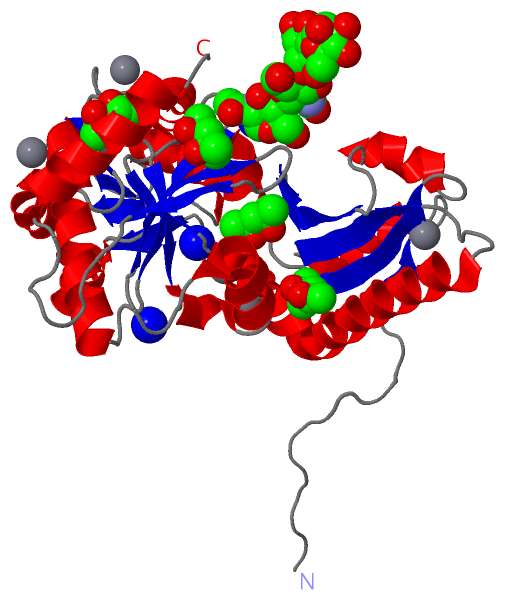
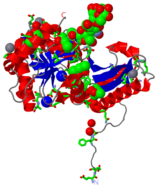
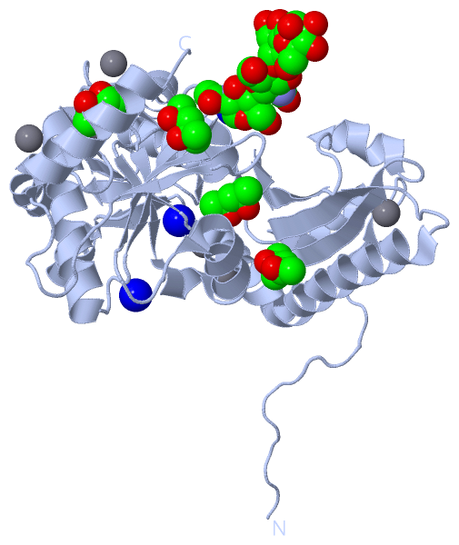
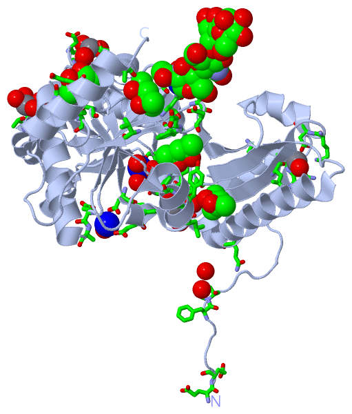
Show PDB file:   
         Plain Text   HTML   (compressed file size)
QuickSearch:   
by PDB,NDB,UniProt,PROSITE Code or Search Term(s)  
(-)Asymmetric Unit
(-)Asym. Unit - sites
(-)Biological Unit 1
(-)Biol. Unit 1 - sites
(-)Biological Unit 2
collapse expand < >
Image Asymmetric Unit
Asymmetric Unit  (Jmol Viewer)
Image Asym. Unit - sites
Asym. Unit - sites  (Jmol Viewer)
Image Biological Unit 1
Biological Unit 1  (Jmol Viewer)
Image Biol. Unit 1 - sites
Biol. Unit 1 - sites  (Jmol Viewer)
Image Biological Unit 2
Biological Unit 2  (Jmol Viewer)

(-) Description

Title :  CRYSTAL STRUCTURE OF NATIVE RABBIT SKELETAL CALSEQUESTRIN
 
Authors :  E. J. Sanchez, K. M. Lewis, G. R. Munske, M. S. Nissen, C. Kang
Date :  09 Sep 11  (Deposition) - 21 Dec 11  (Release) - 15 Feb 12  (Revision)
Method :  X-RAY DIFFRACTION
Resolution :  1.76
Chains :  Asym. Unit :  A
Biol. Unit 1:  A  (1x)
Biol. Unit 2:  A  (2x)
Keywords :  Calcium-Binding Protein (Keyword Search: [Gene Ontology, PubMed, Web (Google))
 
Reference :  E. J. Sanchez, K. M. Lewis, G. R. Munske, M. S. Nissen, C. Kang
Glycosylation Of Skeletal Calsequestrin: Implications For Its Function.
J. Biol. Chem. V. 287 3042 2012
PubMed-ID: 22170046  |  Reference-DOI: 10.1074/JBC.M111.326363

(-) Compounds

Molecule 1 - CALSEQUESTRIN-1
    ChainsA
    Organism CommonEUROPEAN RABBIT,JAPANESE WHITE RABBIT,DOMESTIC RABBIT,RABBITS
    Organism ScientificORYCTOLAGUS CUNICULUS
    Organism Taxid9986
    SynonymASPARTACTIN, CALSEQUESTRIN, SKELETAL MUSCLE ISOFORM, LAMININ-BINDING PROTEIN
    TissueSKELETAL MUSCLE

 Structural Features

(-) Chains, Units

  1
Asymmetric Unit A
Biological Unit 1 (1x)A
Biological Unit 2 (2x)A

Summary Information (see also Sequences/Alignments below)

(-) Ligands, Modified Residues, Ions  (7, 17)

Asymmetric Unit (7, 17)
No.NameCountTypeFull Name
1BMA1Ligand/IonBETA-D-MANNOSE
2CA6Ligand/IonCALCIUM ION
3MAN1Ligand/IonALPHA-D-MANNOSE
4MPD2Ligand/Ion(4S)-2-METHYL-2,4-PENTANEDIOL
5MRD2Ligand/Ion(4R)-2-METHYLPENTANE-2,4-DIOL
6NA3Ligand/IonSODIUM ION
7NAG2Ligand/IonN-ACETYL-D-GLUCOSAMINE
Biological Unit 1 (5, 8)
No.NameCountTypeFull Name
1BMA1Ligand/IonBETA-D-MANNOSE
2CA-1Ligand/IonCALCIUM ION
3MAN1Ligand/IonALPHA-D-MANNOSE
4MPD2Ligand/Ion(4S)-2-METHYL-2,4-PENTANEDIOL
5MRD2Ligand/Ion(4R)-2-METHYLPENTANE-2,4-DIOL
6NA-1Ligand/IonSODIUM ION
7NAG2Ligand/IonN-ACETYL-D-GLUCOSAMINE
Biological Unit 2 (5, 16)
No.NameCountTypeFull Name
1BMA2Ligand/IonBETA-D-MANNOSE
2CA-1Ligand/IonCALCIUM ION
3MAN2Ligand/IonALPHA-D-MANNOSE
4MPD4Ligand/Ion(4S)-2-METHYL-2,4-PENTANEDIOL
5MRD4Ligand/Ion(4R)-2-METHYLPENTANE-2,4-DIOL
6NA-1Ligand/IonSODIUM ION
7NAG4Ligand/IonN-ACETYL-D-GLUCOSAMINE

(-) Sites  (17, 17)

Asymmetric Unit (17, 17)
No.NameEvidenceResiduesDescription
01AC1SOFTWAREASN A:316 , ASP A:319 , HOH A:559 , NAG A:901BINDING SITE FOR RESIDUE NAG A 900
02AC2SOFTWAREGLU A:2 , NAG A:900 , BMA A:902BINDING SITE FOR RESIDUE NAG A 901
03AC3SOFTWARENAG A:901 , MAN A:903BINDING SITE FOR RESIDUE BMA A 902
04AC4SOFTWARELYS A:28 , BMA A:902BINDING SITE FOR RESIDUE MAN A 903
05AC5SOFTWAREASN A:276 , ASN A:279 , GLU A:344 , HOH A:462BINDING SITE FOR RESIDUE MRD A 354
06AC6SOFTWAREPHE A:6 , PRO A:7 , TRP A:242 , TYR A:298 , TRP A:299BINDING SITE FOR RESIDUE MPD A 355
07AC7SOFTWAREGLN A:63 , LEU A:294 , LEU A:295 , HOH A:619 , HOH A:804 , HOH A:834BINDING SITE FOR RESIDUE MRD A 356
08AC8SOFTWAREASN A:316 , ASP A:321 , VAL A:323 , GLU A:350BINDING SITE FOR RESIDUE MPD A 357
09AC9SOFTWAREASP A:210 , PRO A:212 , GLU A:217 , HOH A:553 , HOH A:706BINDING SITE FOR RESIDUE CA A 358
10BC1SOFTWAREASN A:17 , VAL A:18 , LEU A:74 , LYS A:79 , ASP A:80 , HOH A:606BINDING SITE FOR RESIDUE CA A 359
11BC2SOFTWAREGLU A:199 , THR A:229 , THR A:277 , HOH A:552BINDING SITE FOR RESIDUE CA A 360
12BC3SOFTWARETHR A:189 , HOH A:554 , HOH A:573 , HOH A:633 , HOH A:711BINDING SITE FOR RESIDUE CA A 361
13BC4SOFTWAREGLU A:109 , ASP A:291 , HOH A:369BINDING SITE FOR RESIDUE CA A 362
14BC5SOFTWAREPHE A:118 , ASP A:121 , GLU A:236 , HOH A:920BINDING SITE FOR RESIDUE CA A 363
15BC6SOFTWAREGLU A:257 , ALA A:258 , ASP A:288 , ASP A:290 , HOH A:498BINDING SITE FOR RESIDUE NA A 364
16BC7SOFTWAREPRO A:172 , HOH A:492 , HOH A:555 , HOH A:556 , HOH A:557 , HOH A:558BINDING SITE FOR RESIDUE NA A 365
17BC8SOFTWAREGLU A:1 , GLY A:248 , ASN A:316 , THR A:318 , HOH A:559BINDING SITE FOR RESIDUE NA A 366

(-) SS Bonds  (0, 0)

(no "SS Bond" information available for 3TRQ)

(-) Cis Peptide Bonds  (2, 2)

Asymmetric Unit
No.Residues
1His A:171 -Pro A:172
2Lys A:211 -Pro A:212

 Sequence-Structure Mapping

(-) SAPs(SNPs)/Variants  (0, 0)

(no "SAP(SNP)/Variant" information available for 3TRQ)

(-) PROSITE Motifs  (2, 2)

Asymmetric Unit (2, 2)
 PROSITEUniProtKBPDB
No.IDACDescriptionIDLocationCountLocation
1CALSEQUESTRIN_1PS00863 Calsequestrin signature 1.CASQ1_RABIT29-43  1A:1-15
2CALSEQUESTRIN_2PS00864 Calsequestrin signature 2.CASQ1_RABIT366-385  1A:338-353
Biological Unit 1 (2, 2)
 PROSITEUniProtKBPDB
No.IDACDescriptionIDLocationCountLocation
1CALSEQUESTRIN_1PS00863 Calsequestrin signature 1.CASQ1_RABIT29-43  1A:1-15
2CALSEQUESTRIN_2PS00864 Calsequestrin signature 2.CASQ1_RABIT366-385  1A:338-353
Biological Unit 2 (2, 4)
 PROSITEUniProtKBPDB
No.IDACDescriptionIDLocationCountLocation
1CALSEQUESTRIN_1PS00863 Calsequestrin signature 1.CASQ1_RABIT29-43  2A:1-15
2CALSEQUESTRIN_2PS00864 Calsequestrin signature 2.CASQ1_RABIT366-385  2A:338-353

(-) Exons   (0, 0)

(no "Exon" information available for 3TRQ)

(-) Sequences/Alignments

Asymmetric Unit
   Reformat: Number of residues per line =  ('0' or empty: single-line sequence representation)
  Number of residues per labelling interval =   
  UniProt sequence: complete  aligned part    
   Show mapping: SCOP domains CATH domains Pfam domains Secondary structure (by author)
SAPs(SNPs) PROSITE motifs Exons
(details for a mapped element are shown in a popup box when the mouse pointer rests over it)
Chain A from PDB  Type:PROTEIN  Length:353
 aligned with CASQ1_RABIT | P07221 from UniProtKB/Swiss-Prot  Length:395

    Alignment length:353
                                    38        48        58        68        78        88        98       108       118       128       138       148       158       168       178       188       198       208       218       228       238       248       258       268       278       288       298       308       318       328       338       348       358       368       378   
          CASQ1_RABIT    29 EEGLDFPEYDGVDRVINVNAKNYKNVFKKYEVLALLYHEPPEDDKASQRQFEMEELILELAAQVLEDKGVGFGLVDSEKDAAVAKKLGLTEEDSIYVFKEDEVIEYDGEFSADTLVEFLLDVLEDPVELIEGERELQAFENIEDEIKLIGYFKNKDSEHYKAFKEAAEEFHPYIPFFATFDSKVAKKLTLKLNEIDFYEAFMEEPVTIPDKPNSEEEIVNFVEEHRRSTLRKLKPESMYETWEDDMDGIHIVAFAEEADPDGYEFLEILKSVAQDNTDNPDLSIIWIDPDDFPLLVPYWEKTFDIDLSAPQIGVVNVTDADSVWMEMDDEEDLPSAEELEDWLEDVLEGEINT 381
               SCOP domains ----------------------------------------------------------------------------------------------------------------------------------------------------------------------------------------------------------------------------------------------------------------------------------------------------------------------------------------------------------------- SCOP domains
               CATH domains ----------------------------------------------------------------------------------------------------------------------------------------------------------------------------------------------------------------------------------------------------------------------------------------------------------------------------------------------------------------- CATH domains
               Pfam domains ----------------------------------------------------------------------------------------------------------------------------------------------------------------------------------------------------------------------------------------------------------------------------------------------------------------------------------------------------------------- Pfam domains
         Sec.struct. author ...............ee....hhhhhhhhh.eeeeeee.....hhhhhhhhhhhhhhhhhhhhhhhhh.eeeeeee...hhhhhhhhh.....eeeeee..eeee.....hhhhhhhhhhhhhh..eee..hhhhhhhhhhh....eeeee.....hhhhhhhhhhhhhhh....eeee.hhhhhhhhh.....eeee.......ee......hhhhhhhhhhhh....eee.hhhhhhhhhhh....eeeeee....hhhhhhhhhhhhhhhhhh.......eeeehhhhh..hhhhhhhhhh......eeeeee.....eee..........hhhhhhhhhhhhhhhh... Sec.struct. author
                 SAPs(SNPs) ----------------------------------------------------------------------------------------------------------------------------------------------------------------------------------------------------------------------------------------------------------------------------------------------------------------------------------------------------------------- SAPs(SNPs)
                    PROSITE CALSEQUESTRIN_1----------------------------------------------------------------------------------------------------------------------------------------------------------------------------------------------------------------------------------------------------------------------------------------------------------------------------------CALSEQUESTRIN_2  PROSITE
                 Transcript ----------------------------------------------------------------------------------------------------------------------------------------------------------------------------------------------------------------------------------------------------------------------------------------------------------------------------------------------------------------- Transcript
                 3trq A   1 EEGLDFPEYDGVDRVINVNAKNYKNVFKKYEVLALLYHEPPEDDKASQRQFEMEELILELAAQVLEDKGVGFGLVDSEKDAAVAKKLGLTEEDSIYVFKEDEVIEYDGEFSADTLVEFLLDVLEDPVELIEGERELQAFENIEDEIKLIGYFKNKDSEHYKAFKEAAEEFHPYIPFFATFDSKVAKKLTLKLNEIDFYEAFMEEPVTIPDKPNSEEEIVNFVEEHRRSTLRKLKPESMYETWEDDMDGIHIVAFAEEADPDGYEFLEILKSVAQDNTDNPDLSIIWIDPDDFPLLVPYWEKTFDIDLSAPQIGVVNVTDADSVWMEMDDEEDLPSAEELEDWLEDVLEGEINT 353
                                    10        20        30        40        50        60        70        80        90       100       110       120       130       140       150       160       170       180       190       200       210       220       230       240       250       260       270       280       290       300       310       320       330       340       350   

   Legend:   → Mismatch (orange background)
  - → Gap (green background, '-', border residues have a numbering label)
    → Modified Residue (blue background, lower-case, 'x' indicates undefined single-letter code, labelled with number + name)
  x → Chemical Group (purple background, 'x', labelled with number + name, e.g. ACE or NH2)
  extra numbering lines below/above indicate numbering irregularities and modified residue names etc., number ends below/above '|'

 Classification and Annotation

(-) SCOP Domains  (0, 0)

(no "SCOP Domain" information available for 3TRQ)

(-) CATH Domains  (0, 0)

(no "CATH Domain" information available for 3TRQ)

(-) Pfam Domains  (0, 0)

(no "Pfam Domain" information available for 3TRQ)

(-) Gene Ontology  (11, 11)

Asymmetric Unit(hide GO term definitions)
Chain A   (CASQ1_RABIT | P07221)
molecular function
    GO:0005509    calcium ion binding    Interacting selectively and non-covalently with calcium ions (Ca2+).
    GO:0046872    metal ion binding    Interacting selectively and non-covalently with any metal ion.
biological process
    GO:0051258    protein polymerization    The process of creating protein polymers, compounds composed of a large number of component monomers; polymeric proteins may be made up of different or identical monomers. Polymerization occurs by the addition of extra monomers to an existing poly- or oligomeric protein.
    GO:0014809    regulation of skeletal muscle contraction by regulation of release of sequestered calcium ion    Any process that modulates the frequency, rate or extent of skeletal muscle contraction via the regulation of the release of sequestered calcium ion by sarcoplasmic reticulum into cytosol. The sarcoplasmic reticulum is the endoplasmic reticulum of striated muscle, specialised for the sequestration of calcium ions that are released upon receipt of a signal relayed by the T tubules from the neuromuscular junction.
    GO:0045214    sarcomere organization    The myofibril assembly process that results in the organization of muscle actomyosin into sarcomeres. The sarcomere is the repeating unit of a myofibril in a muscle cell, composed of an array of overlapping thick and thin filaments between two adjacent Z discs.
cellular component
    GO:0016020    membrane    A lipid bilayer along with all the proteins and protein complexes embedded in it an attached to it.
    GO:0005759    mitochondrial matrix    The gel-like material, with considerable fine structure, that lies in the matrix space, or lumen, of a mitochondrion. It contains the enzymes of the tricarboxylic acid cycle and, in some organisms, the enzymes concerned with fatty acid oxidation.
    GO:0005739    mitochondrion    A semiautonomous, self replicating organelle that occurs in varying numbers, shapes, and sizes in the cytoplasm of virtually all eukaryotic cells. It is notably the site of tissue respiration.
    GO:0016529    sarcoplasmic reticulum    A fine reticular network of membrane-limited elements that pervades the sarcoplasm of a muscle cell; continuous over large portions of the cell and with the nuclear envelope; that part of the endoplasmic reticulum specialized for calcium release, uptake and storage.
    GO:0033018    sarcoplasmic reticulum lumen    The volume enclosed by the membranes of the sarcoplasmic reticulum.
    GO:0033017    sarcoplasmic reticulum membrane    The lipid bilayer surrounding the sarcoplasmic reticulum.

 Visualization

(-) Interactive Views

Asymmetric Unit
  Complete Structure
    Jena3D(integrated viewing of ligand, site, SAP, PROSITE, SCOP information)
    WebMol | AstexViewer[tm]@PDBe
(Java Applets, require no local installation except for Java; loading may be slow)
    STRAP
(Java WebStart application, automatic local installation, requires Java; full application with system access!)
    RasMol
(require local installation)
    Molscript (VRML)
(requires installation of a VRML viewer; select preferred view via VRML and generate a mono or stereo PDF format file)
 
  Ligands, Modified Residues, Ions
    BMA  [ RasMol | Jena3D ]  +environment [ RasMol | Jena3D ]
    CA  [ RasMol | Jena3D ]  +environment [ RasMol | Jena3D ]
    MAN  [ RasMol | Jena3D ]  +environment [ RasMol | Jena3D ]
    MPD  [ RasMol | Jena3D ]  +environment [ RasMol | Jena3D ]
    MRD  [ RasMol | Jena3D ]  +environment [ RasMol | Jena3D ]
    NA  [ RasMol | Jena3D ]  +environment [ RasMol | Jena3D ]
    NAG  [ RasMol | Jena3D ]  +environment [ RasMol | Jena3D ]
 
  Sites
    AC1  [ RasMol ]  +environment [ RasMol ]
    AC2  [ RasMol ]  +environment [ RasMol ]
    AC3  [ RasMol ]  +environment [ RasMol ]
    AC4  [ RasMol ]  +environment [ RasMol ]
    AC5  [ RasMol ]  +environment [ RasMol ]
    AC6  [ RasMol ]  +environment [ RasMol ]
    AC7  [ RasMol ]  +environment [ RasMol ]
    AC8  [ RasMol ]  +environment [ RasMol ]
    AC9  [ RasMol ]  +environment [ RasMol ]
    BC1  [ RasMol ]  +environment [ RasMol ]
    BC2  [ RasMol ]  +environment [ RasMol ]
    BC3  [ RasMol ]  +environment [ RasMol ]
    BC4  [ RasMol ]  +environment [ RasMol ]
    BC5  [ RasMol ]  +environment [ RasMol ]
    BC6  [ RasMol ]  +environment [ RasMol ]
    BC7  [ RasMol ]  +environment [ RasMol ]
    BC8  [ RasMol ]  +environment [ RasMol ]
 
  Cis Peptide Bonds
    His A:171 - Pro A:172   [ RasMol ]  
    Lys A:211 - Pro A:212   [ RasMol ]  
 
Biological Units
  Complete Structure
    Biological Unit 1  [ Jena3D ]
    Biological Unit 2  [ Jena3D ]

(-) Still Images

Jmol
  protein: cartoon or spacefill or dots and stick; nucleic acid: cartoon and stick; ligands: spacefill; active site: stick
Molscript
  protein, nucleic acid: cartoon; ligands: spacefill; active site: ball and stick

 Databases and Analysis Tools

(-) Databases

Access by PDB/NDB ID
  3trq
    Family and Domain InformationProDom | SYSTERS
    General Structural InformationGlycoscienceDB | MMDB | NDB | OCA | PDB | PDBe | PDBj | PDBsum | PDBWiki | PQS | PROTEOPEDIA
    Orientation in MembranesOPM
    Protein SurfaceSURFACE
    Secondary StructureDSSP (structure derived) | HSSP (homology derived)
    Structural GenomicsGeneCensus
    Structural NeighboursCE | VAST
    Structure ClassificationCATH | Dali | SCOP
    Validation and Original DataBMRB Data View | BMRB Restraints Grid | EDS | PROCHECK | RECOORD | WHAT_CHECK
 
Access by UniProt ID/Accession number
  CASQ1_RABIT | P07221
    Comparative Protein Structure ModelsModBase
    Genomic InformationEnsembl
    Protein-protein InteractionDIP
    Sequence, Family and Domain InformationInterPro | Pfam | SMART | UniProtKB/SwissProt
 
Access by Enzyme Classificator   (EC Number)
  (no 'Enzyme Classificator' available)
    General Enzyme InformationBRENDA | EC-PDB | Enzyme | IntEnz
    PathwayKEGG | MetaCyc
 
Access by Disease Identifier   (MIM ID)
  (no 'MIM ID' available)
    Disease InformationOMIM
 
Access by GenAge ID
  (no 'GenAge ID' available)
    Age Related InformationGenAge

(-) Analysis Tools

Access by PDB/NDB ID
    Domain InformationXDom
    Interatomic Contacts of Structural UnitsCSU
    Ligand-protein ContactsLPC
    Protein CavitiescastP
    Sequence and Secondary StructurePDBCartoon
    Structure AlignmentSTRAP(Java WebStart application, automatic local installation, requires Java; full application with system access!)
    Structure and Sequence BrowserSTING
 
Access by UniProt ID/Accession number
  CASQ1_RABIT | P07221
    Protein Disorder PredictionDisEMBL | FoldIndex | GLOBPLOT (for more information see DisProt)

 Related Entries

(-) Entries Sharing at Least One Protein Chain (UniProt ID)

UniProtKB/Swiss-Prot
        CASQ1_RABIT | P072211a8y 3trp 3us3 3v1w

(-) Related Entries Specified in the PDB File

3trp RECOMBINANT FORM OF PROTEIN