Show PDB file:   
         Plain Text   HTML   (compressed file size)
QuickSearch:   
by PDB,NDB,UniProt,PROSITE Code or Search Term(s)  
(-)Asymmetric Unit
(-)Asym. Unit - sites
(-)Biological Unit 1
(-)Biol. Unit 1 - sites
(-)Biological Unit 2
collapse expand < >
Image Asymmetric Unit
Asymmetric Unit  (Jmol Viewer)
Image Asym. Unit - sites
Asym. Unit - sites  (Jmol Viewer)
Image Biological Unit 1
Biological Unit 1  (Jmol Viewer)
Image Biol. Unit 1 - sites
Biol. Unit 1 - sites  (Jmol Viewer)
Image Biological Unit 2
Biological Unit 2  (Jmol Viewer)

(-) Description

Title :  CRYSTAL STRUCTURE OF H107R MUTANT OF EXTRACELLULAR DOMAIN OF MOUSE RECEPTOR NKR-P1A
 
Authors :  P. Kolenko, D. Rozbesky, K. Bezouska, J. Hasek, J. Dohnalek
Date :  25 Jul 11  (Deposition) - 10 Aug 11  (Release) - 28 Mar 12  (Revision)
Method :  X-RAY DIFFRACTION
Resolution :  2.30
Chains :  Asym. Unit :  A,B
Biol. Unit 1:  A,B  (1x)
Biol. Unit 2:  A,B  (2x)
Keywords :  C-Type Lectin-Like Domain, Domain Swapping, Twinning, Natural Killer Cell Receptor, Transmembrane Receptor, Signaling Protein (Keyword Search: [Gene Ontology, PubMed, Web (Google))
 
Reference :  P. Kolenko, D. Rozbesky, O. Vanek, K. Bezouska, J. Hasek, J. Dohnalek
Structure Of The H107R Variant Of The Extracellular Domain Of Mouse Nkr-P1A At 2. 3 A Resolution.
Acta Crystallogr. , Sect. F V. 67 1519 2011
PubMed-ID: 22139156  |  Reference-DOI: 10.1107/S1744309111046203

(-) Compounds

Molecule 1 - KILLER CELL LECTIN-LIKE RECEPTOR SUBFAMILY B MEMBER 1A
    ChainsA, B
    EngineeredYES
    Expression SystemESCHERICHIA COLI
    Expression System PlasmidPET-30A
    Expression System StrainBL21-(DE3) RIL
    Expression System Taxid562
    Expression System Vector TypePLASMID
    FragmentEXTRACELLULAR DOMAIN (UNP RESIDUES 89-227)
    GeneKLRB1A, LY55, LY55A, NKRP1A
    MutationYES
    Organism CommonMOUSE
    Organism ScientificMUS MUSCULUS
    Organism Taxid10090
    StrainC57BL/6
    SynonymNKR-P1A, CD161 ANTIGEN-LIKE FAMILY MEMBER A, LYMPHOCYTE ANTIGEN 55A, LY-55A, NKR-P1.7, NATURAL KILLER CELL SURFACE PROTEIN P1-2, NKR-P1 2

 Structural Features

(-) Chains, Units

  12
Asymmetric Unit AB
Biological Unit 1 (1x)AB
Biological Unit 2 (2x)AB

Summary Information (see also Sequences/Alignments below)

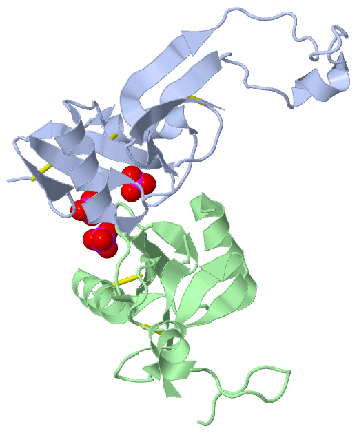
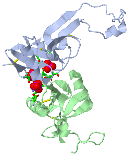
(-) Ligands, Modified Residues, Ions  (1, 3)

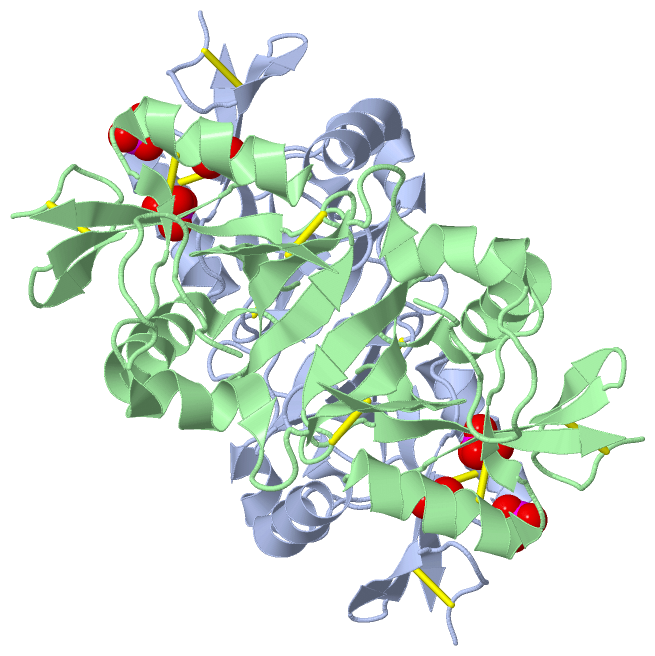
Asymmetric Unit (1, 3)
No.NameCountTypeFull Name
1PO43Ligand/IonPHOSPHATE ION
Biological Unit 1 (1, 3)
No.NameCountTypeFull Name
1PO43Ligand/IonPHOSPHATE ION
Biological Unit 2 (1, 6)
No.NameCountTypeFull Name
1PO46Ligand/IonPHOSPHATE ION

(-) Sites  (3, 3)

Asymmetric Unit (3, 3)
No.NameEvidenceResiduesDescription
1AC1SOFTWAREGLN A:110 , ASP A:121 , LYS A:125 , ASP B:97BINDING SITE FOR RESIDUE PO4 A 301
2AC2SOFTWAREASP A:97 , ARG A:107 , HOH A:467 , GLN B:96 , ARG B:107BINDING SITE FOR RESIDUE PO4 A 303
3AC3SOFTWAREASP A:97 , GLN B:110 , ASP B:121 , LYS B:125BINDING SITE FOR RESIDUE PO4 B 302

(-) SS Bonds  (6, 6)

Asymmetric Unit
No.Residues
1A:94 -A:105
2A:122 -A:210
3A:189 -A:202
4B:94 -B:105
5B:122 -B:210
6B:189 -B:202

(-) Cis Peptide Bonds  (0, 0)

(no "Cis Peptide Bond" information available for 3T3A)

 Sequence-Structure Mapping

(-) SAPs(SNPs)/Variants  (0, 0)

(no "SAP(SNP)/Variant" information available for 3T3A)

(-) PROSITE Motifs  (1, 2)

Asymmetric Unit (1, 2)
 PROSITEUniProtKBPDB
No.IDACDescriptionIDLocationCountLocation
1C_TYPE_LECTIN_2PS50041 C-type lectin domain profile.KLRBA_MOUSE101-211
 
  2A:101-211
B:101-211
Biological Unit 1 (1, 2)
 PROSITEUniProtKBPDB
No.IDACDescriptionIDLocationCountLocation
1C_TYPE_LECTIN_2PS50041 C-type lectin domain profile.KLRBA_MOUSE101-211
 
  2A:101-211
B:101-211
Biological Unit 2 (1, 4)
 PROSITEUniProtKBPDB
No.IDACDescriptionIDLocationCountLocation
1C_TYPE_LECTIN_2PS50041 C-type lectin domain profile.KLRBA_MOUSE101-211
 
  4A:101-211
B:101-211

(-) Exons   (0, 0)

(no "Exon" information available for 3T3A)

(-) Sequences/Alignments

Asymmetric Unit
   Reformat: Number of residues per line =  ('0' or empty: single-line sequence representation)
  Number of residues per labelling interval =   
  UniProt sequence: complete  aligned part    
   Show mapping: SCOP domains CATH domains Pfam domains Secondary structure (by author)
SAPs(SNPs) PROSITE motifs Exons
(details for a mapped element are shown in a popup box when the mouse pointer rests over it)
Chain A from PDB  Type:PROTEIN  Length:122
 aligned with KLRBA_MOUSE | P27811 from UniProtKB/Swiss-Prot  Length:227

    Alignment length:122
                                   102       112       122       132       142       152       162       172       182       192       202       212  
          KLRBA_MOUSE    93 ECPQDWLSHRDKCFHVSHVSNTWEEGLVDCDGKGATLMLIQDQEELRFLLDSIKEKYNSFWIGLRYTLPDMNWKWINGSTLNSDVLKITDDTENDSCAAISGDKVTFESCNSDNRWICQKEL 214
               SCOP domains d3t3aa_ A: automated matches                                                                                               SCOP domains
               CATH domains -------------------------------------------------------------------------------------------------------------------------- CATH domains
               Pfam domains -------------------------------------------------------------------------------------------------------------------------- Pfam domains
         Sec.struct. author ......eee..eeeee.....hhhhhhhhhhhh..ee....hhhhhhhhhhhhh....eeeeeeeee..............hhhhhh.......eeeeeee..eeeeee.....eeeeeee. Sec.struct. author
                 SAPs(SNPs) -------------------------------------------------------------------------------------------------------------------------- SAPs(SNPs)
                    PROSITE --------C_TYPE_LECTIN_2  PDB: A:101-211 UniProt: 101-211                                                               --- PROSITE
                 Transcript -------------------------------------------------------------------------------------------------------------------------- Transcript
                 3t3a A  93 ECPQDWLSHRDKCFRVSQVSNTWEEGLVDCDGKGATLMLIQDQEELRFLLDSIKEKYNSFWIGLRYTLPDMNWKWINGSTLNSDVLKITGDTENDSCAAISGDKVTFESCNSDNRWICQKEL 214
                                   102       112       122       132       142       152       162       172       182       192       202       212  

Chain B from PDB  Type:PROTEIN  Length:119
 aligned with KLRBA_MOUSE | P27811 from UniProtKB/Swiss-Prot  Length:227

    Alignment length:122
                                   102       112       122       132       142       152       162       172       182       192       202       212  
          KLRBA_MOUSE    93 ECPQDWLSHRDKCFHVSHVSNTWEEGLVDCDGKGATLMLIQDQEELRFLLDSIKEKYNSFWIGLRYTLPDMNWKWINGSTLNSDVLKITDDTENDSCAAISGDKVTFESCNSDNRWICQKEL 214
               SCOP domains d3t3ab_ B: automated matches                                                                                               SCOP domains
               CATH domains -------------------------------------------------------------------------------------------------------------------------- CATH domains
               Pfam domains -------------------------------------------------------------------------------------------------------------------------- Pfam domains
         Sec.struct. author ......eee..eeeee.....hhhhhhhhhhhh..ee....hhhhhhhhhhhhh..-..eeeeeeee.--...........hhhhhhhhh....eeeeeee..eeeee......eeeeeee. Sec.struct. author
                 SAPs(SNPs) -------------------------------------------------------------------------------------------------------------------------- SAPs(SNPs)
                    PROSITE --------C_TYPE_LECTIN_2  PDB: B:101-211 UniProt: 101-211                                                               --- PROSITE
                 Transcript -------------------------------------------------------------------------------------------------------------------------- Transcript
                 3t3a B  93 ECPQDWLSHRDKCFRVSQVSNTWEEGLVDCDGKGATLMLIQDQEELRFLLDSIKEK-NSFWIGLRYTL--MNWKWINGSTLNSDVLKITGDTENDSCAAISGDKVTFESCNSDNRWICQKEL 214
                                   102       112       122       132       142     | 152       | -|      172       182       192       202       212  
                                                                                 148 |       160  |                                                   
                                                                                   150          163                                                   

   Legend:   → Mismatch (orange background)
  - → Gap (green background, '-', border residues have a numbering label)
    → Modified Residue (blue background, lower-case, 'x' indicates undefined single-letter code, labelled with number + name)
  x → Chemical Group (purple background, 'x', labelled with number + name, e.g. ACE or NH2)
  extra numbering lines below/above indicate numbering irregularities and modified residue names etc., number ends below/above '|'

 Classification and Annotation

(-) SCOP Domains  (1, 2)

Asymmetric Unit

(-) CATH Domains  (0, 0)

(no "CATH Domain" information available for 3T3A)

(-) Pfam Domains  (0, 0)

(no "Pfam Domain" information available for 3T3A)

(-) Gene Ontology  (5, 5)

Asymmetric Unit(hide GO term definitions)
Chain A,B   (KLRBA_MOUSE | P27811)
molecular function
    GO:0030246    carbohydrate binding    Interacting selectively and non-covalently with any carbohydrate, which includes monosaccharides, oligosaccharides and polysaccharides as well as substances derived from monosaccharides by reduction of the carbonyl group (alditols), by oxidation of one or more hydroxy groups to afford the corresponding aldehydes, ketones, or carboxylic acids, or by replacement of one or more hydroxy group(s) by a hydrogen atom. Cyclitols are generally not regarded as carbohydrates.
biological process
    GO:0008150    biological_process    Any process specifically pertinent to the functioning of integrated living units: cells, tissues, organs, and organisms. A process is a collection of molecular events with a defined beginning and end.
cellular component
    GO:0016021    integral component of membrane    The component of a membrane consisting of the gene products and protein complexes having at least some part of their peptide sequence embedded in the hydrophobic region of the membrane.
    GO:0016020    membrane    A lipid bilayer along with all the proteins and protein complexes embedded in it an attached to it.
    GO:0005886    plasma membrane    The membrane surrounding a cell that separates the cell from its external environment. It consists of a phospholipid bilayer and associated proteins.

 Visualization

(-) Interactive Views

Asymmetric Unit
  Complete Structure
    Jena3D(integrated viewing of ligand, site, SAP, PROSITE, SCOP information)
    WebMol | AstexViewer[tm]@PDBe
(Java Applets, require no local installation except for Java; loading may be slow)
    STRAP
(Java WebStart application, automatic local installation, requires Java; full application with system access!)
    RasMol
(require local installation)
    Molscript (VRML)
(requires installation of a VRML viewer; select preferred view via VRML and generate a mono or stereo PDF format file)
 
  Ligands, Modified Residues, Ions
    PO4  [ RasMol | Jena3D ]  +environment [ RasMol | Jena3D ]
 
  Sites
    AC1  [ RasMol ]  +environment [ RasMol ]
    AC2  [ RasMol ]  +environment [ RasMol ]
    AC3  [ RasMol ]  +environment [ RasMol ]
 
  Cis Peptide Bonds
(no "Cis Peptide Bonds" information available for 3t3a)
 
Biological Units
  Complete Structure
    Biological Unit 1  [ Jena3D ]
    Biological Unit 2  [ Jena3D ]

(-) Still Images

Jmol
  protein: cartoon or spacefill or dots and stick; nucleic acid: cartoon and stick; ligands: spacefill; active site: stick
Molscript
  protein, nucleic acid: cartoon; ligands: spacefill; active site: ball and stick

 Databases and Analysis Tools

(-) Databases

Access by PDB/NDB ID
  3t3a
    Family and Domain InformationProDom | SYSTERS
    General Structural InformationGlycoscienceDB | MMDB | NDB | OCA | PDB | PDBe | PDBj | PDBsum | PDBWiki | PQS | PROTEOPEDIA
    Orientation in MembranesOPM
    Protein SurfaceSURFACE
    Secondary StructureDSSP (structure derived) | HSSP (homology derived)
    Structural GenomicsGeneCensus
    Structural NeighboursCE | VAST
    Structure ClassificationCATH | Dali | SCOP
    Validation and Original DataBMRB Data View | BMRB Restraints Grid | EDS | PROCHECK | RECOORD | WHAT_CHECK
 
Access by UniProt ID/Accession number
  KLRBA_MOUSE | P27811
    Comparative Protein Structure ModelsModBase
    Genomic InformationEnsembl
    Protein-protein InteractionDIP
    Sequence, Family and Domain InformationInterPro | Pfam | SMART | UniProtKB/SwissProt
 
Access by Enzyme Classificator   (EC Number)
  (no 'Enzyme Classificator' available)
    General Enzyme InformationBRENDA | EC-PDB | Enzyme | IntEnz
    PathwayKEGG | MetaCyc
 
Access by Disease Identifier   (MIM ID)
  (no 'MIM ID' available)
    Disease InformationOMIM
 
Access by GenAge ID
  (no 'GenAge ID' available)
    Age Related InformationGenAge

(-) Analysis Tools

Access by PDB/NDB ID
    Domain InformationXDom
    Interatomic Contacts of Structural UnitsCSU
    Ligand-protein ContactsLPC
    Protein CavitiescastP
    Sequence and Secondary StructurePDBCartoon
    Structure AlignmentSTRAP(Java WebStart application, automatic local installation, requires Java; full application with system access!)
    Structure and Sequence BrowserSTING
 
Access by UniProt ID/Accession number
  KLRBA_MOUSE | P27811
    Protein Disorder PredictionDisEMBL | FoldIndex | GLOBPLOT (for more information see DisProt)

 Related Entries

(-) Entries Sharing at Least One Protein Chain (UniProt ID)

UniProtKB/Swiss-Prot
        KLRBA_MOUSE | P278112m94 3m9z

(-) Related Entries Specified in the PDB File

3m9z WILD TYPE