|
|
|
|
 Description
|
|
Compounds
|
||||||||||||||||||||||||||||||||||||||||||
Chains, Units
Summary Information (see also Sequences/Alignments below) |
Ligands, Modified Residues, Ions (2, 2)| Asymmetric/Biological Unit (2, 2) |
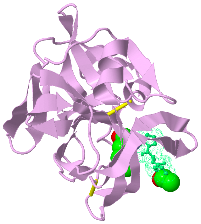
Sites (1, 1)
Asymmetric Unit (1, 1)
|
SS Bonds (2, 2)
Asymmetric/Biological Unit
|
||||||||||||
Cis Peptide Bonds (1, 1)
Asymmetric/Biological Unit
|
||||||||
SAPs(SNPs)/Variants (0, 0)| (no "SAP(SNP)/Variant" information available for 3SGA) |
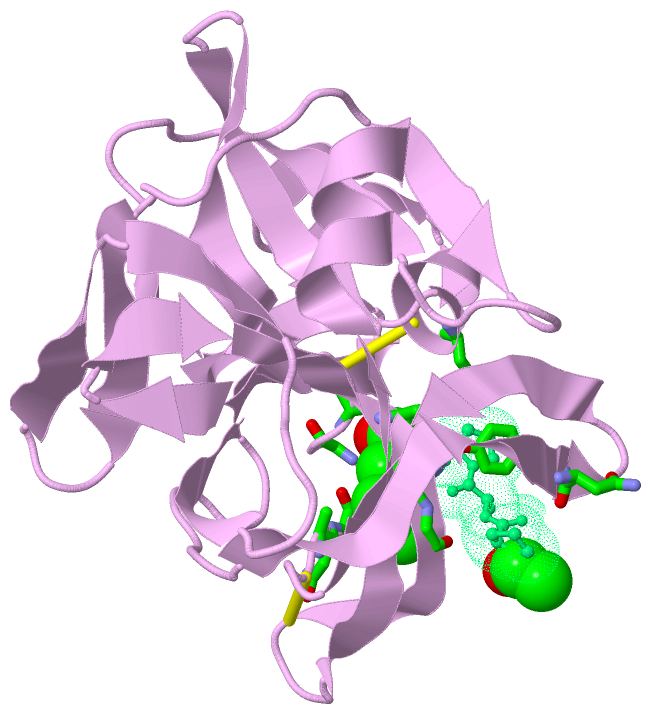
PROSITE Motifs (2, 2)
Asymmetric/Biological Unit (2, 2)
|
||||||||||||||||||||||||||||||||
Exons (0, 0)| (no "Exon" information available for 3SGA) |
Sequences/Alignments
Asymmetric/Biological UnitChain E from PDB Type:PROTEIN Length:181 aligned with PRTA_STRGR | P00776 from UniProtKB/Swiss-Prot Length:297 Alignment length:181 126 136 146 156 166 176 186 196 206 216 226 236 246 256 266 276 286 296 PRTA_STRGR 117 IAGGEAITTGGSRCSLGFNVSVNGVAHALTAGHCTNISASWSIGTRTGTSFPNNDYGIIRHSNPAAADGRVYLYNGSYQDITTAGNAFVGQAVQRSGSTTGLRSGSVTGLNATVNYGSSGIVYGMIQTNVCAEPGDSGGSLFAGSTALGLTSGGSGNCRTGGTTFYQPVTEALSAYGATVL 297 SCOP domains d3sgae_ E: Protease A SCOP domains CATH domains 3sgaE01 E:16-115,E:231-242 Trypsin-like serine proteases 3sgaE02 E:116-230 Trypsin-like serine proteases 3sgaE01 CATH domains Pfam domains --------------------------------------Trypsin-3sgaE01 E:65-236 ------ Pfam domains SAPs(SNPs) ------------------------------------------------------------------------------------------------------------------------------------------------------------------------------------- SAPs(SNPs) PROSITE ----------------------------TRYPSI------------------------------------------------------------------------------------------------TRYPSIN_SER --------------------------------------- PROSITE Transcript ------------------------------------------------------------------------------------------------------------------------------------------------------------------------------------- Transcript 3sga E 16 IAGGEAITTGGSRCSLGFNVSVNGVAHALTAGHCTNISASWSIGTRTGTSFPNNDYGIIRHSNPAAADGRVYLYNGSYQDITTAGNAFVGQAVQRSGSTTGLRSGSVTGLNATVNYGSSGIVYGMIQTNVCAQPGDSGGSLFAGSTALGLTSGGSGNCRTGGTTFYQPVTEALSAYGATVL 242 || 34| 48|||| 54 || 65A|| 93|| 107 117 ||||123 133 143| 165 175 190 || 198 || 212 222 232 | 241 19| 34| 48A||| 59| 65A|| 91||| 120A||| 143| 184| || 202| 235A 29 39 48B|| 62 66| 93|| 120B|| 156 190 || 207 48C| 84 94| 120C| 192A| 48D 99A 120D 192B
Chain P from PDB Type:PROTEIN Length:5
SCOP domains ----- SCOP domains
CATH domains ----- CATH domains
Pfam domains ----- Pfam domains
SAPs(SNPs) ----- SAPs(SNPs)
PROSITE ----- PROSITE
Transcript ----- Transcript
3sga P 5 xPAPf 9
| |
5-ACE
9-PHL
|
||||||||||||||||||||
SCOP Domains (1, 1)
Asymmetric/Biological Unit
 |
CATH Domains (1, 2)
Asymmetric/Biological Unit
|
Pfam Domains (1, 1)
Asymmetric/Biological Unit
|
Gene Ontology (6, 6)|
Asymmetric/Biological Unit(hide GO term definitions) Chain E (PRTA_STRGR | P00776)
|
||||||||||||||||||||||||||||||||||||||||||||||||||||||
Interactive Views
|
||||||||||||||||||||||||||||||||||||||||||||||||||||||||||||||||||||||||||||||||||||||||||||||||||||||||||||||||||||||||||||||
Still Images
|
||||||||||||||||
Databases
|
||||||||||||||||||||||||||||||||||||||||||||||||||||||||||||||||||||||||||||||||||||||||||||||||||||||||||||||||||||||||||||||||||||||||||||||||||||||||||||||||
Analysis Tools
|
|||||||||||||||||||||||||||||||||||||||||||||||||||||||||||||
Entries Sharing at Least One Protein Chain (UniProt ID)
Related Entries Specified in the PDB File
|
|