|
|
|
|
 Description
|
|
Compounds
|
||||||||||||||||||||||||||||||||||||||||||||||||||||||||
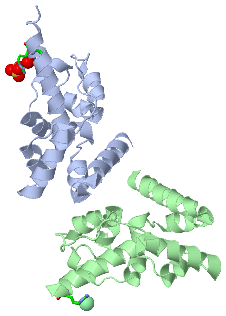
Chains, Units
Summary Information (see also Sequences/Alignments below) |
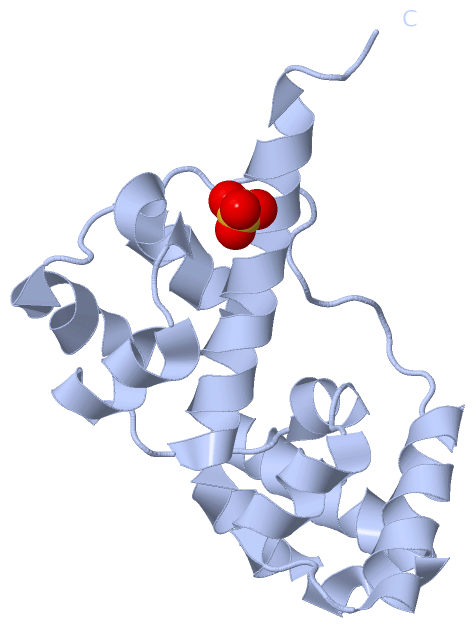
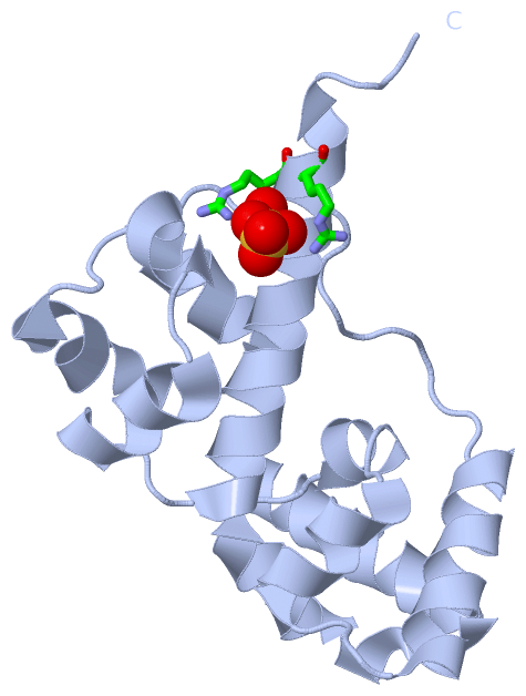
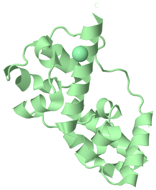
Ligands, Modified Residues, Ions (2, 2)| Asymmetric Unit (2, 2) Biological Unit 1 (1, 1) Biological Unit 2 (0, 0) |
Sites (2, 2)
Asymmetric Unit (2, 2)
|
SS Bonds (0, 0)| (no "SS Bond" information available for 3SEI) |
Cis Peptide Bonds (0, 0)| (no "Cis Peptide Bond" information available for 3SEI) |
SAPs(SNPs)/Variants (0, 0)| (no "SAP(SNP)/Variant" information available for 3SEI) |
PROSITE Motifs (1, 4)
Asymmetric Unit (1, 4)
|
||||||||||||||||||||||||||||||||||||||||||||||||||||||||||||||||||||||||
Exons (0, 0)| (no "Exon" information available for 3SEI) |
Sequences/Alignments
Asymmetric UnitChain A from PDB Type:PROTEIN Length:146 aligned with CSKI1_HUMAN | Q8WXD9 from UniProtKB/Swiss-Prot Length:1431 Alignment length:146 477 487 497 507 517 527 537 547 557 567 577 587 597 607 CSKI1_HUMAN 468 ASEGKSSEAVSQWLTAFQLQLYAPNFISAGYDLPTISRMTPEDLTAIGVTKPGHRKKIAAEISGLSIPDWLPEHKPANLAVWLSMIGLAQYYKVLVDNGYENIDFITDITWEDLQEIGITKLGHQKKLMLAVRKLAELQKAEYAKY 613 SCOP domains d3seia1 A:1-71 automated matches d3seia2 A:72-146 automated matches SCOP domains CATH domains -------------------------------------------------------------------------------------------------------------------------------------------------- CATH domains Pfam domains -------------------------------------------------------------------------------------------------------------------------------------------------- Pfam domains SAPs(SNPs) -------------------------------------------------------------------------------------------------------------------------------------------------- SAPs(SNPs) PROSITE ----SAM_DOMAIN PDB: A:5-68 UniProt: 472-535 -----SAM_DOMAIN PDB: A:74-138 UniProt: 541-605 -------- PROSITE Transcript -------------------------------------------------------------------------------------------------------------------------------------------------- Transcript 3sei A 1 TREGKSSEAVSQWLTAFQLQLYAPNFISAGYDLPTISRMTPEDLTAIGVTKPGHRKKIAAEISGLSIPDWLPEHKPANLAVWLSMIGLAQYYKVLVDNGYENIDFITDITWEDLQEIGITKLGHQKKLMLAVRKLAELRRHHHHHH 146 10 20 30 40 50 60 70 80 90 100 110 120 130 140 Chain B from PDB Type:PROTEIN Length:142 aligned with CSKI1_HUMAN | Q8WXD9 from UniProtKB/Swiss-Prot Length:1431 Alignment length:142 477 487 497 507 517 527 537 547 557 567 577 587 597 607 CSKI1_HUMAN 468 ASEGKSSEAVSQWLTAFQLQLYAPNFISAGYDLPTISRMTPEDLTAIGVTKPGHRKKIAAEISGLSIPDWLPEHKPANLAVWLSMIGLAQYYKVLVDNGYENIDFITDITWEDLQEIGITKLGHQKKLMLAVRKLAELQKAE 609 SCOP domains d3seib1 B:1-71 automated matches d3seib2 B:72-142 automated matches SCOP domains CATH domains ---------------------------------------------------------------------------------------------------------------------------------------------- CATH domains Pfam domains ---------------------------------------------------------------------------------------------------------------------------------------------- Pfam domains SAPs(SNPs) ---------------------------------------------------------------------------------------------------------------------------------------------- SAPs(SNPs) PROSITE ----SAM_DOMAIN PDB: B:5-68 UniProt: 472-535 -----SAM_DOMAIN PDB: B:74-138 UniProt: 541-605 ---- PROSITE Transcript ---------------------------------------------------------------------------------------------------------------------------------------------- Transcript 3sei B 1 TREGKSSEAVSQWLTAFQLQLYAPNFISAGYDLPTISRMTPEDLTAIGVTKPGHRKKIAAEISGLSIPDWLPEHKPANLAVWLSMIGLAQYYKVLVDNGYENIDFITDITWEDLQEIGITKLGHQKKLMLAVRKLAELRRHH 142 10 20 30 40 50 60 70 80 90 100 110 120 130 140
|
||||||||||||||||||||
SCOP Domains (1, 4)
Asymmetric Unit
 |
CATH Domains (0, 0)| (no "CATH Domain" information available for 3SEI) |
Pfam Domains (0, 0)| (no "Pfam Domain" information available for 3SEI) |
Gene Ontology (4, 4)|
Asymmetric Unit(hide GO term definitions) Chain A,B (CSKI1_HUMAN | Q8WXD9)
|
||||||||||||||||||||||||||||||||||||||||||
Interactive Views
|
|||||||||||||||||||||||||||||||||||||||||||||||||||||||||||||||||||||||||||||||||||||||||||||||||||||||||||||||||||||||||||||||||||||||||||||||||||||||||||
Still Images
|
||||||||||||||||
Databases
|
||||||||||||||||||||||||||||||||||||||||||||||||||||||||||||||||||||||||||||||||||||||||||||||||||||||||||||||||||||||||||||||||||||||||||||||||||||||||||||||||
Analysis Tools
|
|||||||||||||||||||||||||||||||||||||||||||||||||||||||||||||
Entries Sharing at Least One Protein Chain (UniProt ID)
Related Entries Specified in the PDB File
|
|