|
|
|
|
 Description
|
|
Compounds
|
||||||||||||||||||||||||||||||||||||||||||||||||
Chains, Units
Summary Information (see also Sequences/Alignments below) |
Ligands, Modified Residues, Ions (0, 0)| (no "Ligand,Modified Residues,Ions" information available for 3RFI) |
Sites (0, 0)| (no "Site" information available for 3RFI) |
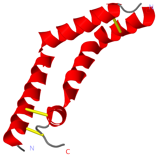
SS Bonds (3, 3)
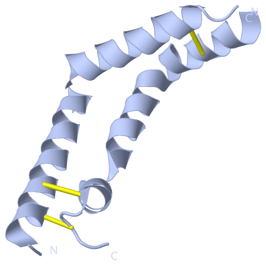
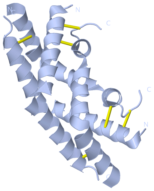
Asymmetric Unit
|
||||||||||||||||
Cis Peptide Bonds (0, 0)| (no "Cis Peptide Bond" information available for 3RFI) |
SAPs(SNPs)/Variants (0, 0)| (no "SAP(SNP)/Variant" information available for 3RFI) |
PROSITE Motifs (0, 0)| (no "PROSITE Motif" information available for 3RFI) |
Exons (0, 0)| (no "Exon" information available for 3RFI) |
Sequences/Alignments
Asymmetric UnitChain A from PDB Type:PROTEIN Length:80 aligned with Q6B9W9_SOLTU | Q6B9W9 from UniProtKB/TrEMBL Length:497 Alignment length:104 309 319 329 339 349 359 369 379 389 399 Q6B9W9_SOLTU 300 GIVSMECKTIVSQYGEMIWDLLVSGVRPDQVCSQAGLCFVDGAQHVSSNIKTVVERETEGSSVGEAPLCTACEMAVVWMQNQLKQEGTKEKVLEYVNQLCEKIP 403 SCOP domains -------------------------------------------------------------------------------------------------------- SCOP domains CATH domains -------------------------------------------------------------------------------------------------------- CATH domains Pfam domains (1) -Asp-3rfiA01 A:1-103 Pfam domains (1) Pfam domains (2) ---SapB_2-3rfiA03 A:3-37 ---------------------------SapB_1-3rfiA02 A:65-103 Pfam domains (2) SAPs(SNPs) -------------------------------------------------------------------------------------------------------- SAPs(SNPs) PROSITE -------------------------------------------------------------------------------------------------------- PROSITE Transcript -------------------------------------------------------------------------------------------------------- Transcript 3rfi A 0 AIVSMECKTIVSQYGEMIWDLLVSGVRPDQVCSQAGLCFV------------------------EAPLCTACEMAVVWMQNQLKQEGTKEKVLEYVNQLCEKIP 103 9 19 29 39 - - | 69 79 89 99 39 64
|
||||||||||||||||||||
SCOP Domains (0, 0)| (no "SCOP Domain" information available for 3RFI) |
CATH Domains (0, 0)| (no "CATH Domain" information available for 3RFI) |
Pfam Domains (3, 3)
Asymmetric Unit

|
Gene Ontology (5, 5)|
Asymmetric Unit(hide GO term definitions) Chain A (Q6B9W9_SOLTU | Q6B9W9)
|
||||||||||||||||||||||||||||||||||||||||||
Interactive Views
|
|||||||||||||||||||||||||||||||||||||||||||||||||||||||||||||||||||||||||||||||||||||||||||||||||||||||||||||||||||||||||||||||||||||||||||
Still Images
|
||||||||||||||||
Databases
|
||||||||||||||||||||||||||||||||||||||||||||||||||||||||||||||||||||||||||||||||||||||||||||||||||||||||||||||||||||||||||||||||||||||||||||||||||||||||||||||||
Analysis Tools
|
|||||||||||||||||||||||||||||||||||||||||||||||||||||||||||||
Entries Sharing at Least One Protein Chain (UniProt ID)
Related Entries Specified in the PDB File
|
|