|
|
|
|
 Description
|
|
Compounds
|
||||||||||||||||||||||||||||||||||||||||||||||||
Chains, Units
Summary Information (see also Sequences/Alignments below) |
Ligands, Modified Residues, Ions (1, 1)
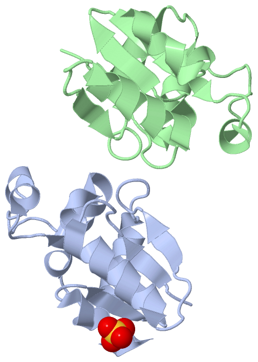
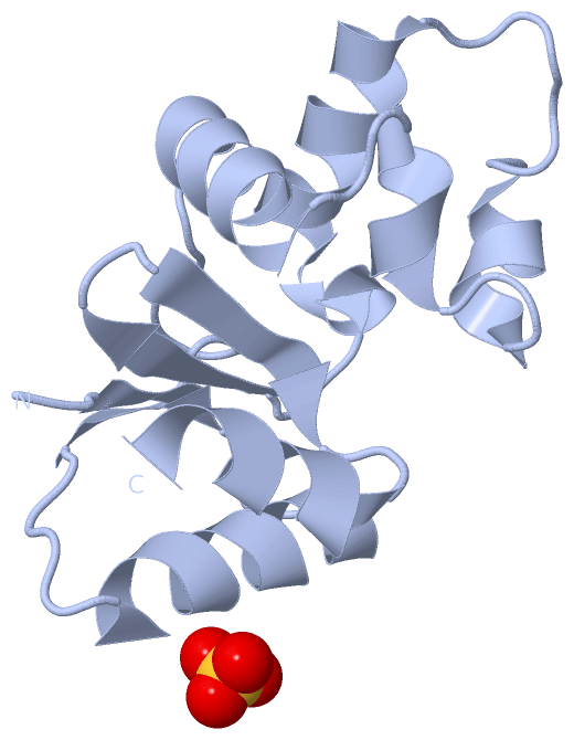
Asymmetric Unit (1, 1)
|
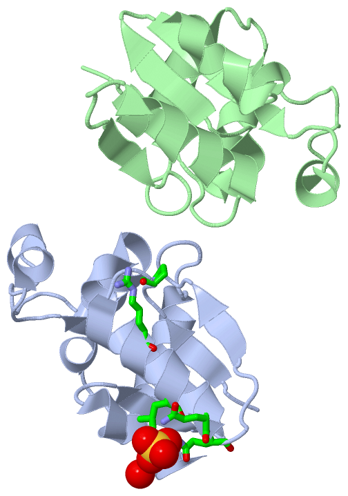
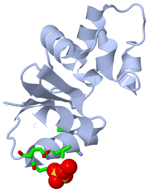
Sites (1, 1)
Asymmetric Unit (1, 1)
|
SS Bonds (0, 0)| (no "SS Bond" information available for 3RDW) |
Cis Peptide Bonds (4, 4)
Asymmetric Unit
|
||||||||||||||||||||
SAPs(SNPs)/Variants (0, 0)| (no "SAP(SNP)/Variant" information available for 3RDW) |
PROSITE Motifs (0, 0)| (no "PROSITE Motif" information available for 3RDW) |
Exons (0, 0)| (no "Exon" information available for 3RDW) |
Sequences/Alignments
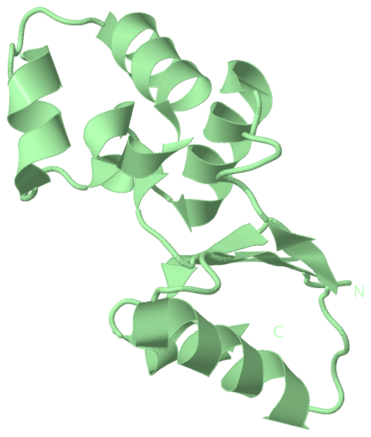
Asymmetric UnitChain A from PDB Type:PROTEIN Length:115 aligned with Q0WCJ8_YERPE | Q0WCJ8 from UniProtKB/TrEMBL Length:118 Alignment length:115 12 22 32 42 52 62 72 82 92 102 112 Q0WCJ8_YERPE 3 DVTIYHNPRCSKSRETLALVEQQGITPQVVLYLETPPSVDKLKELLQQLGFSDARQLMRTKEDLYKTLNLDDRGLTQDQLLQAMADNPKLIERPIVVTQGKARIGRPPEQVLEIL 117 SCOP domains d3rdwa_ A: automated matches SCOP domains CATH domains ------------------------------------------------------------------------------------------------------------------- CATH domains Pfam domains ------------------------------------------------------------------------------------------------------------------- Pfam domains SAPs(SNPs) ------------------------------------------------------------------------------------------------------------------- SAPs(SNPs) PROSITE ------------------------------------------------------------------------------------------------------------------- PROSITE Transcript ------------------------------------------------------------------------------------------------------------------- Transcript 3rdw A 3 DVTIYHNPRCSKSRETLALVEQQGITPQVVLYLETPPSVDKLKELLQQLGFSDARQLMRTKEDLYKTLNLDDRGLTQDQLLQAMADNPKLIERPIVVTQGKARIGRPPEQVLEIL 117 12 22 32 42 52 62 72 82 92 102 112 Chain B from PDB Type:PROTEIN Length:114 aligned with Q0WCJ8_YERPE | Q0WCJ8 from UniProtKB/TrEMBL Length:118 Alignment length:114 12 22 32 42 52 62 72 82 92 102 112 Q0WCJ8_YERPE 3 DVTIYHNPRCSKSRETLALVEQQGITPQVVLYLETPPSVDKLKELLQQLGFSDARQLMRTKEDLYKTLNLDDRGLTQDQLLQAMADNPKLIERPIVVTQGKARIGRPPEQVLEI 116 SCOP domains d3rdwb_ B: automated matches SCOP domains CATH domains ------------------------------------------------------------------------------------------------------------------ CATH domains Pfam domains (1) ----ArsC-3rdwB01 B:7-116 Pfam domains (1) Pfam domains (2) ----ArsC-3rdwB02 B:7-116 Pfam domains (2) SAPs(SNPs) ------------------------------------------------------------------------------------------------------------------ SAPs(SNPs) PROSITE ------------------------------------------------------------------------------------------------------------------ PROSITE Transcript ------------------------------------------------------------------------------------------------------------------ Transcript 3rdw B 3 DVTIYHNPRCSKSRETLALVEQQGITPQVVLYLETPPSVDKLKELLQQLGFSDARQLMRTKEDLYKTLNLDDRGLTQDQLLQAMADNPKLIERPIVVTQGKARIGRPPEQVLEI 116 12 22 32 42 52 62 72 82 92 102 112
|
||||||||||||||||||||
SCOP Domains (1, 2)
Asymmetric Unit
 |
CATH Domains (0, 0)| (no "CATH Domain" information available for 3RDW) |
Pfam Domains (1, 2)
Asymmetric Unit
|
Gene Ontology (3, 3)|
Asymmetric Unit(hide GO term definitions) Chain A,B (Q0WCJ8_YERPE | Q0WCJ8)
|
||||||||||||||||||||||||||||||
Interactive Views
|
|||||||||||||||||||||||||||||||||||||||||||||||||||||||||||||||||||||||||||||||||||||||||||||||||||||||||||||||||||||||||||||||||||||||||||||||||||||||||||||||||||
Still Images
|
||||||||||||||||
Databases
|
||||||||||||||||||||||||||||||||||||||||||||||||||||||||||||||||||||||||||||||||||||||||||||||||||||||||||||||||||||||||||||||||||||||||||||||||||||||||||||||||
Analysis Tools
|
|||||||||||||||||||||||||||||||||||||||||||||||||||||||||||||
Entries Sharing at Least One Protein Chain (UniProt ID)
Related Entries Specified in the PDB File
|
|