Show PDB file:   
         Plain Text   HTML   (compressed file size)
QuickSearch:   
by PDB,NDB,UniProt,PROSITE Code or Search Term(s)  
(-)Asymmetric Unit
(-)Asym. Unit - sites
(-)Biological Unit 1
(-)Biol. Unit 1 - sites
(-)Biological Unit 2
(-)Biological Unit 3
(-)Biological Unit 4
collapse expand < >
Image Asymmetric Unit
Asymmetric Unit  (Jmol Viewer)
Image Asym. Unit - sites
Asym. Unit - sites  (Jmol Viewer)
Image Biological Unit 1
Biological Unit 1  (Jmol Viewer)
Image Biol. Unit 1 - sites
Biol. Unit 1 - sites  (Jmol Viewer)
Image Biological Unit 2
Biological Unit 2  (Jmol Viewer)
Image Biological Unit 3
Biological Unit 3  (Jmol Viewer)
Image Biological Unit 4
Biological Unit 4  (Jmol Viewer)

(-) Description

Title :  STRUCTURE OF THE SLAIN2C-CLIPCG1 COMPLEX
 
Authors :  C. Manatschal, V. Olieric, M. O. Steinmetz
Date :  01 Apr 11  (Deposition) - 29 Jun 11  (Release) - 29 Jun 11  (Revision)
Method :  X-RAY DIFFRACTION
Resolution :  1.75
Chains :  Asym. Unit :  A,B,C,D,E,F,G,H
Biol. Unit 1:  A,E  (1x)
Biol. Unit 2:  B,F  (1x)
Biol. Unit 3:  C,G  (1x)
Biol. Unit 4:  D,H  (1x)
Keywords :  Cytoskeletal Protein, Cap Gly Protein Complex, Structural Protein (Keyword Search: [Gene Ontology, PubMed, Web (Google))
 
Reference :  B. Van Der Vaart, C. Manatschal, I. Grigoriev, V. Olieric, S. M. Gouveia, S. Bjelic, J. Demmers, I. Vorobjev, C. C. Hoogenraad, M. O. Steinmetz, A. Akhmanova
Slain2 Links Microtubule Plus End-Tracking Proteins And Controls Microtubule Growth In Interphase
J. Cell Biol. V. 193 1083 2011
PubMed-ID: 21646404  |  Reference-DOI: 10.1083/JCB.201012179

(-) Compounds

Molecule 1 - CAP-GLY DOMAIN-CONTAINING LINKER PROTEIN 1
    ChainsA, B, C, D
    EngineeredYES
    Expression SystemESCHERICHIA COLI
    Expression System PlasmidPDEST17-OI
    Expression System StrainBL21 (DE3)
    Expression System Taxid562
    Expression System Vector TypePLASMID
    FragmentCAP-GLY 1 DOMAIN, RESIDUES 56-127
    GeneCLIP-170
    Organism CommonHUMAN
    Organism ScientificHOMO SAPIENS
    Organism Taxid9606
    SynonymCYTOPLASMIC LINKER PROTEIN 1, CYTOPLASMIC LINKER PROTEIN 170 ALPHA-2, CLIP-170, REED-STERNBERG INTERMEDIATE FILAMENT-ASSOCIATED PROTEIN, RESTIN
 
Molecule 2 - SLAIN MOTIF-CONTAINING PROTEIN 2
    ChainsE, F, G, H
    EngineeredYES
    FragmentC-TERMINAL DOMAIN, RESIDUES 574-581
    Organism CommonHUMAN
    Organism ScientificHOMO SAPIENS
    Organism Taxid9606
    Other DetailsPOLYPEPTIDE SYNTHESIS
    SyntheticYES

 Structural Features

(-) Chains, Units

  12345678
Asymmetric Unit ABCDEFGH
Biological Unit 1 (1x)A   E   
Biological Unit 2 (1x) B   F  
Biological Unit 3 (1x)  C   G 
Biological Unit 4 (1x)   D   H

Summary Information (see also Sequences/Alignments below)

(-) Ligands, Modified Residues, Ions  (2, 2)

Asymmetric Unit (2, 2)
No.NameCountTypeFull Name
1BME1Ligand/IonBETA-MERCAPTOETHANOL
2NA1Ligand/IonSODIUM ION
Biological Unit 1 (0, 0)
No.NameCountTypeFull Name
1BME-1Ligand/IonBETA-MERCAPTOETHANOL
2NA-1Ligand/IonSODIUM ION
Biological Unit 2 (0, 0)
No.NameCountTypeFull Name
1BME-1Ligand/IonBETA-MERCAPTOETHANOL
2NA-1Ligand/IonSODIUM ION
Biological Unit 3 (1, 1)
No.NameCountTypeFull Name
1BME1Ligand/IonBETA-MERCAPTOETHANOL
2NA-1Ligand/IonSODIUM ION
Biological Unit 4 (0, 0)
No.NameCountTypeFull Name
1BME-1Ligand/IonBETA-MERCAPTOETHANOL
2NA-1Ligand/IonSODIUM ION

(-) Sites  (2, 2)

Asymmetric Unit (2, 2)
No.NameEvidenceResiduesDescription
1AC1SOFTWARETRP A:65 , HOH A:141 , TRP B:65 , HOH B:182 , HOH D:147BINDING SITE FOR RESIDUE NA A 1
2AC2SOFTWAREARG C:120 , PRO C:121 , SER C:122BINDING SITE FOR RESIDUE BME C 1

(-) SS Bonds  (0, 0)

(no "SS Bond" information available for 3RDV)

(-) Cis Peptide Bonds  (0, 0)

(no "Cis Peptide Bond" information available for 3RDV)

 Sequence-Structure Mapping

(-) SAPs(SNPs)/Variants  (0, 0)

(no "SAP(SNP)/Variant" information available for 3RDV)

(-) PROSITE Motifs  (2, 8)

Asymmetric Unit (2, 8)
 PROSITEUniProtKBPDB
No.IDACDescriptionIDLocationCountLocation
1CAP_GLY_1PS00845 CAP-Gly domain signature.CLIP1_HUMAN78-109


232-263
  4A:78-109
B:78-109
C:78-109
D:78-109
-
2CAP_GLY_2PS50245 CAP-Gly domain profile.CLIP1_HUMAN78-120


232-274
  4A:78-120
B:78-120
C:78-120
D:78-120
-
Biological Unit 1 (2, 2)
 PROSITEUniProtKBPDB
No.IDACDescriptionIDLocationCountLocation
1CAP_GLY_1PS00845 CAP-Gly domain signature.CLIP1_HUMAN78-109


232-263
  1A:78-109
-
-
-
-
2CAP_GLY_2PS50245 CAP-Gly domain profile.CLIP1_HUMAN78-120


232-274
  1A:78-120
-
-
-
-
Biological Unit 2 (2, 2)
 PROSITEUniProtKBPDB
No.IDACDescriptionIDLocationCountLocation
1CAP_GLY_1PS00845 CAP-Gly domain signature.CLIP1_HUMAN78-109


232-263
  1-
B:78-109
-
-
-
2CAP_GLY_2PS50245 CAP-Gly domain profile.CLIP1_HUMAN78-120


232-274
  1-
B:78-120
-
-
-
Biological Unit 3 (2, 2)
 PROSITEUniProtKBPDB
No.IDACDescriptionIDLocationCountLocation
1CAP_GLY_1PS00845 CAP-Gly domain signature.CLIP1_HUMAN78-109


232-263
  1-
-
C:78-109
-
-
2CAP_GLY_2PS50245 CAP-Gly domain profile.CLIP1_HUMAN78-120


232-274
  1-
-
C:78-120
-
-
Biological Unit 4 (2, 2)
 PROSITEUniProtKBPDB
No.IDACDescriptionIDLocationCountLocation
1CAP_GLY_1PS00845 CAP-Gly domain signature.CLIP1_HUMAN78-109


232-263
  1-
-
-
D:78-109
-
2CAP_GLY_2PS50245 CAP-Gly domain profile.CLIP1_HUMAN78-120


232-274
  1-
-
-
D:78-120
-

(-) Exons   (0, 0)

(no "Exon" information available for 3RDV)

(-) Sequences/Alignments

Asymmetric Unit
   Reformat: Number of residues per line =  ('0' or empty: single-line sequence representation)
  Number of residues per labelling interval =   
  UniProt sequence: complete  aligned part    
   Show mapping: SCOP domains CATH domains Pfam domains Secondary structure (by author)
SAPs(SNPs) PROSITE motifs Exons
(details for a mapped element are shown in a popup box when the mouse pointer rests over it)
Chain A from PDB  Type:PROTEIN  Length:70
 aligned with CLIP1_HUMAN | P30622 from UniProtKB/Swiss-Prot  Length:1438

    Alignment length:70
                                    66        76        86        96       106       116       126
          CLIP1_HUMAN    57 DFRVGERVWVNGNKPGFIQFLGETQFAPGQWAGIVLDEPIGKNDGSVAGVRYFQCEPLKGIFTRPSKLTR 126
               SCOP domains ---------------------------------------------------------------------- SCOP domains
               CATH domains ---------------------------------------------------------------------- CATH domains
               Pfam domains ---------------------------------------------------------------------- Pfam domains
         Sec.struct. author ......eeee...eeeeeeeee........eeeeee.........ee..ee........eeeehhh.ee. Sec.struct. author
                 SAPs(SNPs) ---------------------------------------------------------------------- SAPs(SNPs)
                PROSITE (1) ---------------------CAP_GLY_2  PDB: A:78-120 UniProt: 78-120   ------ PROSITE (1)
                PROSITE (2) ---------------------CAP_GLY_1  PDB: A:78-109        ----------------- PROSITE (2)
                 Transcript ---------------------------------------------------------------------- Transcript
                 3rdv A  57 DFRVGERVWVNGNKPGFIQFLGETQFAPGQWAGIVLDEPIGKNDGSVAGVRYFQCEPLKGIFTRPSKLTR 126
                                    66        76        86        96       106       116       126

Chain B from PDB  Type:PROTEIN  Length:71
 aligned with CLIP1_HUMAN | P30622 from UniProtKB/Swiss-Prot  Length:1438

    Alignment length:71
                                    66        76        86        96       106       116       126 
          CLIP1_HUMAN    57 DFRVGERVWVNGNKPGFIQFLGETQFAPGQWAGIVLDEPIGKNDGSVAGVRYFQCEPLKGIFTRPSKLTRK 127
               SCOP domains ----------------------------------------------------------------------- SCOP domains
               CATH domains ----------------------------------------------------------------------- CATH domains
               Pfam domains ----------------------------------------------------------------------- Pfam domains
         Sec.struct. author ......eeee...eeeeeeeee........eeeeee.........ee..ee........eeeehhh.eee. Sec.struct. author
                 SAPs(SNPs) ----------------------------------------------------------------------- SAPs(SNPs)
                PROSITE (1) ---------------------CAP_GLY_2  PDB: B:78-120 UniProt: 78-120   ------- PROSITE (1)
                PROSITE (2) ---------------------CAP_GLY_1  PDB: B:78-109        ------------------ PROSITE (2)
                 Transcript ----------------------------------------------------------------------- Transcript
                 3rdv B  57 DFRVGERVWVNGNKPGFIQFLGETQFAPGQWAGIVLDEPIGKNDGSVAGVRYFQCEPLKGIFTRPSKLTRK 127
                                    66        76        86        96       106       116       126 

Chain C from PDB  Type:PROTEIN  Length:71
 aligned with CLIP1_HUMAN | P30622 from UniProtKB/Swiss-Prot  Length:1438

    Alignment length:71
                                    66        76        86        96       106       116       126 
          CLIP1_HUMAN    57 DFRVGERVWVNGNKPGFIQFLGETQFAPGQWAGIVLDEPIGKNDGSVAGVRYFQCEPLKGIFTRPSKLTRK 127
               SCOP domains ----------------------------------------------------------------------- SCOP domains
               CATH domains ----------------------------------------------------------------------- CATH domains
               Pfam domains ----------------------------------------------------------------------- Pfam domains
         Sec.struct. author ......eeee...eeeeeeeee........eeeeee.........ee..ee........eeeehhh.ee.. Sec.struct. author
                 SAPs(SNPs) ----------------------------------------------------------------------- SAPs(SNPs)
                PROSITE (1) ---------------------CAP_GLY_2  PDB: C:78-120 UniProt: 78-120   ------- PROSITE (1)
                PROSITE (2) ---------------------CAP_GLY_1  PDB: C:78-109        ------------------ PROSITE (2)
                 Transcript ----------------------------------------------------------------------- Transcript
                 3rdv C  57 DFRVGERVWVNGNKPGFIQFLGETQFAPGQWAGIVLDEPIGKNDGSVAGVRYFQCEPLKGIFTRPSKLTRK 127
                                    66        76        86        96       106       116       126 

Chain D from PDB  Type:PROTEIN  Length:72
 aligned with CLIP1_HUMAN | P30622 from UniProtKB/Swiss-Prot  Length:1438

    Alignment length:72
                                    65        75        85        95       105       115       125  
          CLIP1_HUMAN    56 DDFRVGERVWVNGNKPGFIQFLGETQFAPGQWAGIVLDEPIGKNDGSVAGVRYFQCEPLKGIFTRPSKLTRK 127
               SCOP domains ------------------------------------------------------------------------ SCOP domains
               CATH domains ------------------------------------------------------------------------ CATH domains
           Pfam domains (1) ----CAP_GLY-3rdvD01 D:60-125                                          -- Pfam domains (1)
           Pfam domains (2) ----CAP_GLY-3rdvD02 D:60-125                                          -- Pfam domains (2)
           Pfam domains (3) ----CAP_GLY-3rdvD03 D:60-125                                          -- Pfam domains (3)
           Pfam domains (4) ----CAP_GLY-3rdvD04 D:60-125                                          -- Pfam domains (4)
         Sec.struct. author .......eeee...eeeeeeeee........eeeeee.........ee..ee........eeeehhh.eee. Sec.struct. author
                 SAPs(SNPs) ------------------------------------------------------------------------ SAPs(SNPs)
                PROSITE (1) ----------------------CAP_GLY_2  PDB: D:78-120 UniProt: 78-120   ------- PROSITE (1)
                PROSITE (2) ----------------------CAP_GLY_1  PDB: D:78-109        ------------------ PROSITE (2)
                 Transcript ------------------------------------------------------------------------ Transcript
                 3rdv D  56 DDFRVGERVWVNGNKPGFIQFLGETQFAPGQWAGIVLDEPIGKNDGSVAGVRYFQCEPLKGIFTRPSKLTRK 127
                                    65        75        85        95       105       115       125  

Chain E from PDB  Type:PROTEIN  Length:8
 aligned with SLAI2_HUMAN | Q9P270 from UniProtKB/Swiss-Prot  Length:581

    Alignment length:8
          SLAI2_HUMAN   574 DSWKDGCY 581
               SCOP domains -------- SCOP domains
               CATH domains -------- CATH domains
               Pfam domains -------- Pfam domains
         Sec.struct. author hhhhh... Sec.struct. author
                 SAPs(SNPs) -------- SAPs(SNPs)
                    PROSITE -------- PROSITE
                 Transcript -------- Transcript
                 3rdv E 574 DSWKDGCY 581

Chain F from PDB  Type:PROTEIN  Length:8
 aligned with SLAI2_HUMAN | Q9P270 from UniProtKB/Swiss-Prot  Length:581

    Alignment length:8
          SLAI2_HUMAN   574 DSWKDGCY 581
               SCOP domains -------- SCOP domains
               CATH domains -------- CATH domains
               Pfam domains -------- Pfam domains
         Sec.struct. author hhhhh... Sec.struct. author
                 SAPs(SNPs) -------- SAPs(SNPs)
                    PROSITE -------- PROSITE
                 Transcript -------- Transcript
                 3rdv F 574 DSWKDGCY 581

Chain G from PDB  Type:PROTEIN  Length:7
 aligned with SLAI2_HUMAN | Q9P270 from UniProtKB/Swiss-Prot  Length:581

    Alignment length:7
          SLAI2_HUMAN   575 SWKDGCY 581
               SCOP domains ------- SCOP domains
               CATH domains ------- CATH domains
               Pfam domains ------- Pfam domains
         Sec.struct. author ....... Sec.struct. author
                 SAPs(SNPs) ------- SAPs(SNPs)
                    PROSITE ------- PROSITE
                 Transcript ------- Transcript
                 3rdv G 575 SWKDGCY 581

Chain H from PDB  Type:PROTEIN  Length:8
 aligned with SLAI2_HUMAN | Q9P270 from UniProtKB/Swiss-Prot  Length:581

    Alignment length:8
          SLAI2_HUMAN   574 DSWKDGCY 581
               SCOP domains -------- SCOP domains
               CATH domains -------- CATH domains
               Pfam domains -------- Pfam domains
         Sec.struct. author ........ Sec.struct. author
                 SAPs(SNPs) -------- SAPs(SNPs)
                    PROSITE -------- PROSITE
                 Transcript -------- Transcript
                 3rdv H 574 DSWKDGCY 581

   Legend:   → Mismatch (orange background)
  - → Gap (green background, '-', border residues have a numbering label)
    → Modified Residue (blue background, lower-case, 'x' indicates undefined single-letter code, labelled with number + name)
  x → Chemical Group (purple background, 'x', labelled with number + name, e.g. ACE or NH2)
  extra numbering lines below/above indicate numbering irregularities and modified residue names etc., number ends below/above '|'

 Classification and Annotation

(-) SCOP Domains  (0, 0)

(no "SCOP Domain" information available for 3RDV)

(-) CATH Domains  (0, 0)

(no "CATH Domain" information available for 3RDV)

(-) Pfam Domains  (1, 4)

Asymmetric Unit

(-) Gene Ontology  (33, 40)

Asymmetric Unit(hide GO term definitions)
Chain A,B,C,D   (CLIP1_HUMAN | P30622)
molecular function
    GO:0046872    metal ion binding    Interacting selectively and non-covalently with any metal ion.
    GO:0008017    microtubule binding    Interacting selectively and non-covalently with microtubules, filaments composed of tubulin monomers.
    GO:0051010    microtubule plus-end binding    Interacting selectively and non-covalently with the plus end of a microtubule.
    GO:0005515    protein binding    Interacting selectively and non-covalently with any protein or protein complex (a complex of two or more proteins that may include other nonprotein molecules).
    GO:0042803    protein homodimerization activity    Interacting selectively and non-covalently with an identical protein to form a homodimer.
    GO:0015631    tubulin binding    Interacting selectively and non-covalently with monomeric or multimeric forms of tubulin, including microtubules.
    GO:0008270    zinc ion binding    Interacting selectively and non-covalently with zinc (Zn) ions.
biological process
    GO:0001578    microtubule bundle formation    A process that results in a parallel arrangement of microtubules.
    GO:0031116    positive regulation of microtubule polymerization    Any process that activates or increases the frequency, rate or extent of microtubule polymerization.
    GO:0044861    protein transport into plasma membrane raft    The directed movement of a protein into a plasma membrane raft.
    GO:0007062    sister chromatid cohesion    The cell cycle process in which the sister chromatids of a replicated chromosome become tethered to each other.
    GO:0006810    transport    The directed movement of substances (such as macromolecules, small molecules, ions) or cellular components (such as complexes and organelles) into, out of or within a cell, or between cells, or within a multicellular organism by means of some agent such as a transporter, pore or motor protein.
cellular component
    GO:0042995    cell projection    A prolongation or process extending from a cell, e.g. a flagellum or axon.
    GO:0005813    centrosome    A structure comprised of a core structure (in most organisms, a pair of centrioles) and peripheral material from which a microtubule-based structure, such as a spindle apparatus, is organized. Centrosomes occur close to the nucleus during interphase in many eukaryotic cells, though in animal cells it changes continually during the cell-division cycle.
    GO:0005737    cytoplasm    All of the contents of a cell excluding the plasma membrane and nucleus, but including other subcellular structures.
    GO:0005881    cytoplasmic microtubule    Any microtubule in the cytoplasm of a cell.
    GO:0031410    cytoplasmic vesicle    A vesicle found in the cytoplasm of a cell.
    GO:0030659    cytoplasmic vesicle membrane    The lipid bilayer surrounding a cytoplasmic vesicle.
    GO:0005856    cytoskeleton    Any of the various filamentous elements that form the internal framework of cells, and typically remain after treatment of the cells with mild detergent to remove membrane constituents and soluble components of the cytoplasm. The term embraces intermediate filaments, microfilaments, microtubules, the microtrabecular lattice, and other structures characterized by a polymeric filamentous nature and long-range order within the cell. The various elements of the cytoskeleton not only serve in the maintenance of cellular shape but also have roles in other cellular functions, including cellular movement, cell division, endocytosis, and movement of organelles.
    GO:0005829    cytosol    The part of the cytoplasm that does not contain organelles but which does contain other particulate matter, such as protein complexes.
    GO:0005768    endosome    A vacuole to which materials ingested by endocytosis are delivered.
    GO:0005882    intermediate filament    A cytoskeletal structure that forms a distinct elongated structure, characteristically 10 nm in diameter, that occurs in the cytoplasm of eukaryotic cells. Intermediate filaments form a fibrous system, composed of chemically heterogeneous subunits and involved in mechanically integrating the various components of the cytoplasmic space. Intermediate filaments may be divided into five chemically distinct classes: Type I, acidic keratins; Type II, basic keratins; Type III, including desmin, vimentin and others; Type IV, neurofilaments and related filaments; and Type V, lamins.
    GO:0000776    kinetochore    A multisubunit complex that is located at the centromeric region of DNA and provides an attachment point for the spindle microtubules.
    GO:0044354    macropinosome    A membrane-bounded, uncoated intracellular vesicle formed by the process of macropinocytosis.
    GO:0016020    membrane    A lipid bilayer along with all the proteins and protein complexes embedded in it an attached to it.
    GO:0005874    microtubule    Any of the long, generally straight, hollow tubes of internal diameter 12-15 nm and external diameter 24 nm found in a wide variety of eukaryotic cells; each consists (usually) of 13 protofilaments of polymeric tubulin, staggered in such a manner that the tubulin monomers are arranged in a helical pattern on the microtubular surface, and with the alpha/beta axes of the tubulin subunits parallel to the long axis of the tubule; exist in equilibrium with pool of tubulin monomers and can be rapidly assembled or disassembled in response to physiological stimuli; concerned with force generation, e.g. in the spindle.
    GO:0015630    microtubule cytoskeleton    The part of the cytoskeleton (the internal framework of a cell) composed of microtubules and associated proteins.
    GO:1990752    microtubule end    Any end of a microtubule. Microtubule ends differ in that the so-called microtubule plus-end is the one that preferentially grows by polymerization, with respect to the minus-end.
    GO:0035371    microtubule plus-end    The growing (plus) end of a microtubule. In vitro, microtubules polymerize more quickly at the plus end than at the minus end. In vivo, microtubule growth occurs only at the plus end, and the plus end switches between periods of growth and shortening, a behavior known as dynamic instability.
    GO:0001726    ruffle    Projection at the leading edge of a crawling cell; the protrusions are supported by a microfilament meshwork.
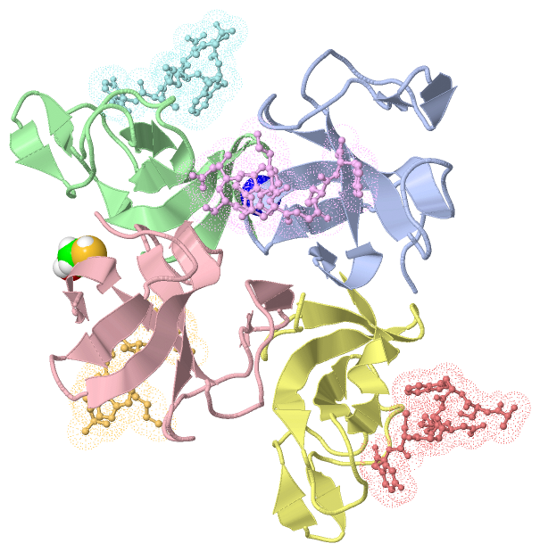
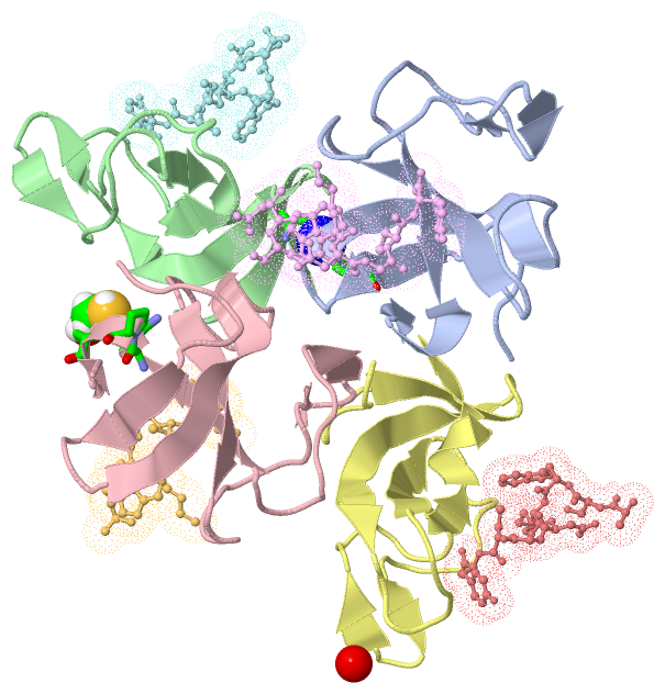
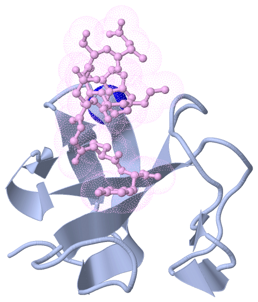
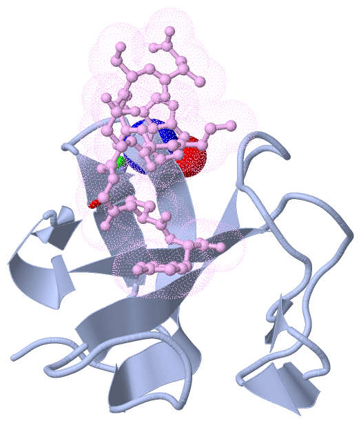
Chain E,F,G,H   (SLAI2_HUMAN | Q9P270)
molecular function
    GO:0005515    protein binding    Interacting selectively and non-covalently with any protein or protein complex (a complex of two or more proteins that may include other nonprotein molecules).
biological process
    GO:0031122    cytoplasmic microtubule organization    A process that is carried out at the cellular level which results in the assembly, arrangement of constituent parts, or disassembly of structures formed of microtubules and associated proteins in the cytoplasm of a cell.
    GO:0007020    microtubule nucleation    The process in which tubulin alpha-beta heterodimers begin aggregation to form an oligomeric tubulin structure (a microtubule seed). Microtubule nucleation is the initiating step in the formation of a microtubule in the absence of any existing microtubules ('de novo' microtubule formation).
    GO:0031116    positive regulation of microtubule polymerization    Any process that activates or increases the frequency, rate or extent of microtubule polymerization.
    GO:0031113    regulation of microtubule polymerization    Any process that modulates the frequency, rate or extent of microtubule polymerization.
cellular component
    GO:0005813    centrosome    A structure comprised of a core structure (in most organisms, a pair of centrioles) and peripheral material from which a microtubule-based structure, such as a spindle apparatus, is organized. Centrosomes occur close to the nucleus during interphase in many eukaryotic cells, though in animal cells it changes continually during the cell-division cycle.
    GO:0005737    cytoplasm    All of the contents of a cell excluding the plasma membrane and nucleus, but including other subcellular structures.
    GO:0005856    cytoskeleton    Any of the various filamentous elements that form the internal framework of cells, and typically remain after treatment of the cells with mild detergent to remove membrane constituents and soluble components of the cytoplasm. The term embraces intermediate filaments, microfilaments, microtubules, the microtrabecular lattice, and other structures characterized by a polymeric filamentous nature and long-range order within the cell. The various elements of the cytoskeleton not only serve in the maintenance of cellular shape but also have roles in other cellular functions, including cellular movement, cell division, endocytosis, and movement of organelles.
    GO:0015630    microtubule cytoskeleton    The part of the cytoskeleton (the internal framework of a cell) composed of microtubules and associated proteins.
    GO:0035371    microtubule plus-end    The growing (plus) end of a microtubule. In vitro, microtubules polymerize more quickly at the plus end than at the minus end. In vivo, microtubule growth occurs only at the plus end, and the plus end switches between periods of growth and shortening, a behavior known as dynamic instability.

 Visualization

(-) Interactive Views

Asymmetric Unit
  Complete Structure
    Jena3D(integrated viewing of ligand, site, SAP, PROSITE, SCOP information)
    WebMol | AstexViewer[tm]@PDBe
(Java Applets, require no local installation except for Java; loading may be slow)
    STRAP
(Java WebStart application, automatic local installation, requires Java; full application with system access!)
    RasMol
(require local installation)
    Molscript (VRML)
(requires installation of a VRML viewer; select preferred view via VRML and generate a mono or stereo PDF format file)
 
  Ligands, Modified Residues, Ions
    BME  [ RasMol | Jena3D ]  +environment [ RasMol | Jena3D ]
    NA  [ RasMol | Jena3D ]  +environment [ RasMol | Jena3D ]
 
  Sites
    AC1  [ RasMol ]  +environment [ RasMol ]
    AC2  [ RasMol ]  +environment [ RasMol ]
 
  Cis Peptide Bonds
(no "Cis Peptide Bonds" information available for 3rdv)
 
Biological Units
  Complete Structure
    Biological Unit 1  [ Jena3D ]
    Biological Unit 2  [ Jena3D ]
    Biological Unit 3  [ Jena3D ]
    Biological Unit 4  [ Jena3D ]

(-) Still Images

Jmol
  protein: cartoon or spacefill or dots and stick; nucleic acid: cartoon and stick; ligands: spacefill; active site: stick
Molscript
  protein, nucleic acid: cartoon; ligands: spacefill; active site: ball and stick

 Databases and Analysis Tools

(-) Databases

Access by PDB/NDB ID
  3rdv
    Family and Domain InformationProDom | SYSTERS
    General Structural InformationGlycoscienceDB | MMDB | NDB | OCA | PDB | PDBe | PDBj | PDBsum | PDBWiki | PQS | PROTEOPEDIA
    Orientation in MembranesOPM
    Protein SurfaceSURFACE
    Secondary StructureDSSP (structure derived) | HSSP (homology derived)
    Structural GenomicsGeneCensus
    Structural NeighboursCE | VAST
    Structure ClassificationCATH | Dali | SCOP
    Validation and Original DataBMRB Data View | BMRB Restraints Grid | EDS | PROCHECK | RECOORD | WHAT_CHECK
 
Access by UniProt ID/Accession number
  CLIP1_HUMAN | P30622
    Comparative Protein Structure ModelsModBase
    Genomic InformationEnsembl
    Protein-protein InteractionDIP
    Sequence, Family and Domain InformationInterPro | Pfam | SMART | UniProtKB/SwissProt
  SLAI2_HUMAN | Q9P270
    Comparative Protein Structure ModelsModBase
    Genomic InformationEnsembl
    Protein-protein InteractionDIP
    Sequence, Family and Domain InformationInterPro | Pfam | SMART | UniProtKB/SwissProt
 
Access by Enzyme Classificator   (EC Number)
  (no 'Enzyme Classificator' available)
    General Enzyme InformationBRENDA | EC-PDB | Enzyme | IntEnz
    PathwayKEGG | MetaCyc
 
Access by Disease Identifier   (MIM ID)
  (no 'MIM ID' available)
    Disease InformationOMIM
 
Access by GenAge ID
  (no 'GenAge ID' available)
    Age Related InformationGenAge

(-) Analysis Tools

Access by PDB/NDB ID
    Domain InformationXDom
    Interatomic Contacts of Structural UnitsCSU
    Ligand-protein ContactsLPC
    Protein CavitiescastP
    Sequence and Secondary StructurePDBCartoon
    Structure AlignmentSTRAP(Java WebStart application, automatic local installation, requires Java; full application with system access!)
    Structure and Sequence BrowserSTING
 
Access by UniProt ID/Accession number
  CLIP1_HUMAN | P30622
    Protein Disorder PredictionDisEMBL | FoldIndex | GLOBPLOT (for more information see DisProt)
  SLAI2_HUMAN | Q9P270
    Protein Disorder PredictionDisEMBL | FoldIndex | GLOBPLOT (for more information see DisProt)

 Related Entries

(-) Entries Sharing at Least One Protein Chain (UniProt ID)

UniProtKB/Swiss-Prot
        CLIP1_HUMAN | P306222cp5 2cp6 2e3h 2e3i 2e4h 2hqh 2qk0 3e2u

(-) Related Entries Specified in the PDB File

(no "Related Entries Specified in the PDB File" available for 3RDV)