|
|
|
|
 Description
|
|
Compounds
|
||||||||||||||||||||||||||||||||||||
Chains, Units
Summary Information (see also Sequences/Alignments below) |
Ligands, Modified Residues, Ions (1, 1)
Asymmetric/Biological Unit (1, 1)
|
Sites (1, 1)
Asymmetric Unit (1, 1)
|
SS Bonds (0, 0)| (no "SS Bond" information available for 3R26) |
Cis Peptide Bonds (0, 0)| (no "Cis Peptide Bond" information available for 3R26) |
SAPs(SNPs)/Variants (0, 0)| (no "SAP(SNP)/Variant" information available for 3R26) |
PROSITE Motifs (0, 0)| (no "PROSITE Motif" information available for 3R26) |
Exons (0, 0)| (no "Exon" information available for 3R26) |
Sequences/Alignments
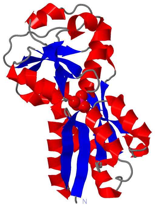
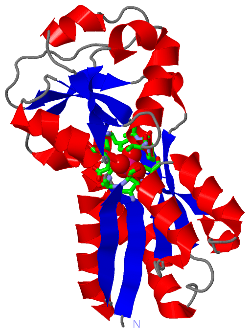
Asymmetric/Biological UnitChain A from PDB Type:PROTEIN Length:231 aligned with MODA_ECOLI | P37329 from UniProtKB/Swiss-Prot Length:257 Alignment length:231 36 46 56 66 76 86 96 106 116 126 136 146 156 166 176 186 196 206 216 226 236 246 256 MODA_ECOLI 27 GKITVFAAASLTNAMQDIATQFKKEKGVDVVSSFASSSTLARQIEAGAPADLFISADQKWMDYAVDKKAIDTATRQTLLGNSLVVVAPKASVQKDFTIDSKTNWTSLLNGGRLAVGDPEHVPAGIYAKEALQKLGAWDTLSPKLAPAEDVRGALALVERNEAPLGIVYGSDAVASKGVKVVATFPEDSHKKVEYPVAVVEGHNNATVKAFYDYLKGPQAAEIFKRYGFTIK 257 SCOP domains d3r26a_ A: automated matches SCOP domains CATH domains --------------------------------------------------------------------------------------------------------------------------------------------------------------------------------------------------------------------------------------- CATH domains Pfam domains --------------------------------------------------------------------------------------------------------------------------------------------------------------------------------------------------------------------------------------- Pfam domains SAPs(SNPs) --------------------------------------------------------------------------------------------------------------------------------------------------------------------------------------------------------------------------------------- SAPs(SNPs) PROSITE --------------------------------------------------------------------------------------------------------------------------------------------------------------------------------------------------------------------------------------- PROSITE Transcript --------------------------------------------------------------------------------------------------------------------------------------------------------------------------------------------------------------------------------------- Transcript 3r26 A 3 GKITVFAAASLTNAMQDIATQFKKEKGVDVVSSFASSSTLARQIEAGAPADLFISADQKWMDYAVDKKAIDTATRQTLLGNSLVVVAPKASVQKDFTIDSKTNWTSLLNGGRLAVGDPEHVPAGIYAKEALQKLGAWDTLSPKLAPAEDVRGALALVERNEAPLGIVYGSDAVASKGVKVVATFPEDSHKKVEYPVAVVEGHNNATVKAFYDYLKGPQAAEIFKRYGFTIK 233 12 22 32 42 52 62 72 82 92 102 112 122 132 142 152 162 172 182 192 202 212 222 232
|
||||||||||||||||||||
SCOP Domains (1, 1)
Asymmetric/Biological Unit
 |
CATH Domains (0, 0)| (no "CATH Domain" information available for 3R26) |
Pfam Domains (0, 0)| (no "Pfam Domain" information available for 3R26) |
Gene Ontology (5, 5)|
Asymmetric/Biological Unit(hide GO term definitions) Chain A (MODA_ECOLI | P37329)
|
||||||||||||||||||||||||||||||||||||||||||||||||
Interactive Views
|
||||||||||||||||||||||||||||||||||||||||||||||||||||||||||||||||||||||||||||||||||||||||||||||||||||||||||||||||||||||
Still Images
|
||||||||||||||||
Databases
|
||||||||||||||||||||||||||||||||||||||||||||||||||||||||||||||||||||||||||||||||||||||||||||||||||||||||||||||||||||||||||||||||||||||||||||||||||||||||||||||||
Analysis Tools
|
|||||||||||||||||||||||||||||||||||||||||||||||||||||||||||||
Entries Sharing at Least One Protein Chain (UniProt ID)
Related Entries Specified in the PDB File
|
|