Show PDB file:   
         Plain Text   HTML   (compressed file size)
QuickSearch:   
by PDB,NDB,UniProt,PROSITE Code or Search Term(s)  
(-)Asymmetric Unit
(-)Asym. Unit - sites
(-)Biological Unit 1
(-)Biol. Unit 1 - sites
(-)Biological Unit 2
(-)Biological Unit 3
(-)Biological Unit 4
collapse expand < >
Image Asymmetric Unit
Asymmetric Unit  (Jmol Viewer)
Image Asym. Unit - sites
Asym. Unit - sites  (Jmol Viewer)
Image Biological Unit 1
Biological Unit 1  (Jmol Viewer)
Image Biol. Unit 1 - sites
Biol. Unit 1 - sites  (Jmol Viewer)
Image Biological Unit 2
Biological Unit 2  (Jmol Viewer)
Image Biological Unit 3
Biological Unit 3  (Jmol Viewer)
Image Biological Unit 4
Biological Unit 4  (Jmol Viewer)

(-) Description

Title :  CRYSTAL STRUCTURE OF PYROPHOSPHATASE FROM BACTEROIDES THETAIOTAOMICRON COMPLEXED WITH CALCIUM, A CLOSED CAP CONFORMATION
 
Authors :  Y. Patskovsky, H. Huang, R. Toro, J. A. Gerlt, S. K. Burley, D. Dunaway-M S. C. Almo, New York Sgx Research Center For Structural Genomi (Nysgxrc), Enzyme Function Initiative (Efi)
Date :  01 Mar 11  (Deposition) - 27 Apr 11  (Release) - 19 Sep 12  (Revision)
Method :  X-RAY DIFFRACTION
Resolution :  1.24
Chains :  Asym. Unit :  A,B
Biol. Unit 1:  A,B  (2x)
Biol. Unit 2:  A (1x),B (1x)
Biol. Unit 3:  A  (1x)
Biol. Unit 4:  B  (1x)
Keywords :  Hydrolase, Pyrophosphatase, Magnesium Binding Site, New York Sgx Research Center For Structural Genomics, Enzyme Function Initiative, Efi, Psi-2, Protein Structure Initiative, Nysgxrc (Keyword Search: [Gene Ontology, PubMed, Web (Google))
 
Reference :  H. Huang, Y. Patskovsky, R. Toro, J. D. Farelli, C. Pandya, S. C. Almo, K. N. Allen, D. Dunaway-Mariano
Divergence Of Structure And Function In The Haloacid Dehalogenase Enzyme Superfamily: Bacteroides Thetaiotaomicron Bt2127 Is An Inorganic Pyrophosphatase.
Biochemistry V. 50 8937 2011
PubMed-ID: 21894910  |  Reference-DOI: 10.1021/BI201181Q

(-) Compounds

Molecule 1 - INORGANIC PYROPHOSPHATASE
    ChainsA, B
    EC Number3.6.1.1
    EngineeredYES
    GeneBT_2127
    Organism ScientificBACTEROIDES THETAIOTAOMICRON
    Organism Taxid818

 Structural Features

(-) Chains, Units

  12
Asymmetric Unit AB
Biological Unit 1 (2x)AB
Biological Unit 2 (1x)A (1x)B (1x)
Biological Unit 3 (1x)A 
Biological Unit 4 (1x) B

Summary Information (see also Sequences/Alignments below)

(-) Ligands, Modified Residues, Ions  (5, 15)

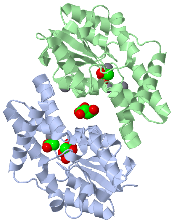
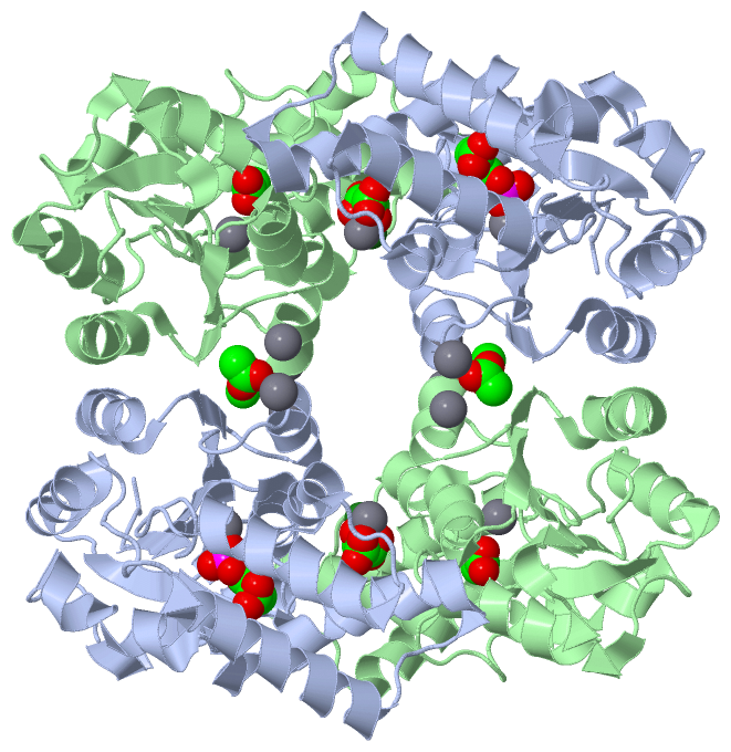
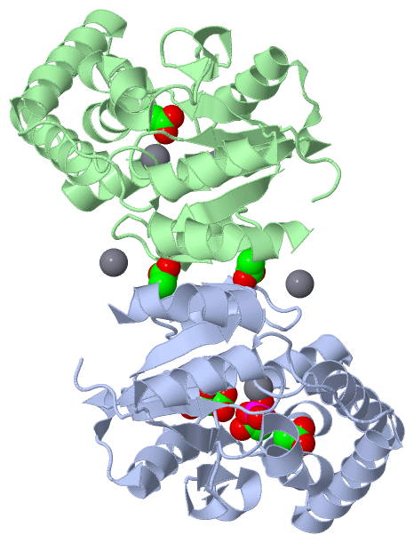
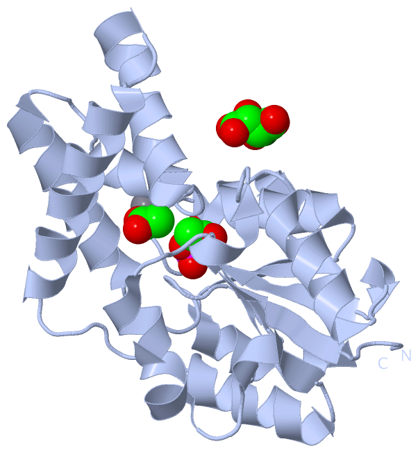
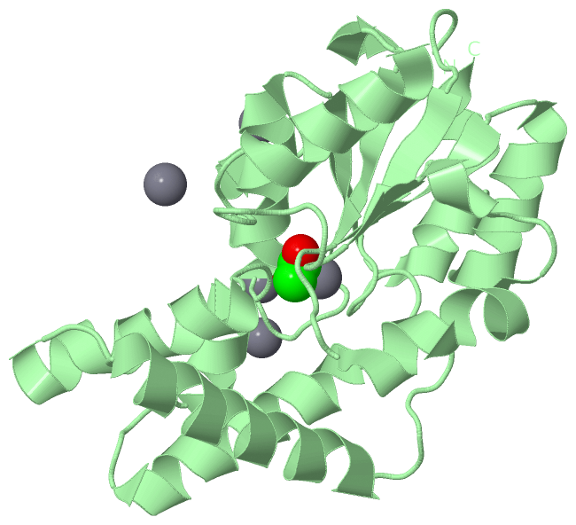
Asymmetric Unit (5, 15)
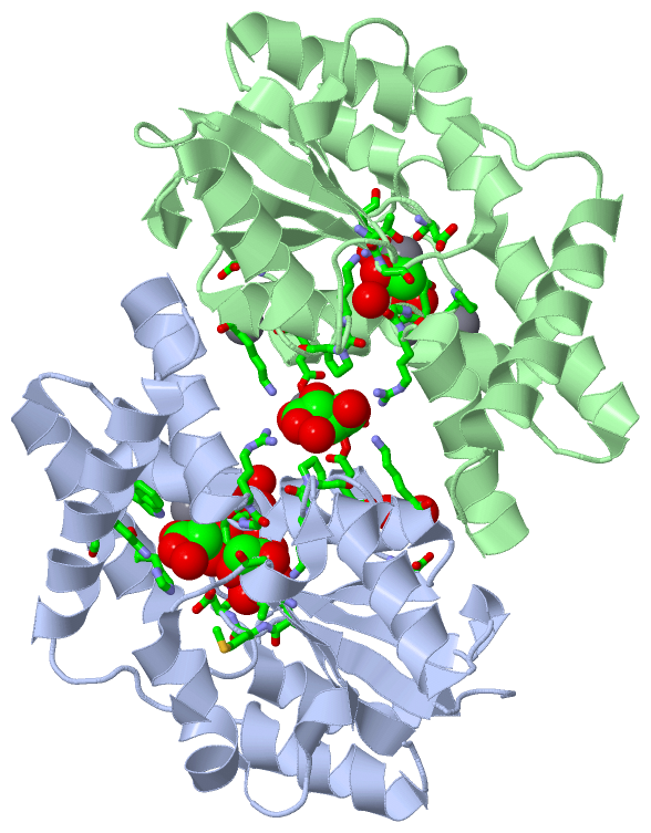
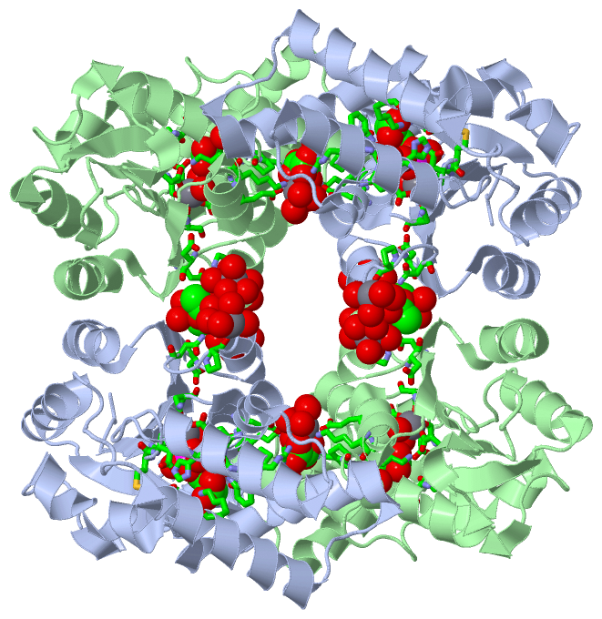
No.NameCountTypeFull Name
1ACT5Ligand/IonACETATE ION
2CA7Ligand/IonCALCIUM ION
3MG1Ligand/IonMAGNESIUM ION
4PO41Ligand/IonPHOSPHATE ION
5TLA1Ligand/IonL(+)-TARTARIC ACID
Biological Unit 1 (3, 14)
No.NameCountTypeFull Name
1ACT10Ligand/IonACETATE ION
2CA-1Ligand/IonCALCIUM ION
3MG-1Ligand/IonMAGNESIUM ION
4PO42Ligand/IonPHOSPHATE ION
5TLA2Ligand/IonL(+)-TARTARIC ACID
Biological Unit 2 (3, 5)
No.NameCountTypeFull Name
1ACT3Ligand/IonACETATE ION
2CA-1Ligand/IonCALCIUM ION
3MG-1Ligand/IonMAGNESIUM ION
4PO41Ligand/IonPHOSPHATE ION
5TLA1Ligand/IonL(+)-TARTARIC ACID
Biological Unit 3 (3, 5)
No.NameCountTypeFull Name
1ACT3Ligand/IonACETATE ION
2CA-1Ligand/IonCALCIUM ION
3MG-1Ligand/IonMAGNESIUM ION
4PO41Ligand/IonPHOSPHATE ION
5TLA1Ligand/IonL(+)-TARTARIC ACID
Biological Unit 4 (1, 2)
No.NameCountTypeFull Name
1ACT2Ligand/IonACETATE ION
2CA-1Ligand/IonCALCIUM ION
3MG-1Ligand/IonMAGNESIUM ION
4PO4-1Ligand/IonPHOSPHATE ION
5TLA-1Ligand/IonL(+)-TARTARIC ACID

(-) Sites  (15, 15)

Asymmetric Unit (15, 15)
No.NameEvidenceResiduesDescription
01AC1SOFTWAREASN A:191 , GLY A:197 , HOH A:269 , HOH A:307 , HOH A:479 , ASP B:206BINDING SITE FOR RESIDUE ACT A 225
02AC2SOFTWAREPRO A:194 , HOH A:375 , HOH A:386 , HOH A:387 , HOH A:388 , HOH A:414BINDING SITE FOR RESIDUE CA A 226
03AC3SOFTWAREASP A:11 , ASP A:13 , GLU A:47 , ASN A:172 , PO4 A:228 , HOH A:239 , HOH A:372BINDING SITE FOR RESIDUE CA A 227
04AC4SOFTWAREASP A:11 , MET A:12 , ASP A:13 , GLU A:47 , THR A:113 , LYS A:147 , CA A:227 , HOH A:239 , HOH A:285 , HOH A:371 , HOH A:372BINDING SITE FOR RESIDUE PO4 A 228
05AC5SOFTWAREARG A:49 , PRO A:148 , GLU A:178 , LYS A:182 , MG A:232 , HOH A:254 , HOH A:400 , HOH A:405 , ARG B:49 , PRO B:148 , GLU B:178 , LYS B:182 , CA B:231 , HOH B:394 , HOH B:395BINDING SITE FOR RESIDUE TLA A 229
06AC6SOFTWAREASP A:11 , GLY A:48 , THR A:113 , GLY A:114 , LYS A:147 , HOH A:372BINDING SITE FOR RESIDUE ACT A 230
07AC7SOFTWAREHIS A:23 , TRP A:27 , ARG A:49 , LYS A:79 , HOH A:292 , HOH A:293BINDING SITE FOR RESIDUE ACT A 231
08AC8SOFTWAREGLU A:178 , TLA A:229 , HOH A:250 , HOH A:254 , HOH A:405 , CA B:231 , HOH B:394BINDING SITE FOR RESIDUE MG A 232
09AC9SOFTWAREASP B:11 , GLY B:48 , THR B:113 , GLY B:114 , LYS B:147 , HOH B:247 , HOH B:333BINDING SITE FOR RESIDUE ACT B 225
10BC1SOFTWAREHOH A:297 , HOH A:307 , HOH A:376 , HOH A:398 , HOH A:437 , HOH B:312 , HOH B:463BINDING SITE FOR RESIDUE CA B 226
11BC2SOFTWAREHOH A:324 , HOH A:423 , HOH B:252 , HOH B:309 , HOH B:319 , HOH B:418BINDING SITE FOR RESIDUE CA B 227
12BC3SOFTWAREASP A:206 , ASN B:191 , GLY B:197 , HOH B:308 , HOH B:309 , HOH B:497BINDING SITE FOR RESIDUE ACT B 228
13BC4SOFTWAREPRO B:194 , HOH B:261 , HOH B:274 , HOH B:287 , HOH B:288 , HOH B:460BINDING SITE FOR RESIDUE CA B 229
14BC5SOFTWAREASP B:11 , ASP B:13 , GLU B:47 , ASN B:172 , HOH B:246 , HOH B:247BINDING SITE FOR RESIDUE CA B 230
15BC6SOFTWARETLA A:229 , MG A:232 , HOH A:405 , GLU B:178 , HOH B:394 , HOH B:395 , HOH B:396BINDING SITE FOR RESIDUE CA B 231

(-) SS Bonds  (0, 0)

(no "SS Bond" information available for 3QXG)

(-) Cis Peptide Bonds  (2, 2)

Asymmetric Unit
No.Residues
1Lys A:147 -Pro A:148
2Lys B:147 -Pro B:148

 Sequence-Structure Mapping

(-) SAPs(SNPs)/Variants  (0, 0)

(no "SAP(SNP)/Variant" information available for 3QXG)

(-) PROSITE Motifs  (0, 0)

(no "PROSITE Motif" information available for 3QXG)

(-) Exons   (0, 0)

(no "Exon" information available for 3QXG)

(-) Sequences/Alignments

Asymmetric Unit
   Reformat: Number of residues per line =  ('0' or empty: single-line sequence representation)
  Number of residues per labelling interval =   
  UniProt sequence: complete  aligned part    
   Show mapping: SCOP domains CATH domains Pfam domains Secondary structure (by author)
SAPs(SNPs) PROSITE motifs Exons
(details for a mapped element are shown in a popup box when the mouse pointer rests over it)
Chain A from PDB  Type:PROTEIN  Length:222
 aligned with Q8A5V9_BACTN | Q8A5V9 from UniProtKB/TrEMBL  Length:224

    Alignment length:222
                                    12        22        32        42        52        62        72        82        92       102       112       122       132       142       152       162       172       182       192       202       212       222  
         Q8A5V9_BACTN     3 KKLKAVLFDMDGVLFNSMPYHSEAWHQVMKTHGLDLSREEAYMHEGRTGASTINIVFQRELGKEATQEEIESIYHEKSILFNSYPEAERMPGAWELLQKVKSEGLTPMVVTGSGQLSLLERLEHNFPGMFHKELMVTAFDVKYGKPNPEPYLMALKKGGLKADEAVVIENAPLGVEAGHKAGIFTIAVNTGPLDGQVLLDAGADLLFPSMQTLCDSWDTIML 224
               SCOP domains d3qxga_ A: automated matches                                                                                                                                                                                                   SCOP domains
               CATH domains ------------------------------------------------------------------------------------------------------------------------------------------------------------------------------------------------------------------------------ CATH domains
               Pfam domains ------------------------------------------------------------------------------------------------------------------------------------------------------------------------------------------------------------------------------ Pfam domains
         Sec.struct. author ....eeee........hhhhhhhhhhhhhhhh....hhhhhhhh...hhhhhhhhhhhhhhh...hhhhhhhhhhhhhhhhhh........hhhhhhhhhhhh..eeeee....hhhhhhhhhhhh....hhh.eehhhhh......hhhhhhhhhhh..hhh.eeeee.hhhhhhhhhhh..eeeee.....hhhhhhhh...eee.hhhhhhhhhhhhh. Sec.struct. author
                 SAPs(SNPs) ------------------------------------------------------------------------------------------------------------------------------------------------------------------------------------------------------------------------------ SAPs(SNPs)
                    PROSITE ------------------------------------------------------------------------------------------------------------------------------------------------------------------------------------------------------------------------------ PROSITE
                 Transcript ------------------------------------------------------------------------------------------------------------------------------------------------------------------------------------------------------------------------------ Transcript
                 3qxg A   3 KKLKAVLFDMDGVLFNSMPYHSEAWHQVMKTHGLDLSREEAYMHEGRTGASTINIVFQRELGKEATQEEIESIYHEKSILFNSYPEAERMPGAWELLQKVKSEGLTPMVVTGSGQLSLLERLEHNFPGMFHKELMVTAFDVKYGKPNPEPYLMALKKGGLKADEAVVIENAPLGVEAGHKAGIFTIAVNTGPLDGQVLLDAGADLLFPSMQTLCDSWDTIML 224
                                    12        22        32        42        52        62        72        82        92       102       112       122       132       142       152       162       172       182       192       202       212       222  

Chain B from PDB  Type:PROTEIN  Length:223
 aligned with Q8A5V9_BACTN | Q8A5V9 from UniProtKB/TrEMBL  Length:224

    Alignment length:223
                                    11        21        31        41        51        61        71        81        91       101       111       121       131       141       151       161       171       181       191       201       211       221   
         Q8A5V9_BACTN     2 RKKLKAVLFDMDGVLFNSMPYHSEAWHQVMKTHGLDLSREEAYMHEGRTGASTINIVFQRELGKEATQEEIESIYHEKSILFNSYPEAERMPGAWELLQKVKSEGLTPMVVTGSGQLSLLERLEHNFPGMFHKELMVTAFDVKYGKPNPEPYLMALKKGGLKADEAVVIENAPLGVEAGHKAGIFTIAVNTGPLDGQVLLDAGADLLFPSMQTLCDSWDTIML 224
               SCOP domains d3qxgb_ B: automated matches                                                                                                                                                                                                    SCOP domains
               CATH domains ------------------------------------------------------------------------------------------------------------------------------------------------------------------------------------------------------------------------------- CATH domains
           Pfam domains (1) ------HAD_2-3qxgB01 B:8-190                                                                                                                                                                  ---------------------------------- Pfam domains (1)
           Pfam domains (2) ------HAD_2-3qxgB02 B:8-190                                                                                                                                                                  ---------------------------------- Pfam domains (2)
         Sec.struct. author .....eeee........hhhhhhhhhhhhhhhh....hhhhhhhh...hhhhhhhhhhhhhhh...hhhhhhhhhhhhhhhhhh........hhhhhhhhhhhh..eeeee....hhhhhhhhhhhh....hhh.ee...........hhhhhhhhhhhh.hhh.eeeee.hhhhhhhhhhh..eeeee.....hhhhhhhh...eee.hhhhhhhhhhhhhh Sec.struct. author
                 SAPs(SNPs) ------------------------------------------------------------------------------------------------------------------------------------------------------------------------------------------------------------------------------- SAPs(SNPs)
                    PROSITE ------------------------------------------------------------------------------------------------------------------------------------------------------------------------------------------------------------------------------- PROSITE
                 Transcript ------------------------------------------------------------------------------------------------------------------------------------------------------------------------------------------------------------------------------- Transcript
                 3qxg B   2 RKKLKAVLFDMDGVLFNSMPYHSEAWHQVMKTHGLDLSREEAYMHEGRTGASTINIVFQRELGKEATQEEIESIYHEKSILFNSYPEAERMPGAWELLQKVKSEGLTPMVVTGSGQLSLLERLEHNFPGMFHKELMVTAFDVKYGKPNPEPYLMALKKGGLKADEAVVIENAPLGVEAGHKAGIFTIAVNTGPLDGQVLLDAGADLLFPSMQTLCDSWDTIML 224
                                    11        21        31        41        51        61        71        81        91       101       111       121       131       141       151       161       171       181       191       201       211       221   

   Legend:   → Mismatch (orange background)
  - → Gap (green background, '-', border residues have a numbering label)
    → Modified Residue (blue background, lower-case, 'x' indicates undefined single-letter code, labelled with number + name)
  x → Chemical Group (purple background, 'x', labelled with number + name, e.g. ACE or NH2)
  extra numbering lines below/above indicate numbering irregularities and modified residue names etc., number ends below/above '|'

 Classification and Annotation

(-) SCOP Domains  (1, 2)

Asymmetric Unit

(-) CATH Domains  (0, 0)

(no "CATH Domain" information available for 3QXG)

(-) Pfam Domains  (1, 2)

Asymmetric Unit
(-)
Clan: HAD (186)

(-) Gene Ontology  (3, 3)

Asymmetric Unit(hide GO term definitions)
Chain A,B   (Q8A5V9_BACTN | Q8A5V9)
molecular function
    GO:0016787    hydrolase activity    Catalysis of the hydrolysis of various bonds, e.g. C-O, C-N, C-C, phosphoric anhydride bonds, etc. Hydrolase is the systematic name for any enzyme of EC class 3.
    GO:0046872    metal ion binding    Interacting selectively and non-covalently with any metal ion.
biological process
    GO:0008152    metabolic process    The chemical reactions and pathways, including anabolism and catabolism, by which living organisms transform chemical substances. Metabolic processes typically transform small molecules, but also include macromolecular processes such as DNA repair and replication, and protein synthesis and degradation.

 Visualization

(-) Interactive Views

Asymmetric Unit
  Complete Structure
    Jena3D(integrated viewing of ligand, site, SAP, PROSITE, SCOP information)
    WebMol | AstexViewer[tm]@PDBe
(Java Applets, require no local installation except for Java; loading may be slow)
    STRAP
(Java WebStart application, automatic local installation, requires Java; full application with system access!)
    RasMol
(require local installation)
    Molscript (VRML)
(requires installation of a VRML viewer; select preferred view via VRML and generate a mono or stereo PDF format file)
 
  Ligands, Modified Residues, Ions
    ACT  [ RasMol | Jena3D ]  +environment [ RasMol | Jena3D ]
    CA  [ RasMol | Jena3D ]  +environment [ RasMol | Jena3D ]
    MG  [ RasMol | Jena3D ]  +environment [ RasMol | Jena3D ]
    PO4  [ RasMol | Jena3D ]  +environment [ RasMol | Jena3D ]
    TLA  [ RasMol | Jena3D ]  +environment [ RasMol | Jena3D ]
 
  Sites
    AC1  [ RasMol ]  +environment [ RasMol ]
    AC2  [ RasMol ]  +environment [ RasMol ]
    AC3  [ RasMol ]  +environment [ RasMol ]
    AC4  [ RasMol ]  +environment [ RasMol ]
    AC5  [ RasMol ]  +environment [ RasMol ]
    AC6  [ RasMol ]  +environment [ RasMol ]
    AC7  [ RasMol ]  +environment [ RasMol ]
    AC8  [ RasMol ]  +environment [ RasMol ]
    AC9  [ RasMol ]  +environment [ RasMol ]
    BC1  [ RasMol ]  +environment [ RasMol ]
    BC2  [ RasMol ]  +environment [ RasMol ]
    BC3  [ RasMol ]  +environment [ RasMol ]
    BC4  [ RasMol ]  +environment [ RasMol ]
    BC5  [ RasMol ]  +environment [ RasMol ]
    BC6  [ RasMol ]  +environment [ RasMol ]
 
  Cis Peptide Bonds
    Lys A:147 - Pro A:148   [ RasMol ]  
    Lys B:147 - Pro B:148   [ RasMol ]  
 
Biological Units
  Complete Structure
    Biological Unit 1  [ Jena3D ]
    Biological Unit 2  [ Jena3D ]
    Biological Unit 3  [ Jena3D ]
    Biological Unit 4  [ Jena3D ]

(-) Still Images

Jmol
  protein: cartoon or spacefill or dots and stick; nucleic acid: cartoon and stick; ligands: spacefill; active site: stick
Molscript
  protein, nucleic acid: cartoon; ligands: spacefill; active site: ball and stick

 Databases and Analysis Tools

(-) Databases

Access by PDB/NDB ID
  3qxg
    Family and Domain InformationProDom | SYSTERS
    General Structural InformationGlycoscienceDB | MMDB | NDB | OCA | PDB | PDBe | PDBj | PDBsum | PDBWiki | PQS | PROTEOPEDIA
    Orientation in MembranesOPM
    Protein SurfaceSURFACE
    Secondary StructureDSSP (structure derived) | HSSP (homology derived)
    Structural GenomicsGeneCensus
    Structural NeighboursCE | VAST
    Structure ClassificationCATH | Dali | SCOP
    Validation and Original DataBMRB Data View | BMRB Restraints Grid | EDS | PROCHECK | RECOORD | WHAT_CHECK
 
Access by UniProt ID/Accession number
  Q8A5V9_BACTN | Q8A5V9
    Comparative Protein Structure ModelsModBase
    Genomic InformationEnsembl
    Protein-protein InteractionDIP
    Sequence, Family and Domain InformationInterPro | Pfam | SMART | UniProtKB/TrEMBL
 
Access by Enzyme Classificator   (EC Number)
  3.6.1.1
    General Enzyme InformationBRENDA | EC-PDB | Enzyme | IntEnz
    PathwayKEGG | MetaCyc
 
Access by Disease Identifier   (MIM ID)
  (no 'MIM ID' available)
    Disease InformationOMIM
 
Access by GenAge ID
  (no 'GenAge ID' available)
    Age Related InformationGenAge

(-) Analysis Tools

Access by PDB/NDB ID
    Domain InformationXDom
    Interatomic Contacts of Structural UnitsCSU
    Ligand-protein ContactsLPC
    Protein CavitiescastP
    Sequence and Secondary StructurePDBCartoon
    Structure AlignmentSTRAP(Java WebStart application, automatic local installation, requires Java; full application with system access!)
    Structure and Sequence BrowserSTING
 
Access by UniProt ID/Accession number
  Q8A5V9_BACTN | Q8A5V9
    Protein Disorder PredictionDisEMBL | FoldIndex | GLOBPLOT (for more information see DisProt)

 Related Entries

(-) Entries Sharing at Least One Protein Chain (UniProt ID)

UniProtKB/TrEMBL
        Q8A5V9_BACTN | Q8A5V93qu2 3qu4 3qu5 3qu7 3qu9 3qub 3quc 3quq 3qut 3qx7 3qyp 3r9k

(-) Related Entries Specified in the PDB File

3qu2 CRYSTAL STRUCTURE OF PYROPHOSPHATASE FROM BACTEROIDES THETAIOTAOMICRON, A CLOSED CAP CONFORMATION, WILD-TYPE PROTEIN
3qu4 CRYSTAL STRUCTURE OF PYROPHOSPHATASE FROM BACTEROIDES THETAIOTAOMICRON, ASP13ALA MUTANT
3qu5 CRYSTAL STRUCTURE OF PYROPHOSPHATASE FROM BACTEROIDES THETAIOTAOMICRON, ASP11ASN MUTANT
3qu7 CRYSTAL STRUCTURE OF PYROPHOSPHATASE FROM BACTEROIDES THETAIOTAOMICRON, ASP13ASN MUTANT COMPLEXED WITH CALCIUM AND PHOSPHATE
3qu9 CRYSTAL STRUCTURE OF PYROPHOSPHATASE FROM BACTEROIDES THETAIOTAOMICRON, ASP13ASN MUTANT COMPLEXED WITH MAGNESIUM AND TARTRATE
3qub CRYSTAL STRUCTURE OF PYROPHOSPHATASE FROM BACTEROIDES THETAIOTAOMICRON, GLU47ALA MUTANT COMPLEXED WITH SULFATE
3quc CRYSTAL STRUCTURE OF PYROPHOSPHATASE FROM BACTEROIDES THETAIOTAOMICRON, GLU47ASN MUTANT COMPLEXED WITH SULFATE
3quq CRYSTAL STRUCTURE OF PYROPHOSPHATASE FROM BACTEROIDES THETAIOTAOMICRON, AN OPEN CAP CONFORMATION, WILD-TYPE PROTEIN
3qut CRYSTAL STRUCTURE OF PYROPHOSPHATASE FROM BACTEROIDES THETAIOTAOMICRON, ASP13ASN MUTANT, AN OPEN CAP CONFORMATION
3qx7 CRYSTAL STRUCTURE OF PYROPHOSPHATASE FROM BACTEROIDES THETAIOTAOMICRON COMPLEXED WITH PHOSPHATE, A CLOSED CAP CONFORMATION, WILD-TYPE PROTEIN
3qyp CRYSTAL STRUCTURE OF PYROPHOSPHATASE FROM BACTEROIDES THETAIOTAOMICRON, GLU47ASN MUTANT COMPLEXED WITH CALCIUM AND PHOSPHATE
3r9k CRYSTAL STRUCTURE OF PYROPHOSPHATASE FROM BACTEROIDES THETAIOTAOMICRON COMPLEXED WITH CALCIUM, GLU47ASP MUTANT COMPLEXED WITH SULFATE, A CLOSED CAP CONFORMATION