Show PDB file:   
         Plain Text   HTML   (compressed file size)
QuickSearch:   
by PDB,NDB,UniProt,PROSITE Code or Search Term(s)  
(-)Asymmetric Unit
(-)Asym. Unit - sites
(-)Biological Unit 1
(-)Biol. Unit 1 - sites
collapse expand < >
Image Asymmetric Unit
Asymmetric Unit  (Jmol Viewer)
Image Asym. Unit - sites
Asym. Unit - sites  (Jmol Viewer)
Image Biological Unit 1
Biological Unit 1  (Jmol Viewer)
Image Biol. Unit 1 - sites
Biol. Unit 1 - sites  (Jmol Viewer)

(-) Description

Title :  HUMAN GLUTATHIONE TRANSFERASE O2 WITH GLUTATHIONE -NEW CRYSTAL FORM
 
Authors :  H. Zhou, P. G. Board, A. J. Oakley
Date :  11 Jan 11  (Deposition) - 11 Jan 12  (Release) - 26 Jun 13  (Revision)
Method :  X-RAY DIFFRACTION
Resolution :  2.00
Chains :  Asym. Unit :  A
Biol. Unit 1:  A  (2x)
Keywords :  Glutathione Transferase, Gst, Dehydroascorbate Reductase, Transferase, Reductase (Keyword Search: [Gene Ontology, PubMed, Web (Google))
 
Reference :  H. Zhou, J. Brock, D. Liu, P. G. Board, A. J. Oakley
Structural Insights Into The Dehydroascorbate Reductase Activity Of Human Omega-Class Glutathione Transferases.
J. Mol. Biol. V. 420 190 2012
PubMed-ID: 22522127  |  Reference-DOI: 10.1016/J.JMB.2012.04.014

(-) Compounds

Molecule 1 - GLUTATHIONE S-TRANSFERASE OMEGA-2
    ChainsA
    EC Number2.5.1.18
    EngineeredYES
    Expression SystemESCHERICHIA COLI
    Expression System PlasmidPQE30
    Expression System StrainM15/REP4
    Expression System Taxid562
    Expression System Vector TypePLASMID
    GeneGSTO2
    MutationYES
    Organism CommonHUMAN
    Organism ScientificHOMO SAPIENS
    Organism Taxid9606
    SynonymGSTO-2

 Structural Features

(-) Chains, Units

  1
Asymmetric Unit A
Biological Unit 1 (2x)A

Summary Information (see also Sequences/Alignments below)

(-) Ligands, Modified Residues, Ions  (3, 4)

Asymmetric Unit (3, 4)
No.NameCountTypeFull Name
1EDO1Ligand/Ion1,2-ETHANEDIOL
2GSH1Ligand/IonGLUTATHIONE
3PEG2Ligand/IonDI(HYDROXYETHYL)ETHER
Biological Unit 1 (3, 8)
No.NameCountTypeFull Name
1EDO2Ligand/Ion1,2-ETHANEDIOL
2GSH2Ligand/IonGLUTATHIONE
3PEG4Ligand/IonDI(HYDROXYETHYL)ETHER

(-) Sites  (3, 3)

Asymmetric Unit (3, 3)
No.NameEvidenceResiduesDescription
1AC1SOFTWARECYS A:32 , TYR A:34 , LYS A:59 , HIS A:71 , ILE A:72 , PRO A:73 , GLU A:85 , SER A:86 , HOH A:249 , HOH A:308 , HOH A:327BINDING SITE FOR RESIDUE GSH A 1001
2AC2SOFTWARELYS A:101 , LEU A:102 , PHE A:103 , GLY A:167 , SER A:172BINDING SITE FOR RESIDUE PEG A 1002
3AC3SOFTWARETRP A:209BINDING SITE FOR RESIDUE PEG A 1003

(-) SS Bonds  (0, 0)

(no "SS Bond" information available for 3QAG)

(-) Cis Peptide Bonds  (1, 1)

Asymmetric Unit
No.Residues
1Ile A:72 -Pro A:73

 Sequence-Structure Mapping

(-) SAPs(SNPs)/Variants  (2, 2)

Asymmetric Unit (2, 2)
  dbSNPPDB
No.SourceVariant IDVariantUniProt IDStatusIDChainVariant
1UniProtVAR_049492C130YGSTO2_HUMANPolymorphism45582439AC130Y
2UniProtVAR_016812N142DGSTO2_HUMANPolymorphism156697AN142D

  SNP/SAP Summary Statistics (UniProtKB/Swiss-Prot)
Biological Unit 1 (2, 4)
  dbSNPPDB
No.SourceVariant IDVariantUniProt IDStatusIDChainVariant
1UniProtVAR_049492C130YGSTO2_HUMANPolymorphism45582439AC130Y
2UniProtVAR_016812N142DGSTO2_HUMANPolymorphism156697AN142D

  SNP/SAP Summary Statistics (UniProtKB/Swiss-Prot)

(-) PROSITE Motifs  (2, 2)

Asymmetric Unit (2, 2)
 PROSITEUniProtKBPDB
No.IDACDescriptionIDLocationCountLocation
1GST_NTERPS50404 Soluble glutathione S-transferase N-terminal domain profile.GSTO2_HUMAN22-101  1A:22-101
2GST_CTERPS50405 Soluble glutathione S-transferase C-terminal domain profile.GSTO2_HUMAN106-231  1A:106-231
Biological Unit 1 (2, 4)
 PROSITEUniProtKBPDB
No.IDACDescriptionIDLocationCountLocation
1GST_NTERPS50404 Soluble glutathione S-transferase N-terminal domain profile.GSTO2_HUMAN22-101  2A:22-101
2GST_CTERPS50405 Soluble glutathione S-transferase C-terminal domain profile.GSTO2_HUMAN106-231  2A:106-231

(-) Exons   (0, 0)

(no "Exon" information available for 3QAG)

(-) Sequences/Alignments

Asymmetric Unit
   Reformat: Number of residues per line =  ('0' or empty: single-line sequence representation)
  Number of residues per labelling interval =   
  UniProt sequence: complete  aligned part    
   Show mapping: SCOP domains CATH domains Pfam domains Secondary structure (by author)
SAPs(SNPs) PROSITE motifs Exons
(details for a mapped element are shown in a popup box when the mouse pointer rests over it)
Chain A from PDB  Type:PROTEIN  Length:238
 aligned with GSTO2_HUMAN | Q9H4Y5 from UniProtKB/Swiss-Prot  Length:243

    Alignment length:238
                                    11        21        31        41        51        61        71        81        91       101       111       121       131       141       151       161       171       181       191       201       211       221       231        
          GSTO2_HUMAN     2 SGDATRTLGKGSQPPGPVPEGLIRIYSMRFCPYSHRTRLVLKAKDIRHEVVNINLRNKPEWYYTKHPFGHIPVLETSQCQLIYESVIACEYLDDAYPGRKLFPYDPYERARQKMLLELFCKVPHLTKECLVALRCGRECTNLKAALRQEFSNLEEILEYQNTTFFGGTCISMIDYLLWPWFERLDVYGILDCVSHTPALRLWISAMKWDPTVCALLMDKSIFQGFLNLYFQNNPNAFD 239
               SCOP domains d3qaga1 A:2-102 automated matches                                                                    d3qaga2 A:103-239 automated matches                                                                                                       SCOP domains
               CATH domains ---------------------------------------------------------------------------------------------------------------------------------------------------------------------------------------------------------------------------------------------- CATH domains
               Pfam domains ---------------------------------------------------------------------------------------------------------------------------------------------------------------------------------------------------------------------------------------------- Pfam domains
         Sec.struct. author ......................eeeee...hhhhhhhhhhhhhh...eeeee......hhhhhhh.......eee.....eeehhhhhhhhhhhhh........hhhhhhhhhhhhhhh.hhhhhhhhhhhhhh....hhhhhhhhhhhhhhhhhhhhhhh.........hhhhhhhhhhhhhhhhhhhhhhhh.hhhhhhhhhhhhhhhhhhhhh.hhhhhhhhhhhhhhhhhhhhh Sec.struct. author
                 SAPs(SNPs) --------------------------------------------------------------------------------------------------------------------------------Y-----------D------------------------------------------------------------------------------------------------- SAPs(SNPs)
                    PROSITE --------------------GST_NTER  PDB: A:22-101 UniProt: 22-101                                         ----GST_CTER  PDB: A:106-231 UniProt: 106-231                                                                                     -------- PROSITE
                 Transcript ---------------------------------------------------------------------------------------------------------------------------------------------------------------------------------------------------------------------------------------------- Transcript
                 3qag A   2 SGDATRTLGKGSQPPGPVPEGLIRIYSMRFCPYSHRTRLVLKAKDIRHEVVNINLRNKPEWYYTKHPFGHIPVLETSQSQLIYESVIACEYLDDAYPGRKLFPYDPYERARQKMLLELFSKVPHLTKECLVALRSGRESTNLKAALRQEFSNLEEILEYQNTTFFGGTSISMIDYLLWPWFERLDVYGILDCVSHTPALRLWISAMKWDPTVSALLMDKSIFQGFLNLYFQNNPNAFD 239
                                    11        21        31        41        51        61        71        81        91       101       111       121       131       141       151       161       171       181       191       201       211       221       231        

   Legend:   → Mismatch (orange background)
  - → Gap (green background, '-', border residues have a numbering label)
    → Modified Residue (blue background, lower-case, 'x' indicates undefined single-letter code, labelled with number + name)
  x → Chemical Group (purple background, 'x', labelled with number + name, e.g. ACE or NH2)
  extra numbering lines below/above indicate numbering irregularities and modified residue names etc., number ends below/above '|'

 Classification and Annotation

(-) SCOP Domains  (2, 2)

Asymmetric Unit

(-) CATH Domains  (0, 0)

(no "CATH Domain" information available for 3QAG)

(-) Pfam Domains  (0, 0)

(no "Pfam Domain" information available for 3QAG)

(-) Gene Ontology  (17, 17)

Asymmetric Unit(hide GO term definitions)
Chain A   (GSTO2_HUMAN | Q9H4Y5)
molecular function
    GO:0045174    glutathione dehydrogenase (ascorbate) activity    Catalysis of the reaction: dehydroascorbate + 2 glutathione = L-ascorbate + glutathione disulfide.
    GO:0004364    glutathione transferase activity    Catalysis of the reaction: R-X + glutathione = H-X + R-S-glutathione. R may be an aliphatic, aromatic or heterocyclic group; X may be a sulfate, nitrile or halide group.
    GO:0050610    methylarsonate reductase activity    Catalysis of the reaction: 2 glutathione + H(+) + methylarsonate = glutathione disulfide + H(2)O + methylarsonous acid.
    GO:0016491    oxidoreductase activity    Catalysis of an oxidation-reduction (redox) reaction, a reversible chemical reaction in which the oxidation state of an atom or atoms within a molecule is altered. One substrate acts as a hydrogen or electron donor and becomes oxidized, while the other acts as hydrogen or electron acceptor and becomes reduced.
    GO:0005515    protein binding    Interacting selectively and non-covalently with any protein or protein complex (a complex of two or more proteins that may include other nonprotein molecules).
    GO:0016740    transferase activity    Catalysis of the transfer of a group, e.g. a methyl group, glycosyl group, acyl group, phosphorus-containing, or other groups, from one compound (generally regarded as the donor) to another compound (generally regarded as the acceptor). Transferase is the systematic name for any enzyme of EC class 2.
biological process
    GO:0019852    L-ascorbic acid metabolic process    The chemical reactions and pathways involving L-ascorbic acid, (2R)-2-[(1S)-1,2-dihydroxyethyl]-4-hydroxy-5-oxo-2,5-dihydrofuran-3-olate; L-ascorbic acid is vitamin C and has co-factor and anti-oxidant activities in many species.
    GO:0098869    cellular oxidant detoxification    Any process carried out at the cellular level that reduces or removes the toxicity superoxide radicals or hydrogen peroxide.
    GO:0071243    cellular response to arsenic-containing substance    Any process that results in a change in state or activity of a cell (in terms of movement, secretion, enzyme production, gene expression, etc.) as a result of an arsenic stimulus from compounds containing arsenic, including arsenates, arsenites, and arsenides.
    GO:1901687    glutathione derivative biosynthetic process    The chemical reactions and pathways resulting in the formation of glutathione derivative.
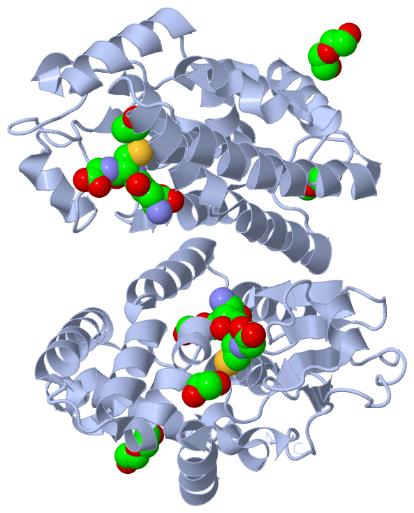
    GO:0006749    glutathione metabolic process    The chemical reactions and pathways involving glutathione, the tripeptide glutamylcysteinylglycine, which acts as a coenzyme for some enzymes and as an antioxidant in the protection of sulfhydryl groups in enzymes and other proteins; it has a specific role in the reduction of hydrogen peroxide (H2O2) and oxidized ascorbate, and it participates in the gamma-glutamyl cycle.
    GO:0008152    metabolic process    The chemical reactions and pathways, including anabolism and catabolism, by which living organisms transform chemical substances. Metabolic processes typically transform small molecules, but also include macromolecular processes such as DNA repair and replication, and protein synthesis and degradation.
    GO:0055114    oxidation-reduction process    A metabolic process that results in the removal or addition of one or more electrons to or from a substance, with or without the concomitant removal or addition of a proton or protons.
    GO:0006805    xenobiotic metabolic process    The chemical reactions and pathways involving a xenobiotic compound, a compound foreign to living organisms. Used of chemical compounds, e.g. a xenobiotic chemical, such as a pesticide.
cellular component
    GO:0005737    cytoplasm    All of the contents of a cell excluding the plasma membrane and nucleus, but including other subcellular structures.
    GO:0005829    cytosol    The part of the cytoplasm that does not contain organelles but which does contain other particulate matter, such as protein complexes.
    GO:0070062    extracellular exosome    A vesicle that is released into the extracellular region by fusion of the limiting endosomal membrane of a multivesicular body with the plasma membrane. Extracellular exosomes, also simply called exosomes, have a diameter of about 40-100 nm.

 Visualization

(-) Interactive Views

Asymmetric Unit
  Complete Structure
    Jena3D(integrated viewing of ligand, site, SAP, PROSITE, SCOP information)
    WebMol | AstexViewer[tm]@PDBe
(Java Applets, require no local installation except for Java; loading may be slow)
    STRAP
(Java WebStart application, automatic local installation, requires Java; full application with system access!)
    RasMol
(require local installation)
    Molscript (VRML)
(requires installation of a VRML viewer; select preferred view via VRML and generate a mono or stereo PDF format file)
 
  Ligands, Modified Residues, Ions
    EDO  [ RasMol | Jena3D ]  +environment [ RasMol | Jena3D ]
    GSH  [ RasMol | Jena3D ]  +environment [ RasMol | Jena3D ]
    PEG  [ RasMol | Jena3D ]  +environment [ RasMol | Jena3D ]
 
  Sites
    AC1  [ RasMol ]  +environment [ RasMol ]
    AC2  [ RasMol ]  +environment [ RasMol ]
    AC3  [ RasMol ]  +environment [ RasMol ]
 
  Cis Peptide Bonds
    Ile A:72 - Pro A:73   [ RasMol ]  
 
Biological Unit
  Complete Structure
    Biological Unit 1  [ Jena3D ]

(-) Still Images

Jmol
  protein: cartoon or spacefill or dots and stick; nucleic acid: cartoon and stick; ligands: spacefill; active site: stick
Molscript
  protein, nucleic acid: cartoon; ligands: spacefill; active site: ball and stick

 Databases and Analysis Tools

(-) Databases

Access by PDB/NDB ID
  3qag
    Family and Domain InformationProDom | SYSTERS
    General Structural InformationGlycoscienceDB | MMDB | NDB | OCA | PDB | PDBe | PDBj | PDBsum | PDBWiki | PQS | PROTEOPEDIA
    Orientation in MembranesOPM
    Protein SurfaceSURFACE
    Secondary StructureDSSP (structure derived) | HSSP (homology derived)
    Structural GenomicsGeneCensus
    Structural NeighboursCE | VAST
    Structure ClassificationCATH | Dali | SCOP
    Validation and Original DataBMRB Data View | BMRB Restraints Grid | EDS | PROCHECK | RECOORD | WHAT_CHECK
 
Access by UniProt ID/Accession number
  GSTO2_HUMAN | Q9H4Y5
    Comparative Protein Structure ModelsModBase
    Genomic InformationEnsembl
    Protein-protein InteractionDIP
    Sequence, Family and Domain InformationInterPro | Pfam | SMART | UniProtKB/SwissProt
 
Access by Enzyme Classificator   (EC Number)
  2.5.1.18
    General Enzyme InformationBRENDA | EC-PDB | Enzyme | IntEnz
    PathwayKEGG | MetaCyc
 
Access by Disease Identifier   (MIM ID)
  (no 'MIM ID' available)
    Disease InformationOMIM
 
Access by GenAge ID
  (no 'GenAge ID' available)
    Age Related InformationGenAge

(-) Analysis Tools

Access by PDB/NDB ID
    Domain InformationXDom
    Interatomic Contacts of Structural UnitsCSU
    Ligand-protein ContactsLPC
    Protein CavitiescastP
    Sequence and Secondary StructurePDBCartoon
    Structure AlignmentSTRAP(Java WebStart application, automatic local installation, requires Java; full application with system access!)
    Structure and Sequence BrowserSTING
 
Access by UniProt ID/Accession number
  GSTO2_HUMAN | Q9H4Y5
    Protein Disorder PredictionDisEMBL | FoldIndex | GLOBPLOT (for more information see DisProt)

 Related Entries

(-) Entries Sharing at Least One Protein Chain (UniProt ID)

UniProtKB/Swiss-Prot
        GSTO2_HUMAN | Q9H4Y53q18 3q19

(-) Related Entries Specified in the PDB File

1eem GSTO1
3q18 GSTO2
3q19 GSTO2 WITH GSH