Show PDB file:   
         Plain Text   HTML   (compressed file size)
QuickSearch:   
by PDB,NDB,UniProt,PROSITE Code or Search Term(s)  
(-)Asymmetric Unit
(-)Asym. Unit - sites
(-)Biological Unit 1
(-)Biol. Unit 1 - sites
(-)Biological Unit 2
(-)Biological Unit 3
collapse expand < >
Image Asymmetric Unit
Asymmetric Unit  (Jmol Viewer)
Image Asym. Unit - sites
Asym. Unit - sites  (Jmol Viewer)
Image Biological Unit 1
Biological Unit 1  (Jmol Viewer)
Image Biol. Unit 1 - sites
Biol. Unit 1 - sites  (Jmol Viewer)
Image Biological Unit 2
Biological Unit 2  (Jmol Viewer)
Image Biological Unit 3
Biological Unit 3  (Jmol Viewer)

(-) Description

Title :  CRYSTAL STRUCTURE OF THE MAIN PROTEASE (3C) FROM HUMAN ENTEROVIRUS B EV93
 
Authors :  L. Costenaro, M. Sola, B. Coutard, H. Norder, B. Canard, M. Coll
Date :  22 Dec 10  (Deposition) - 07 Sep 11  (Release) - 05 Oct 11  (Revision)
Method :  X-RAY DIFFRACTION
Resolution :  1.90
Chains :  Asym. Unit :  A,B
Biol. Unit 1:  A  (1x)
Biol. Unit 2:  B  (1x)
Biol. Unit 3:  A,B  (1x)
Keywords :  Cysteine Trypsin-Like Protease, 3C Cysteine Protease (Picornain 3C), Hydrolase (Keyword Search: [Gene Ontology, PubMed, Web (Google))
 
Reference :  L. Costenaro, Z. Kaczmarska, C. Arnan, R. Janowski, B. Coutard, M. Sola, A. E. Gorbalenya, H. Norder, B. Canard, M. Coll
Structural Basis For Antiviral Inhibition Of The Main Protease, 3C, From Human Enterovirus 93.
J. Virol. V. 85 10764 2011
PubMed-ID: 21835784  |  Reference-DOI: 10.1128/JVI.05062-11

(-) Compounds

Molecule 1 - HEVB EV93 3C PROTEASE
    ChainsA, B
    EC Number3.4.22.28
    EngineeredYES
    Expression SystemESCHERICHIA COLI
    Expression System PlasmidPDEST14
    Expression System StrainBL21
    Expression System Taxid562
    Expression System Vector TypePLASMID
    Gene3C
    MutationYES
    Organism ScientificHUMAN ENTEROVIRUS B
    Organism Taxid138949

 Structural Features

(-) Chains, Units

  12
Asymmetric Unit AB
Biological Unit 1 (1x)A 
Biological Unit 2 (1x) B
Biological Unit 3 (1x)AB

Summary Information (see also Sequences/Alignments below)

(-) Ligands, Modified Residues, Ions  (3, 10)

Asymmetric Unit (3, 10)
No.NameCountTypeFull Name
1GOL6Ligand/IonGLYCEROL
2MG2Ligand/IonMAGNESIUM ION
3OCS2Mod. Amino AcidCYSTEINESULFONIC ACID
Biological Unit 1 (2, 4)
No.NameCountTypeFull Name
1GOL3Ligand/IonGLYCEROL
2MG-1Ligand/IonMAGNESIUM ION
3OCS1Mod. Amino AcidCYSTEINESULFONIC ACID
Biological Unit 2 (2, 4)
No.NameCountTypeFull Name
1GOL3Ligand/IonGLYCEROL
2MG-1Ligand/IonMAGNESIUM ION
3OCS1Mod. Amino AcidCYSTEINESULFONIC ACID
Biological Unit 3 (2, 8)
No.NameCountTypeFull Name
1GOL6Ligand/IonGLYCEROL
2MG-1Ligand/IonMAGNESIUM ION
3OCS2Mod. Amino AcidCYSTEINESULFONIC ACID

(-) Sites  (8, 8)

Asymmetric Unit (8, 8)
No.NameEvidenceResiduesDescription
1AC1SOFTWAREMET A:49 , ASN A:50 , ARG A:79 , LYS A:133 , GOL A:192 , HOH A:203 , HOH A:278 , HOH A:340BINDING SITE FOR RESIDUE GOL A 190
2AC2SOFTWARETYR A:31 , TRP A:34 , LYS A:60 , LEU A:74 , TYR A:178 , PHE A:179 , ASN A:180 , GLU A:181 , HOH A:280BINDING SITE FOR RESIDUE GOL A 191
3AC3SOFTWAREARG A:13 , ASN A:14 , ALA A:15 , ASN A:50 , PHE A:83 , LYS A:133 , GOL A:190 , HOH A:232 , HOH A:258 , HOH A:314BINDING SITE FOR RESIDUE GOL A 192
4AC4SOFTWAREHOH B:241 , HOH B:242 , HOH B:243 , HOH B:256 , HOH B:258 , HOH B:265BINDING SITE FOR RESIDUE MG B 190
5AC5SOFTWAREGLU B:99 , HOH B:199 , HOH B:205 , HOH B:207 , HOH B:323 , HOH B:344BINDING SITE FOR RESIDUE MG B 191
6AC6SOFTWAREMET B:49 , ASN B:50 , VAL B:54 , ASP B:66 , ARG B:79 , LYS B:133 , HOH B:309 , HOH B:317 , HOH B:320BINDING SITE FOR RESIDUE GOL B 192
7AC7SOFTWARELYS B:19 , THR B:20 , GLU B:21 , THR B:46 , LEU B:48 , HOH B:337BINDING SITE FOR RESIDUE GOL B 193
8AC8SOFTWARETYR B:31 , TRP B:34 , LEU B:74 , TYR B:178 , PHE B:179 , ASN B:180BINDING SITE FOR RESIDUE GOL B 194

(-) SS Bonds  (0, 0)

(no "SS Bond" information available for 3Q3X)

(-) Cis Peptide Bonds  (0, 0)

(no "Cis Peptide Bond" information available for 3Q3X)

 Sequence-Structure Mapping

(-) SAPs(SNPs)/Variants  (0, 0)

(no "SAP(SNP)/Variant" information available for 3Q3X)

(-) PROSITE Motifs  (0, 0)

(no "PROSITE Motif" information available for 3Q3X)

(-) Exons   (0, 0)

(no "Exon" information available for 3Q3X)

(-) Sequences/Alignments

Asymmetric Unit
   Reformat: Number of residues per line =  ('0' or empty: single-line sequence representation)
  Number of residues per labelling interval =   
  UniProt sequence: complete  aligned part    
   Show mapping: SCOP domains CATH domains Pfam domains Secondary structure (by author)
SAPs(SNPs) PROSITE motifs Exons
(details for a mapped element are shown in a popup box when the mouse pointer rests over it)
Chain A from PDB  Type:PROTEIN  Length:183
 aligned with Q5DSM6_9ENTO | Q5DSM6 from UniProtKB/TrEMBL  Length:2200

    Alignment length:183
                                  1563      1573      1583      1593      1603      1613      1623      1633      1643      1653      1663      1673      1683      1693      1703      1713      1723      1733   
        Q5DSM6_9ENTO   1554 VQGPAFEFAVAMMKRNASTVKTEYGEFTMLGIYDRWAVLPRHAKPGPTILMNDQEVGVLDAKELVDKDGTNLELTLLKLNRNEKFRDIRGFLAREEVEVNEAVLAINTSKFPNMYIPVGQVTDYGFLNLGGTPTKRMLMYNFPTRAGQCGGVLMSTGKVLGIHVGGNGHQGFSAALLRHYFNE 1736
               SCOP domains d3q3xa_ A: 3C cysteine protease (picornain 3C)                                                                                                                                          SCOP domains
               CATH domains --------------------------------------------------------------------------------------------------------------------------------------------------------------------------------------- CATH domains
               Pfam domains --------------------------------------------------------------------------------------------------------------------------------------------------------------------------------------- Pfam domains
         Sec.struct. author .hhhhhhhhhhhhhhheeeeee..eeeeeeeee..eeeee.hhh...eeee..eeeeeeeeeeee.....eeeeeeeee........hhhhh......eeeeeeee.......eeeeeeeeeeeeeeee..eeeeeeeeee..........eeee..eeeeeeeeee..eeeeee.hhhhhh. Sec.struct. author
                 SAPs(SNPs) --------------------------------------------------------------------------------------------------------------------------------------------------------------------------------------- SAPs(SNPs)
                    PROSITE --------------------------------------------------------------------------------------------------------------------------------------------------------------------------------------- PROSITE
                 Transcript --------------------------------------------------------------------------------------------------------------------------------------------------------------------------------------- Transcript
                3q3x A   -1 MKGPAFEFAVAMMKRNASTVKTEYGEFTMLGIYDRWAVLPRHAKPGPTILMNDQEVGVLDAKELVDKDGTNLELTLLKLNRNEKFRDIRGFLAREEVEVNEAVLAINTSKFPNMYIPVGQVTDYGFLNLGGTPTKRMLVYNFPTRAGQcGGVLMSTGKVLGIHVGGNGHQGFSAALLRHYFNE  181
                                     8        18        28        38        48        58        68        78        88        98       108       118       128       138       148       158       168       178   
                                                                                                                                                                              147-OCS                              

Chain B from PDB  Type:PROTEIN  Length:182
 aligned with Q5DSM6_9ENTO | Q5DSM6 from UniProtKB/TrEMBL  Length:2200

    Alignment length:182
                                  1563      1573      1583      1593      1603      1613      1623      1633      1643      1653      1663      1673      1683      1693      1703      1713      1723      1733  
        Q5DSM6_9ENTO   1554 VQGPAFEFAVAMMKRNASTVKTEYGEFTMLGIYDRWAVLPRHAKPGPTILMNDQEVGVLDAKELVDKDGTNLELTLLKLNRNEKFRDIRGFLAREEVEVNEAVLAINTSKFPNMYIPVGQVTDYGFLNLGGTPTKRMLMYNFPTRAGQCGGVLMSTGKVLGIHVGGNGHQGFSAALLRHYFN 1735
               SCOP domains d3q3xb_ B: 3C cysteine protease (picornain 3C)                                                                                                                                         SCOP domains
               CATH domains -------------------------------------------------------------------------------------------------------------------------------------------------------------------------------------- CATH domains
               Pfam domains -------------------------------------------------------------------------------------------------------------------------------------------------------------------------------------- Pfam domains
         Sec.struct. author .hhhhhhhhhhhhhhheeeeee..eeeeeeeee..eeeee.hhh...eeee..eeeeeeeeeeee.....eeeeeeeee........hhhhh......eeeeeeee.......eeeeeeeeeeeeeeee..eeeeeeeeee..........eeee..eeeeeeeeee..eeeeee.hhhhh. Sec.struct. author
                 SAPs(SNPs) -------------------------------------------------------------------------------------------------------------------------------------------------------------------------------------- SAPs(SNPs)
                    PROSITE -------------------------------------------------------------------------------------------------------------------------------------------------------------------------------------- PROSITE
                 Transcript -------------------------------------------------------------------------------------------------------------------------------------------------------------------------------------- Transcript
                3q3x B   -1 MKGPAFEFAVAMMKRNASTVKTEYGEFTMLGIYDRWAVLPRHAKPGPTILMNDQEVGVLDAKELVDKDGTNLELTLLKLNRNEKFRDIRGFLAREEVEVNEAVLAINTSKFPNMYIPVGQVTDYGFLNLGGTPTKRMLVYNFPTRAGQcGGVLMSTGKVLGIHVGGNGHQGFSAALLRHYFN  180
                                     8        18        28        38        48        58        68        78        88        98       108       118       128       138       148       158       168       178  
                                                                                                                                                                              147-OCS                             

   Legend:   → Mismatch (orange background)
  - → Gap (green background, '-', border residues have a numbering label)
    → Modified Residue (blue background, lower-case, 'x' indicates undefined single-letter code, labelled with number + name)
  x → Chemical Group (purple background, 'x', labelled with number + name, e.g. ACE or NH2)
  extra numbering lines below/above indicate numbering irregularities and modified residue names etc., number ends below/above '|'

 Classification and Annotation

(-) SCOP Domains  (1, 2)

Asymmetric Unit

(-) CATH Domains  (0, 0)

(no "CATH Domain" information available for 3Q3X)

(-) Pfam Domains  (0, 0)

(no "Pfam Domain" information available for 3Q3X)

(-) Gene Ontology  (36, 36)

Asymmetric Unit(hide GO term definitions)
Chain A,B   (Q5DSM6_9ENTO | Q5DSM6)
molecular function
    GO:0005524    ATP binding    Interacting selectively and non-covalently with ATP, adenosine 5'-triphosphate, a universally important coenzyme and enzyme regulator.
    GO:0003723    RNA binding    Interacting selectively and non-covalently with an RNA molecule or a portion thereof.
    GO:0003724    RNA helicase activity    Catalysis of the reaction: NTP + H2O = NDP + phosphate, to drive the unwinding of a RNA helix.
    GO:0003968    RNA-directed 5'-3' RNA polymerase activity    Catalysis of the reaction: nucleoside triphosphate + RNA(n) = diphosphate + RNA(n+1); uses an RNA template, i.e. the catalysis of RNA-template-directed extension of the 3'-end of an RNA strand by one nucleotide at a time.
    GO:0004197    cysteine-type endopeptidase activity    Catalysis of the hydrolysis of internal, alpha-peptide bonds in a polypeptide chain by a mechanism in which the sulfhydryl group of a cysteine residue at the active center acts as a nucleophile.
    GO:0008234    cysteine-type peptidase activity    Catalysis of the hydrolysis of peptide bonds in a polypeptide chain by a mechanism in which the sulfhydryl group of a cysteine residue at the active center acts as a nucleophile.
    GO:0004386    helicase activity    Catalysis of the reaction: NTP + H2O = NDP + phosphate, to drive the unwinding of a DNA or RNA helix.
    GO:0016787    hydrolase activity    Catalysis of the hydrolysis of various bonds, e.g. C-O, C-N, C-C, phosphoric anhydride bonds, etc. Hydrolase is the systematic name for any enzyme of EC class 3.
    GO:0005216    ion channel activity    Enables the facilitated diffusion of an ion (by an energy-independent process) by passage through a transmembrane aqueous pore or channel without evidence for a carrier-mediated mechanism. May be either selective (it enables passage of a specific ion only) or non-selective (it enables passage of two or more ions of same charge but different size).
    GO:0017111    nucleoside-triphosphatase activity    Catalysis of the reaction: a nucleoside triphosphate + H2O = nucleoside diphosphate + phosphate.
    GO:0000166    nucleotide binding    Interacting selectively and non-covalently with a nucleotide, any compound consisting of a nucleoside that is esterified with (ortho)phosphate or an oligophosphate at any hydroxyl group on the ribose or deoxyribose.
    GO:0016779    nucleotidyltransferase activity    Catalysis of the transfer of a nucleotidyl group to a reactant.
    GO:0008233    peptidase activity    Catalysis of the hydrolysis of a peptide bond. A peptide bond is a covalent bond formed when the carbon atom from the carboxyl group of one amino acid shares electrons with the nitrogen atom from the amino group of a second amino acid.
    GO:0005198    structural molecule activity    The action of a molecule that contributes to the structural integrity of a complex or its assembly within or outside a cell.
    GO:0016740    transferase activity    Catalysis of the transfer of a group, e.g. a methyl group, glycosyl group, acyl group, phosphorus-containing, or other groups, from one compound (generally regarded as the donor) to another compound (generally regarded as the acceptor). Transferase is the systematic name for any enzyme of EC class 2.
biological process
    GO:0018144    RNA-protein covalent cross-linking    The formation of a covalent cross-link between RNA and a protein.
    GO:0034220    ion transmembrane transport    A process in which an ion is transported from one side of a membrane to the other by means of some agent such as a transporter or pore.
    GO:0006811    ion transport    The directed movement of charged atoms or small charged molecules into, out of or within a cell, or between cells, by means of some agent such as a transporter or pore.
    GO:0039707    pore formation by virus in membrane of host cell    The aggregation, arrangement and bonding together of a set of components by a virus to form a pore complex in a membrane of a host organism.
    GO:0051259    protein oligomerization    The process of creating protein oligomers, compounds composed of a small number, usually between three and ten, of component monomers; protein oligomers may be composed of different or identical monomers. Oligomers may be formed by the polymerization of a number of monomers or the depolymerization of a large protein polymer.
    GO:0006508    proteolysis    The hydrolysis of proteins into smaller polypeptides and/or amino acids by cleavage of their peptide bonds.
    GO:0006351    transcription, DNA-templated    The cellular synthesis of RNA on a template of DNA.
    GO:0001172    transcription, RNA-templated    The cellular synthesis of RNA on a template of RNA.
    GO:0006810    transport    The directed movement of substances (such as macromolecules, small molecules, ions) or cellular components (such as complexes and organelles) into, out of or within a cell, or between cells, or within a multicellular organism by means of some agent such as a transporter, pore or motor protein.
    GO:0039694    viral RNA genome replication    The replication of a viral RNA genome.
    GO:0046718    viral entry into host cell    The process that occurs after viral attachment by which a virus, or viral nucleic acid, breaches the plasma membrane or cell envelope and enters the host cell. The process ends when the viral nucleic acid is released into the host cell cytoplasm.
    GO:0016032    viral process    A multi-organism process in which a virus is a participant. The other participant is the host. Includes infection of a host cell, replication of the viral genome, and assembly of progeny virus particles. In some cases the viral genetic material may integrate into the host genome and only subsequently, under particular circumstances, 'complete' its life cycle.
    GO:0019062    virion attachment to host cell    The process by which a virion protein binds to molecules on the host cellular surface or host cell surface projection.
cellular component
    GO:0030430    host cell cytoplasm    The cytoplasm of a host cell.
    GO:0044161    host cell cytoplasmic vesicle    A vesicle formed of membrane or protein, found in the cytoplasm of a host cell.
    GO:0044162    host cell cytoplasmic vesicle membrane    The lipid bilayer surrounding a host cell cytoplasmic vesicle.
    GO:0033644    host cell membrane    Double layer of lipid molecules as it encloses host cells, and, in eukaryotes, many organelles; may be a single or double lipid bilayer; also includes associated proteins. The host is defined as the larger of the organisms involved in a symbiotic interaction.
    GO:0044385    integral to membrane of host cell    Penetrating at least one phospholipid bilayer of a membrane. May also refer to the state of being buried in the bilayer with no exposure outside the bilayer. When used to describe a protein, indicates that all or part of the peptide sequence is embedded in the membrane. Occurring in a host cell.
    GO:0016020    membrane    A lipid bilayer along with all the proteins and protein complexes embedded in it an attached to it.
    GO:0019028    viral capsid    The protein coat that surrounds the infective nucleic acid in some virus particles. It comprises numerous regularly arranged subunits, or capsomeres.
    GO:0019012    virion    The complete fully infectious extracellular virus particle.
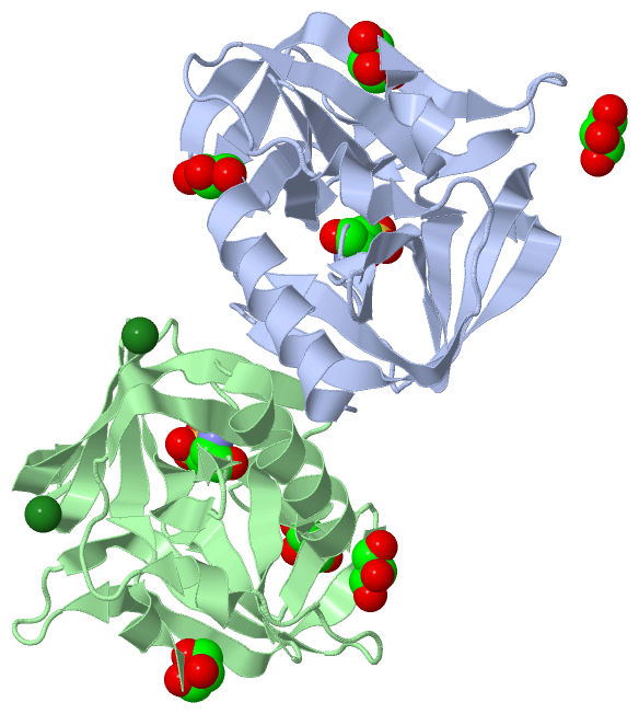
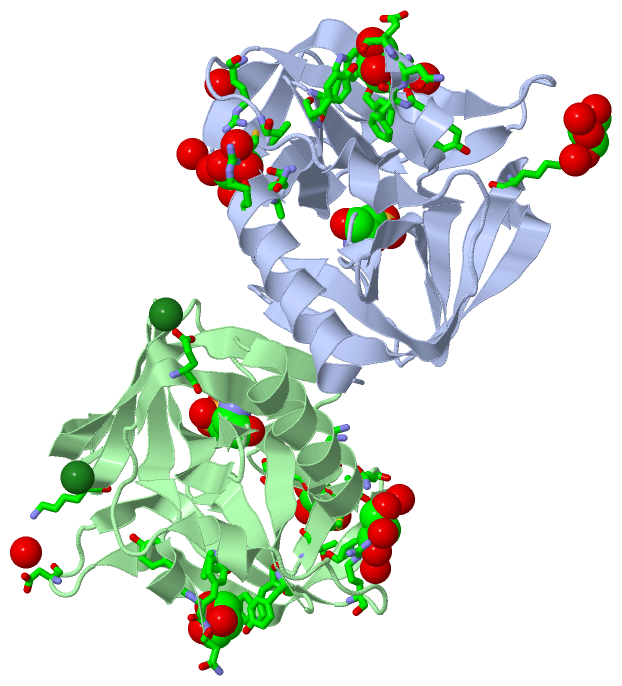
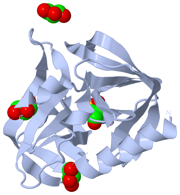
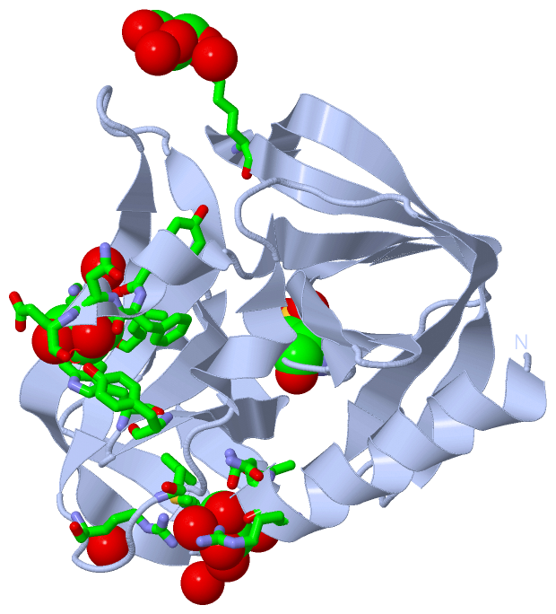
 Visualization

(-) Interactive Views

Asymmetric Unit
  Complete Structure
    Jena3D(integrated viewing of ligand, site, SAP, PROSITE, SCOP information)
    WebMol | AstexViewer[tm]@PDBe
(Java Applets, require no local installation except for Java; loading may be slow)
    STRAP
(Java WebStart application, automatic local installation, requires Java; full application with system access!)
    RasMol
(require local installation)
    Molscript (VRML)
(requires installation of a VRML viewer; select preferred view via VRML and generate a mono or stereo PDF format file)
 
  Ligands, Modified Residues, Ions
    GOL  [ RasMol | Jena3D ]  +environment [ RasMol | Jena3D ]
    MG  [ RasMol | Jena3D ]  +environment [ RasMol | Jena3D ]
    OCS  [ RasMol | Jena3D ]  +environment [ RasMol | Jena3D ]
 
  Sites
    AC1  [ RasMol ]  +environment [ RasMol ]
    AC2  [ RasMol ]  +environment [ RasMol ]
    AC3  [ RasMol ]  +environment [ RasMol ]
    AC4  [ RasMol ]  +environment [ RasMol ]
    AC5  [ RasMol ]  +environment [ RasMol ]
    AC6  [ RasMol ]  +environment [ RasMol ]
    AC7  [ RasMol ]  +environment [ RasMol ]
    AC8  [ RasMol ]  +environment [ RasMol ]
 
  Cis Peptide Bonds
(no "Cis Peptide Bonds" information available for 3q3x)
 
Biological Units
  Complete Structure
    Biological Unit 1  [ Jena3D ]
    Biological Unit 2  [ Jena3D ]
    Biological Unit 3  [ Jena3D ]

(-) Still Images

Jmol
  protein: cartoon or spacefill or dots and stick; nucleic acid: cartoon and stick; ligands: spacefill; active site: stick
Molscript
  protein, nucleic acid: cartoon; ligands: spacefill; active site: ball and stick

 Databases and Analysis Tools

(-) Databases

Access by PDB/NDB ID
  3q3x
    Family and Domain InformationProDom | SYSTERS
    General Structural InformationGlycoscienceDB | MMDB | NDB | OCA | PDB | PDBe | PDBj | PDBsum | PDBWiki | PQS | PROTEOPEDIA
    Orientation in MembranesOPM
    Protein SurfaceSURFACE
    Secondary StructureDSSP (structure derived) | HSSP (homology derived)
    Structural GenomicsGeneCensus
    Structural NeighboursCE | VAST
    Structure ClassificationCATH | Dali | SCOP
    Validation and Original DataBMRB Data View | BMRB Restraints Grid | EDS | PROCHECK | RECOORD | WHAT_CHECK
 
Access by UniProt ID/Accession number
  Q5DSM6_9ENTO | Q5DSM6
    Comparative Protein Structure ModelsModBase
    Genomic InformationEnsembl
    Protein-protein InteractionDIP
    Sequence, Family and Domain InformationInterPro | Pfam | SMART | UniProtKB/TrEMBL
 
Access by Enzyme Classificator   (EC Number)
  3.4.22.28
    General Enzyme InformationBRENDA | EC-PDB | Enzyme | IntEnz
    PathwayKEGG | MetaCyc
 
Access by Disease Identifier   (MIM ID)
  (no 'MIM ID' available)
    Disease InformationOMIM
 
Access by GenAge ID
  (no 'GenAge ID' available)
    Age Related InformationGenAge

(-) Analysis Tools

Access by PDB/NDB ID
    Domain InformationXDom
    Interatomic Contacts of Structural UnitsCSU
    Ligand-protein ContactsLPC
    Protein CavitiescastP
    Sequence and Secondary StructurePDBCartoon
    Structure AlignmentSTRAP(Java WebStart application, automatic local installation, requires Java; full application with system access!)
    Structure and Sequence BrowserSTING
 
Access by UniProt ID/Accession number
  Q5DSM6_9ENTO | Q5DSM6
    Protein Disorder PredictionDisEMBL | FoldIndex | GLOBPLOT (for more information see DisProt)

 Related Entries

(-) Entries Sharing at Least One Protein Chain (UniProt ID)

UniProtKB/TrEMBL
        Q5DSM6_9ENTO | Q5DSM63q3y 3ruo

(-) Related Entries Specified in the PDB File

3q3y