|
|
|
|
 Description
|
|
Compounds
|
||||||||||||||||||||||||||||||||||||||||||||||||
Chains, Units
Summary Information (see also Sequences/Alignments below) |
Ligands, Modified Residues, Ions (1, 5)
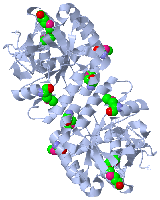
Asymmetric Unit (1, 5)
|
Sites (0, 0)| (no "Site" information available for 3PFM) |
SS Bonds (0, 0)| (no "SS Bond" information available for 3PFM) |
Cis Peptide Bonds (0, 0)| (no "Cis Peptide Bond" information available for 3PFM) |
SAPs(SNPs)/Variants (0, 0)| (no "SAP(SNP)/Variant" information available for 3PFM) |
PROSITE Motifs (0, 0)| (no "PROSITE Motif" information available for 3PFM) |
Exons (0, 0)| (no "Exon" information available for 3PFM) |
Sequences/Alignments
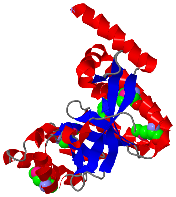
Asymmetric UnitChain A from PDB Type:PROTEIN Length:243 aligned with Q4KKF5_PSEF5 | Q4KKF5 from UniProtKB/TrEMBL Length:648 Alignment length:243 415 425 435 445 455 465 475 485 495 505 515 525 535 545 555 565 575 585 595 605 615 625 635 645 Q4KKF5_PSEF5 406 VGDDHHAWHRLLDRALSEQHFQLYFQPVVAARDTHLVLHYKVLSRLLDEQGQTIPAGRFLPWLERFGWTSRLDLLMLEQVLRQMASHEDCLALNLSAATLADPHALNRVFEILRQHSDLGPRLTLEIGEEQLPEQAMLEQLTRRLRELGFSLSLQRFGGRFSMIGNLARLGLAYLKIDGSYIRDIDQESDKRLFIEAIQRAAHSIDLPLIAERVETEGELQVIREMGLYGVQGQLFGEPAPWG 648 SCOP domains --------------------------------------------------------------------------------------------------------------------------------------------------------------------------------------------------------------------------------------------------- SCOP domains CATH domains --------------------------------------------------------------------------------------------------------------------------------------------------------------------------------------------------------------------------------------------------- CATH domains Pfam domains --------EAL-3pfmA01 A:414-644 ---- Pfam domains SAPs(SNPs) --------------------------------------------------------------------------------------------------------------------------------------------------------------------------------------------------------------------------------------------------- SAPs(SNPs) PROSITE --------------------------------------------------------------------------------------------------------------------------------------------------------------------------------------------------------------------------------------------------- PROSITE Transcript --------------------------------------------------------------------------------------------------------------------------------------------------------------------------------------------------------------------------------------------------- Transcript 3pfm A 406 SNADHHAWHRLLDRALSEQHFQLYFQPVVAARDTHLVLHYKVLSRLLDEQGQTIPAGRFLPWLERFGWTSRLDLLmLEQVLRQmASHEDCLALNLSAATLADPHALNRVFEILRQHSDLGPRLTLEIGEEQLPEQAmLEQLTRRLRELGFSLSLQRFGGRFSmIGNLARLGLAYLKIDGSYIRDIDQESDKRLFIEAIQRAAHSIDLPLIAERVETEGELQVIREmGLYGVQGQLFGEPAPWG 648 415 425 435 445 455 465 475 | 485 | 495 505 515 525 535 |545 555 565 | 575 585 595 605 615 625 | 635 645 481-MSE 489-MSE 542-MSE 568-MSE 631-MSE
|
||||||||||||||||||||
SCOP Domains (0, 0)| (no "SCOP Domain" information available for 3PFM) |
CATH Domains (0, 0)| (no "CATH Domain" information available for 3PFM) |
Pfam Domains (1, 1)
Asymmetric Unit
|
Gene Ontology (4, 4)|
Asymmetric Unit(hide GO term definitions) Chain A (Q4KKF5_PSEF5 | Q4KKF5)
|
||||||||||||||||||||||||||||||||||||||||||
Interactive Views
|
|||||||||||||||||||||||||||||||||||||||||||||||||||||||||||||||||||||||||||||||||||||||||||||||||||||||||||||||||||||||||||||||||||||||
Still Images
|
||||||||||||||||
Databases
|
||||||||||||||||||||||||||||||||||||||||||||||||||||||||||||||||||||||||||||||||||||||||||||||||||||||||||||||||||||||||||||||||||||||||||||||||||||||||||||||||
Analysis Tools
|
|||||||||||||||||||||||||||||||||||||||||||||||||||||||||||||
Entries Sharing at Least One Protein Chain (UniProt ID)
Related Entries Specified in the PDB File
|
|