|
|
|
|
 Description
|
|
Compounds
|
||||||||||||||||||||||||||||||||||||||||||||||||||||
Chains, Units
Summary Information (see also Sequences/Alignments below) |
Ligands, Modified Residues, Ions (4, 16)| Asymmetric/Biological Unit (4, 16) |
Sites (13, 13)
Asymmetric Unit (13, 13)
|
SS Bonds (0, 0)| (no "SS Bond" information available for 3PEB) |
Cis Peptide Bonds (0, 0)| (no "Cis Peptide Bond" information available for 3PEB) |
SAPs(SNPs)/Variants (0, 0)| (no "SAP(SNP)/Variant" information available for 3PEB) |
PROSITE Motifs (0, 0)| (no "PROSITE Motif" information available for 3PEB) |
Exons (0, 0)| (no "Exon" information available for 3PEB) |
Sequences/Alignments
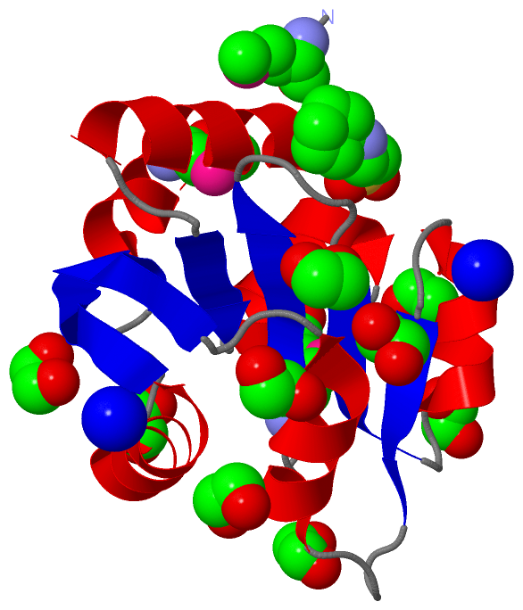
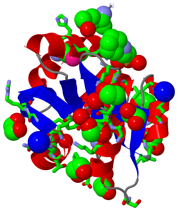
Asymmetric/Biological UnitChain A from PDB Type:PROTEIN Length:132 aligned with Q5M567_STRT2 | Q5M567 from UniProtKB/TrEMBL Length:361 Alignment length:132 1 | 9 19 29 39 49 59 69 79 89 99 109 119 129 Q5M567_STRT2 - -MSKLDRIRHFLNENKSGLAIVSDPVTVNYLTGFDCDPHERQMFLFVYENREPALFVPALEVARASSVLDFPVFGYVDSENPWQKIKAGLASTDIPIIYAEFDNLNVTKFQGLQTVFEGRFENLTPFIHKMR 131 SCOP domains ------------------------------------------------------------------------------------------------------------------------------------ SCOP domains CATH domains ------------------------------------------------------------------------------------------------------------------------------------ CATH domains Pfam domains ---Creatinase_N-3pebA01 A:3-131 Pfam domains SAPs(SNPs) ------------------------------------------------------------------------------------------------------------------------------------ SAPs(SNPs) PROSITE ------------------------------------------------------------------------------------------------------------------------------------ PROSITE Transcript ------------------------------------------------------------------------------------------------------------------------------------ Transcript 3peb A 0 AmSKLDRIRHFLNENKSGLAIVSDPVTVNYLTGFDCDPHERQmFLFVYENREPALFVPALEVARASSVLDFPVFGYVDSENPWQKIKAGLASTDIPIIYAEFDNLNVTKFQGLQTVFEGRFENLTPFIHKmR 131 | 9 19 29 39 | 49 59 69 79 89 99 109 119 129| | 42-MSE 130-MSE 1-MSE
|
||||||||||||||||||||
SCOP Domains (0, 0)| (no "SCOP Domain" information available for 3PEB) |
CATH Domains (0, 0)| (no "CATH Domain" information available for 3PEB) |
Pfam Domains (1, 1)
Asymmetric/Biological Unit
 |
Gene Ontology (2, 2)|
Asymmetric/Biological Unit(hide GO term definitions) Chain A (Q5M567_STRT2 | Q5M567)
|
||||||||||||||||||
Interactive Views
|
|||||||||||||||||||||||||||||||||||||||||||||||||||||||||||||||||||||||||||||||||||||||||||||||||||||||||||||||||||||||||||||||||||||||||||||||||||||||||||||||||||||||||||||||||||||||||||||||||||||||||||||||||||||||||||||||
Still Images
|
||||||||||||||||
Databases
|
||||||||||||||||||||||||||||||||||||||||||||||||||||||||||||||||||||||||||||||||||||||||||||||||||||||||||||||||||||||||||||||||||||||||||||||||||||||||||||||||
Analysis Tools
|
|||||||||||||||||||||||||||||||||||||||||||||||||||||||||||||
Entries Sharing at Least One Protein Chain (UniProt ID)
Related Entries Specified in the PDB File
|
|