Show PDB file:   
         Plain Text   HTML   (compressed file size)
QuickSearch:   
by PDB,NDB,UniProt,PROSITE Code or Search Term(s)  
(-)Asymmetric Unit
(-)Asym. Unit - sites
(-)Biological Unit 1
(-)Biol. Unit 1 - sites
(-)Biological Unit 2
(-)Biological Unit 3
collapse expand < >
Image Asymmetric Unit
Asymmetric Unit  (Jmol Viewer)
Image Asym. Unit - sites
Asym. Unit - sites  (Jmol Viewer)
Image Biological Unit 1
Biological Unit 1  (Jmol Viewer)
Image Biol. Unit 1 - sites
Biol. Unit 1 - sites  (Jmol Viewer)
Image Biological Unit 2
Biological Unit 2  (Jmol Viewer)
Image Biological Unit 3
Biological Unit 3  (Jmol Viewer)

(-) Description

Title :  HYDROLYTIC CATALYSIS AND STRUCTURAL STABILIZATION IN A DESIGNED METALLOPROTEIN
 
Authors :  M. L. Zastrow, A. F. A. Peacock, J. A. Stuckey, V. L. Pecoraro
Date :  20 Oct 10  (Deposition) - 30 Nov 11  (Release) - 29 Feb 12  (Revision)
Method :  X-RAY DIFFRACTION
Resolution :  2.20
Chains :  Asym. Unit :  A,B,C,D,E,F
Biol. Unit 1:  A,B,C  (1x)
Biol. Unit 2:  D,E,F  (1x)
Biol. Unit 3:  A (1x),B (1x),C (1x),D (1x),E (1x),F (1x)
Keywords :  De Novo Protein, Parallel Three-Stranded Coiled Coil, Mercury(Ii) Binding Protein, Zinc(Ii) Binding Protein, L-Penicillamine (Keyword Search: [Gene Ontology, PubMed, Web (Google))
 
Reference :  M. L. Zastrow, A. F. Peacock, J. A. Stuckey, V. L. Pecoraro
Hydrolytic Catalysis And Structural Stabilization In A Designed Metalloprotein.
Nat Chem V. 4 118 2012
PubMed-ID: 22270627  |  Reference-DOI: 10.1038/NCHEM.1201

(-) Compounds

Molecule 1 - COIL SER L9L-PEN L23H
    ChainsA, B, C, D, E, F
    EngineeredYES
    Organism ScientificARTIFICIAL GENE
    Organism Taxid32630
    SyntheticYES

 Structural Features

(-) Chains, Units

  123456
Asymmetric Unit ABCDEF
Biological Unit 1 (1x)ABC   
Biological Unit 2 (1x)   DEF
Biological Unit 3 (1x)A (1x)B (1x)C (1x)D (1x)E (1x)F (1x)

Summary Information (see also Sequences/Alignments below)

(-) Ligands, Modified Residues, Ions  (7, 26)

Asymmetric Unit (7, 26)
No.NameCountTypeFull Name
1ACE5Mod. Amino AcidACETYL GROUP
2CL3Ligand/IonCHLORIDE ION
3HG2Ligand/IonMERCURY (II) ION
4LE16Mod. Amino Acid3-SULFANYL-L-VALINE
5NA1Ligand/IonSODIUM ION
6NH22Mod. Amino AcidAMINO GROUP
7ZN7Ligand/IonZINC ION
Biological Unit 1 (2, 5)
No.NameCountTypeFull Name
1ACE2Mod. Amino AcidACETYL GROUP
2CL-1Ligand/IonCHLORIDE ION
3HG-1Ligand/IonMERCURY (II) ION
4LE13Mod. Amino Acid3-SULFANYL-L-VALINE
5NA-1Ligand/IonSODIUM ION
6NH2-1Mod. Amino AcidAMINO GROUP
7ZN-1Ligand/IonZINC ION
Biological Unit 2 (3, 8)
No.NameCountTypeFull Name
1ACE3Mod. Amino AcidACETYL GROUP
2CL-1Ligand/IonCHLORIDE ION
3HG-1Ligand/IonMERCURY (II) ION
4LE13Mod. Amino Acid3-SULFANYL-L-VALINE
5NA-1Ligand/IonSODIUM ION
6NH22Mod. Amino AcidAMINO GROUP
7ZN-1Ligand/IonZINC ION
Biological Unit 3 (3, 8)
No.NameCountTypeFull Name
1ACE3Mod. Amino AcidACETYL GROUP
2CL-1Ligand/IonCHLORIDE ION
3HG-1Ligand/IonMERCURY (II) ION
4LE13Mod. Amino Acid3-SULFANYL-L-VALINE
5NA-1Ligand/IonSODIUM ION
6NH22Mod. Amino AcidAMINO GROUP
7ZN-1Ligand/IonZINC ION

(-) Sites  (13, 13)

Asymmetric Unit (13, 13)
No.NameEvidenceResiduesDescription
01AC1SOFTWAREHIS A:23 , CL A:34 , HIS B:23 , HIS C:23BINDING SITE FOR RESIDUE ZN A 31
02AC2SOFTWAREGLU A:6 , HOH A:37 , GLU C:1 , HOH C:34BINDING SITE FOR RESIDUE ZN A 32
03AC3SOFTWAREGLU A:1 , HOH E:43BINDING SITE FOR RESIDUE ZN A 33
04AC4SOFTWAREHIS A:23 , ZN A:31 , HIS B:23 , HIS C:23BINDING SITE FOR RESIDUE CL A 34
05AC5SOFTWARELE1 A:9 , LE1 B:9 , LE1 C:9BINDING SITE FOR RESIDUE HG B 31
06AC6SOFTWAREGLU B:27 , CL E:33BINDING SITE FOR RESIDUE NA B 32
07AC7SOFTWAREHIS D:23 , HIS E:23 , HIS F:23 , HOH F:32BINDING SITE FOR RESIDUE ZN D 31
08AC8SOFTWAREGLU D:6 , GLU F:1 , HOH F:66BINDING SITE FOR RESIDUE ZN D 32
09AC9SOFTWARELE1 D:9 , LE1 E:9 , LE1 F:9BINDING SITE FOR RESIDUE HG D 33
10BC1SOFTWAREGLU E:1 , CL E:32 , GLU F:6 , HOH F:37BINDING SITE FOR RESIDUE ZN E 31
11BC2SOFTWAREZN E:31BINDING SITE FOR RESIDUE CL E 32
12BC3SOFTWARENA B:32 , HIS E:28BINDING SITE FOR RESIDUE CL E 33
13BC4SOFTWAREGLU B:1 , GLU C:6 , GLU F:3BINDING SITE FOR RESIDUE ZN F 31

(-) SS Bonds  (0, 0)

(no "SS Bond" information available for 3PBJ)

(-) Cis Peptide Bonds  (1, 1)

Asymmetric Unit
No.Residues
1His D:28 -Gly D:29

 Sequence-Structure Mapping

(-) SAPs(SNPs)/Variants  (0, 0)

(no "SAP(SNP)/Variant" information available for 3PBJ)

(-) PROSITE Motifs  (0, 0)

(no "PROSITE Motif" information available for 3PBJ)

(-) Exons   (0, 0)

(no "Exon" information available for 3PBJ)

(-) Sequences/Alignments

Asymmetric Unit
   Reformat: Number of residues per line =  ('0' or empty: single-line sequence representation)
  Number of residues per labelling interval =   
  UniProt sequence: complete  aligned part    
   Show mapping: SCOP domains CATH domains Pfam domains Secondary structure (by author)
SAPs(SNPs) PROSITE motifs Exons
(details for a mapped element are shown in a popup box when the mouse pointer rests over it)
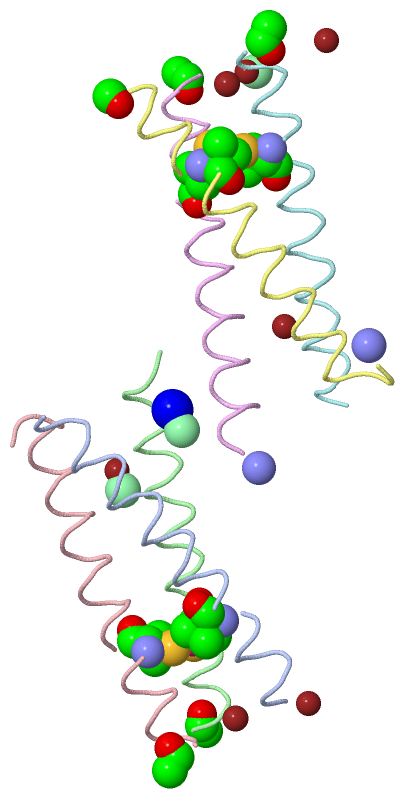
Chain A from PDB  Type:PROTEIN  Length:29
                                                            
               SCOP domains ----------------------------- SCOP domains
               CATH domains ----------------------------- CATH domains
               Pfam domains ----------------------------- Pfam domains
         Sec.struct. author hhhhhhhhhhhhhhhhhhhhhhhhhhhhh Sec.struct. author
                 SAPs(SNPs) ----------------------------- SAPs(SNPs)
                    PROSITE ----------------------------- PROSITE
                 Transcript ----------------------------- Transcript
                  3pbj A  1 EWEALEKKvAALESKLQALEKKHEALEHG 29
                                    10        20         
                                    9-LE1                

Chain B from PDB  Type:PROTEIN  Length:29
                                                            
               SCOP domains ----------------------------- SCOP domains
               CATH domains ----------------------------- CATH domains
               Pfam domains ----------------------------- Pfam domains
         Sec.struct. author .hhhhhhhhhhhhhhhhhhhhhhhhhhh. Sec.struct. author
                 SAPs(SNPs) ----------------------------- SAPs(SNPs)
                    PROSITE ----------------------------- PROSITE
                 Transcript ----------------------------- Transcript
                  3pbj B  0 xEWEALEKKvAALESKLQALEKKHEALEH 28
                            |        9        19         
                            0-ACE    9-LE1               

Chain C from PDB  Type:PROTEIN  Length:30
                                                             
               SCOP domains ------------------------------ SCOP domains
               CATH domains ------------------------------ CATH domains
               Pfam domains ------------------------------ Pfam domains
         Sec.struct. author .hhhhhhhhhhhhhhhhhhhhhhhhhhh.. Sec.struct. author
                 SAPs(SNPs) ------------------------------ SAPs(SNPs)
                    PROSITE ------------------------------ PROSITE
                 Transcript ------------------------------ Transcript
                  3pbj C  0 xEWEALEKKvAALESKLQALEKKHEALEHG 29
                            |        9        19        29
                            0-ACE    9-LE1                

Chain D from PDB  Type:PROTEIN  Length:31
                                                              
               SCOP domains ------------------------------- SCOP domains
               CATH domains ------------------------------- CATH domains
               Pfam domains ------------------------------- Pfam domains
         Sec.struct. author .hhhhhhhhhhhhhhhhhhhhhhhhhhh... Sec.struct. author
                 SAPs(SNPs) ------------------------------- SAPs(SNPs)
                    PROSITE ------------------------------- PROSITE
                 Transcript ------------------------------- Transcript
                  3pbj D  0 xEWEALEKKvAALESKLQALEKKHEALEHGx 30
                            |        9        19        29|
                            0-ACE    9-LE1               30-NH2

Chain E from PDB  Type:PROTEIN  Length:31
                                                              
               SCOP domains ------------------------------- SCOP domains
               CATH domains ------------------------------- CATH domains
               Pfam domains ------------------------------- Pfam domains
         Sec.struct. author .hhhhhhhhhhhhhhhhhhhhhh........ Sec.struct. author
                 SAPs(SNPs) ------------------------------- SAPs(SNPs)
                    PROSITE ------------------------------- PROSITE
                 Transcript ------------------------------- Transcript
                  3pbj E  0 xEWEALEKKvAALESKLQALEKKHEALEHGx 30
                            |        9        19        29|
                            |        9-LE1               30-NH2
                            0-ACE                          

Chain F from PDB  Type:PROTEIN  Length:30
                                                             
               SCOP domains ------------------------------ SCOP domains
               CATH domains ------------------------------ CATH domains
               Pfam domains ------------------------------ Pfam domains
         Sec.struct. author .hhhhhhhhhhhhhhhhhhhhhhhhhh... Sec.struct. author
                 SAPs(SNPs) ------------------------------ SAPs(SNPs)
                    PROSITE ------------------------------ PROSITE
                 Transcript ------------------------------ Transcript
                  3pbj F  0 xEWEALEKKvAALESKLQALEKKHEALEHG 29
                            |        9        19        29
                            |        9-LE1                
                            0-ACE                         

   Legend:   → Mismatch (orange background)
  - → Gap (green background, '-', border residues have a numbering label)
    → Modified Residue (blue background, lower-case, 'x' indicates undefined single-letter code, labelled with number + name)
  x → Chemical Group (purple background, 'x', labelled with number + name, e.g. ACE or NH2)
  extra numbering lines below/above indicate numbering irregularities and modified residue names etc., number ends below/above '|'

 Classification and Annotation

(-) SCOP Domains  (0, 0)

(no "SCOP Domain" information available for 3PBJ)

(-) CATH Domains  (0, 0)

(no "CATH Domain" information available for 3PBJ)

(-) Pfam Domains  (0, 0)

(no "Pfam Domain" information available for 3PBJ)

(-) Gene Ontology  (0, 0)

Asymmetric Unit(hide GO term definitions)
    (no "Gene Ontology" information available for 3PBJ)

 Visualization

(-) Interactive Views

Asymmetric Unit
  Complete Structure
    Jena3D(integrated viewing of ligand, site, SAP, PROSITE, SCOP information)
    WebMol | AstexViewer[tm]@PDBe
(Java Applets, require no local installation except for Java; loading may be slow)
    STRAP
(Java WebStart application, automatic local installation, requires Java; full application with system access!)
    RasMol
(require local installation)
    Molscript (VRML)
(requires installation of a VRML viewer; select preferred view via VRML and generate a mono or stereo PDF format file)
 
  Ligands, Modified Residues, Ions
    ACE  [ RasMol | Jena3D ]  +environment [ RasMol | Jena3D ]
    CL  [ RasMol | Jena3D ]  +environment [ RasMol | Jena3D ]
    HG  [ RasMol | Jena3D ]  +environment [ RasMol | Jena3D ]
    LE1  [ RasMol | Jena3D ]  +environment [ RasMol | Jena3D ]
    NA  [ RasMol | Jena3D ]  +environment [ RasMol | Jena3D ]
    NH2  [ RasMol | Jena3D ]  +environment [ RasMol | Jena3D ]
    ZN  [ RasMol | Jena3D ]  +environment [ RasMol | Jena3D ]
 
  Sites
    AC1  [ RasMol ]  +environment [ RasMol ]
    AC2  [ RasMol ]  +environment [ RasMol ]
    AC3  [ RasMol ]  +environment [ RasMol ]
    AC4  [ RasMol ]  +environment [ RasMol ]
    AC5  [ RasMol ]  +environment [ RasMol ]
    AC6  [ RasMol ]  +environment [ RasMol ]
    AC7  [ RasMol ]  +environment [ RasMol ]
    AC8  [ RasMol ]  +environment [ RasMol ]
    AC9  [ RasMol ]  +environment [ RasMol ]
    BC1  [ RasMol ]  +environment [ RasMol ]
    BC2  [ RasMol ]  +environment [ RasMol ]
    BC3  [ RasMol ]  +environment [ RasMol ]
    BC4  [ RasMol ]  +environment [ RasMol ]
 
  Cis Peptide Bonds
    His D:28 - Gly D:29   [ RasMol ]  
 
Biological Units
  Complete Structure
    Biological Unit 1  [ Jena3D ]
    Biological Unit 2  [ Jena3D ]
    Biological Unit 3  [ Jena3D ]

(-) Still Images

Jmol
  protein: cartoon or spacefill or dots and stick; nucleic acid: cartoon and stick; ligands: spacefill; active site: stick
Molscript
  protein, nucleic acid: cartoon; ligands: spacefill; active site: ball and stick

 Databases and Analysis Tools

(-) Databases

Access by PDB/NDB ID
  3pbj
    Family and Domain InformationProDom | SYSTERS
    General Structural InformationGlycoscienceDB | MMDB | NDB | OCA | PDB | PDBe | PDBj | PDBsum | PDBWiki | PQS | PROTEOPEDIA
    Orientation in MembranesOPM
    Protein SurfaceSURFACE
    Secondary StructureDSSP (structure derived) | HSSP (homology derived)
    Structural GenomicsGeneCensus
    Structural NeighboursCE | VAST
    Structure ClassificationCATH | Dali | SCOP
    Validation and Original DataBMRB Data View | BMRB Restraints Grid | EDS | PROCHECK | RECOORD | WHAT_CHECK
 
Access by UniProt ID/Accession number
  (no 'UniProt ID/Accession number' available) |
    Comparative Protein Structure ModelsModBase
    Genomic InformationEnsembl
    Protein-protein InteractionDIP
    Sequence, Family and Domain InformationInterPro | Pfam | SMART | UniProt
 
Access by Enzyme Classificator   (EC Number)
  (no 'Enzyme Classificator' available)
    General Enzyme InformationBRENDA | EC-PDB | Enzyme | IntEnz
    PathwayKEGG | MetaCyc
 
Access by Disease Identifier   (MIM ID)
  (no 'MIM ID' available)
    Disease InformationOMIM
 
Access by GenAge ID
  (no 'GenAge ID' available)
    Age Related InformationGenAge

(-) Analysis Tools

Access by PDB/NDB ID
    Domain InformationXDom
    Interatomic Contacts of Structural UnitsCSU
    Ligand-protein ContactsLPC
    Protein CavitiescastP
    Sequence and Secondary StructurePDBCartoon
    Structure AlignmentSTRAP(Java WebStart application, automatic local installation, requires Java; full application with system access!)
    Structure and Sequence BrowserSTING
 
Access by UniProt ID/Accession number
  (no 'UniProt ID/Accession number' available) |
    Protein Disorder PredictionDisEMBL | FoldIndex | GLOBPLOT

 Related Entries

(-) Entries Sharing at Least One Protein Chain (UniProt ID)

(no "Entries Sharing at Least One Protein Chain" available for 3PBJ)

(-) Related Entries Specified in the PDB File

2jgo ARSENATED COIL SER L9C
2x6p COIL SER L19C
3h5f COIL SER L16L-PEN
3h5g COIL SER L16D-PEN
3ljm COIL SER L9C