|
|
|
|
 Description
|
|
Compounds
|
||||||||||||||||||||||||||||||||||||||||||||
Chains, Units
Summary Information (see also Sequences/Alignments below) |
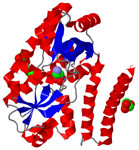
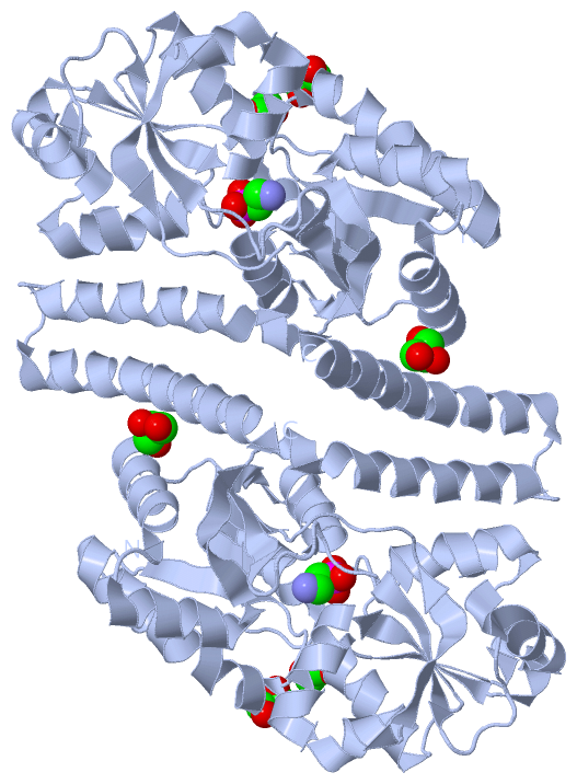
Ligands, Modified Residues, Ions (2, 4)| Asymmetric Unit (2, 4) Biological Unit 1 (2, 8) |
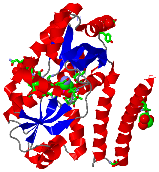
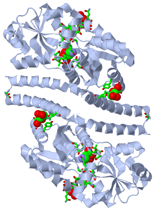
Sites (4, 4)
Asymmetric Unit (4, 4)
|
SS Bonds (0, 0)| (no "SS Bond" information available for 3P7I) |
Cis Peptide Bonds (0, 0)| (no "Cis Peptide Bond" information available for 3P7I) |
SAPs(SNPs)/Variants (0, 0)| (no "SAP(SNP)/Variant" information available for 3P7I) |
PROSITE Motifs (0, 0)| (no "PROSITE Motif" information available for 3P7I) |
Exons (0, 0)| (no "Exon" information available for 3P7I) |
Sequences/Alignments
Asymmetric UnitChain A from PDB Type:PROTEIN Length:300 aligned with Q1R3F7_ECOUT | Q1R3F7 from UniProtKB/TrEMBL Length:338 Alignment length:300 40 50 60 70 80 90 100 110 120 130 140 150 160 170 180 190 200 210 220 230 240 250 260 270 280 290 300 310 320 330 Q1R3F7_ECOUT 31 KALNFGIISTESQQNLKPQWTPFLQDMEKKLGVKVNAFFAPDYAGIIQGMRFNKVDIAWYGNLSAMEAVDRANGQVFAQTVAADGSPGYWSVLIVNKDSPINNLNDLLAKRKDLTFGNGDPNSTSGFLVPGYYVFAKNNISASDFKRTVNAGHETNALAVANKQVDVATNNTENLDKLKTSAPEKLKELKVIWKSPLIPGDPIVWRKNLSETTKDKIYDFFMNYGKTPEEKAVLERLGWAPFRASSDLQLVPIRQLALFKEMQSVKDNKGLNEQDKLAKTTAIQAQLDDLDRLNNALSAM 330 SCOP domains ------------------------------------------------------------------------------------------------------------------------------------------------------------------------------------------------------------------------------------------------------------------------------------------------------------ SCOP domains CATH domains ------------------------------------------------------------------------------------------------------------------------------------------------------------------------------------------------------------------------------------------------------------------------------------------------------------ CATH domains Pfam domains ------------------------------------------------------------------------------------------------------------------------------------------------------------------------------------------------------------------------------------------------------------------------------------------------------------ Pfam domains SAPs(SNPs) ------------------------------------------------------------------------------------------------------------------------------------------------------------------------------------------------------------------------------------------------------------------------------------------------------------ SAPs(SNPs) PROSITE ------------------------------------------------------------------------------------------------------------------------------------------------------------------------------------------------------------------------------------------------------------------------------------------------------------ PROSITE Transcript ------------------------------------------------------------------------------------------------------------------------------------------------------------------------------------------------------------------------------------------------------------------------------------------------------------ Transcript 3p7i A 5 KALNFGIISTESQQNLKPQWTPFLQDMEKKLGVKVNAFFAPDYAGIIQGMRFNKVDIAWYGNLSAMEAVDRANGQVFAQTVAADGSPGYWSVLIVNKDSPINNLNDLLAKRKDLTFGNGDPNSTSGFLVPGYYVFAKNNISASDFKRTVNAGHETNALAVANKQVDVATNNTENLDKLKTSAPEKLKELKVIWKSPLIPGDPIVWRKNLSETTKDKIYDFFMNYGKTPEEKAVLERLGWAPFRASSDLQLVPIRQLALFKEMQSVKDNKGLNEQDKLAKTTAIQAQLDDLDRLNNALSAM 304 14 24 34 44 54 64 74 84 94 104 114 124 134 144 154 164 174 184 194 204 214 224 234 244 254 264 274 284 294 304
|
||||||||||||||||||||
SCOP Domains (0, 0)| (no "SCOP Domain" information available for 3P7I) |
CATH Domains (0, 0)| (no "CATH Domain" information available for 3P7I) |
Pfam Domains (0, 0)| (no "Pfam Domain" information available for 3P7I) |
Gene Ontology (4, 4)|
Asymmetric Unit(hide GO term definitions) Chain A (Q1R3F7_ECOUT | Q1R3F7)
|
||||||||||||||||||||||||||||||||||||||||||
Interactive Views
|
||||||||||||||||||||||||||||||||||||||||||||||||||||||||||||||||||||||||||||||||||||||||||||||||||||||||||||||||||||||||||||||||||||||||||||||||||||||||||||||||||||
Still Images
|
||||||||||||||||
Databases
|
||||||||||||||||||||||||||||||||||||||||||||||||||||||||||||||||||||||||||||||||||||||||||||||||||||||||||||||||||||||||||||||||||||||||||||||||||||||||||||||||
Analysis Tools
|
|||||||||||||||||||||||||||||||||||||||||||||||||||||||||||||
Entries Sharing at Least One Protein Chain (UniProt ID)
Related Entries Specified in the PDB File
|
|