|
|
|
|
 Description
|
|
Compounds
|
||||||||||||||||||||||||||||||||||||||||||||||||||||||||||||
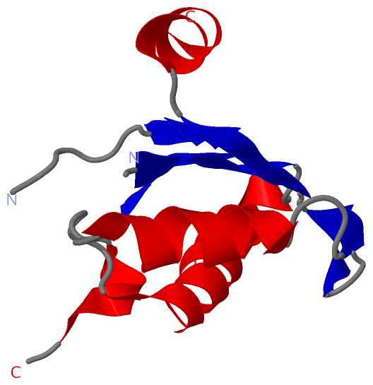
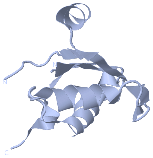
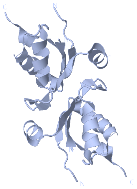
Chains, Units
Summary Information (see also Sequences/Alignments below) |
Ligands, Modified Residues, Ions (0, 0)| (no "Ligand,Modified Residues,Ions" information available for 3P3D) |
Sites (0, 0)| (no "Site" information available for 3P3D) |
SS Bonds (0, 0)| (no "SS Bond" information available for 3P3D) |
Cis Peptide Bonds (0, 0)| (no "Cis Peptide Bond" information available for 3P3D) |
SAPs(SNPs)/Variants (0, 0)| (no "SAP(SNP)/Variant" information available for 3P3D) |
PROSITE Motifs (0, 0)| (no "PROSITE Motif" information available for 3P3D) |
Exons (0, 0)| (no "Exon" information available for 3P3D) |
Sequences/Alignments
Asymmetric UnitChain A from PDB Type:PROTEIN Length:87 aligned with A5DMN1_PICGU | A5DMN1 from UniProtKB/TrEMBL Length:448 Alignment length:98 275 285 295 305 315 325 335 345 355 A5DMN1_PICGU 266 SAILVFGYPETMANQVIAYFQEFGTILEDFEVLRKPQAMTVGLQDKQFVPIFSGNSWTKITYDNPASAVDALLENGAVFNGVLLGVIPYTKDAVERLQ 363 SCOP domains -------------------------------------------------------------------------------------------------- SCOP domains CATH domains -------------------------------------------------------------------------------------------------- CATH domains Pfam domains Nup35_RRM-3p3dA01 A:266-363 Pfam domains SAPs(SNPs) -------------------------------------------------------------------------------------------------- SAPs(SNPs) PROSITE -------------------------------------------------------------------------------------------------- PROSITE Transcript -------------------------------------------------------------------------------------------------- Transcript 3p3d A 266 LAILVFGYPETMANQVIAYFQEFGTILEDFEVLRKP-----------FVPIFSGNSWTKITYDNPASAVDALLENGAVFNGVLLGVIPYTKDAVERLQ 363 275 285 295 | - 315 325 335 345 355 301 313
|
||||||||||||||||||||
SCOP Domains (0, 0)| (no "SCOP Domain" information available for 3P3D) |
CATH Domains (0, 0)| (no "CATH Domain" information available for 3P3D) |
Pfam Domains (1, 1)
Asymmetric Unit
|
Gene Ontology (4, 4)|
Asymmetric Unit(hide GO term definitions) Chain A (A5DMN1_PICGU | A5DMN1)
|
||||||||||||||||||||||||||||||||||||
Interactive Views
|
|||||||||||||||||||||||||||||||||||||||||||||||||||||||||||||||||||||||||||||||||||||||||||||||||||||||||||||||||||||||||||||||||||||||||||
Still Images
|
||||||||||||||||
Databases
|
||||||||||||||||||||||||||||||||||||||||||||||||||||||||||||||||||||||||||||||||||||||||||||||||||||||||||||||||||||||||||||||||||||||||||||||||||||||||||||||||
Analysis Tools
|
|||||||||||||||||||||||||||||||||||||||||||||||||||||||||||||
Entries Sharing at Least One Protein Chain (UniProt ID)
Related Entries Specified in the PDB File
|
|