Show PDB file:   
         Plain Text   HTML   (compressed file size)
QuickSearch:   
by PDB,NDB,UniProt,PROSITE Code or Search Term(s)  
(-)Asymmetric Unit
(-)Asym. Unit - sites
(-)Biological Unit 1
(-)Biol. Unit 1 - sites
(-)Biological Unit 2
(-)Biological Unit 3
collapse expand < >
Image Asymmetric Unit
Asymmetric Unit  (Jmol Viewer)
Image Asym. Unit - sites
Asym. Unit - sites  (Jmol Viewer)
Image Biological Unit 1
Biological Unit 1  (Jmol Viewer)
Image Biol. Unit 1 - sites
Biol. Unit 1 - sites  (Jmol Viewer)
Image Biological Unit 2
Biological Unit 2  (Jmol Viewer)
Image Biological Unit 3
Biological Unit 3  (Jmol Viewer)

(-) Description

Title :  NISCHARIN PX-DOMAIN
 
Authors :  P. Schutz, T. Karlberg, C. H. Arrowsmith, H. Berglund, C. Bountra, R. Co A. M. Edwards, S. Flodin, A. Flores, S. Graslund, M. Hammarstrom, I. Jo S. Kol, T. Kotenyova, E. Kouznetsova, M. Moche, T. Nyman, C. Persson, M. I. Siponen, A. G. Thorsell, L. Tresaugues, S. Van Der Berg, E. Wahl M. Welin, J. Weigelt, P. Nordlund, H. Schuler, Structural Genomics Consortium (Sgc)
Date :  28 Sep 10  (Deposition) - 13 Oct 10  (Release) - 13 Oct 10  (Revision)
Method :  X-RAY DIFFRACTION
Resolution :  2.27
Chains :  Asym. Unit :  A,B
Biol. Unit 1:  A  (1x)
Biol. Unit 2:  B  (1x)
Biol. Unit 3:  A (1x),B (1x)
Keywords :  Structural Genomics, Structural Genomics Consortium, Sgc, Px-Domain, Signaling Protein (Keyword Search: [Gene Ontology, PubMed, Web (Google))
 
Reference :  P. Schutz, T. Karlberg, C. H. Arrowsmith, H. Berglund, C. Bountra, R. Collins, A. M. Edwards, S. Flodin, A. Flores, S. Graslund, M. Hammarstrom, I. Johansson, S. Kol, T. Kotenyova, E. Kouznetsova, M. Moche, T. Nyman, C. Persson, M. I. Siponen, A. G. Thorsell, L. Tresaugues, S. Van Der Berg, E. Wahlberg, M. Welin, J. Weigelt, P. Nordlund, H. Schuler
Crystal Structure Of Nischarin Px-Domain
To Be Published
PubMed: search

(-) Compounds

Molecule 1 - NISCHARIN
    ChainsA, B
    EngineeredYES
    Expression SystemESCHERICHIA COLI
    Expression System PlasmidPNIC-BSA4
    Expression System StrainBL21(DE3) R3 PRARE
    Expression System Taxid562
    Expression System Vector TypePLASMID
    FragmentPX-DOMAIN
    GeneNISCH, IRAS, KIAA0975
    Organism CommonHUMAN
    Organism ScientificHOMO SAPIENS
    Organism Taxid9606

 Structural Features

(-) Chains, Units

  12
Asymmetric Unit AB
Biological Unit 1 (1x)A 
Biological Unit 2 (1x) B
Biological Unit 3 (1x)A (1x)B (1x)

Summary Information (see also Sequences/Alignments below)

(-) Ligands, Modified Residues, Ions  (1, 2)

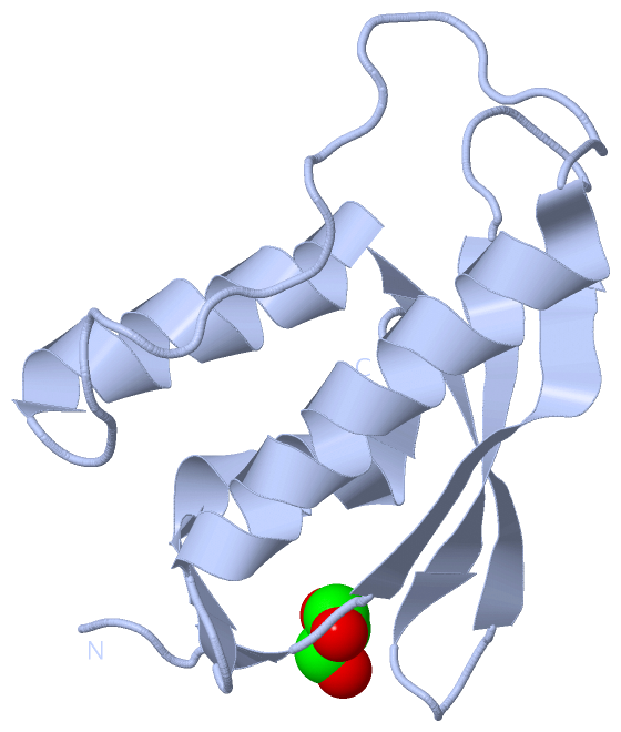
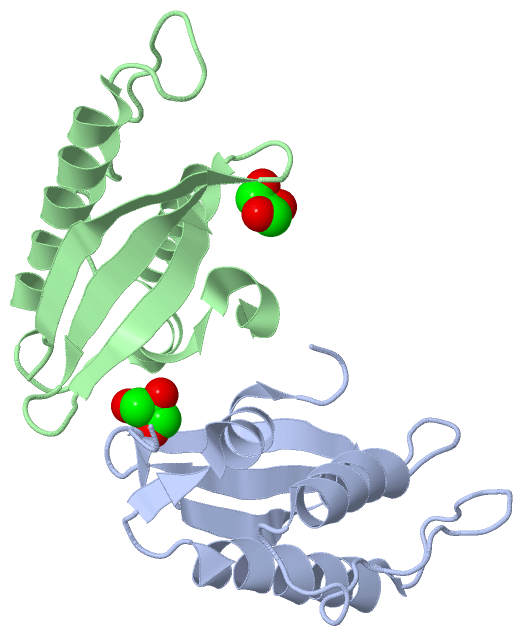
Asymmetric Unit (1, 2)
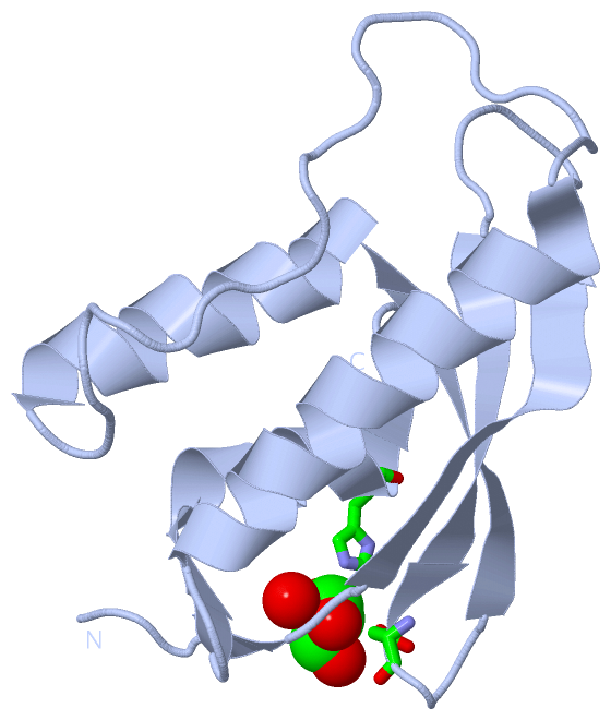
No.NameCountTypeFull Name
1GOL2Ligand/IonGLYCEROL
Biological Unit 1 (1, 1)
No.NameCountTypeFull Name
1GOL1Ligand/IonGLYCEROL
Biological Unit 2 (1, 1)
No.NameCountTypeFull Name
1GOL1Ligand/IonGLYCEROL
Biological Unit 3 (1, 1)
No.NameCountTypeFull Name
1GOL1Ligand/IonGLYCEROL

(-) Sites  (2, 2)

Asymmetric Unit (2, 2)
No.NameEvidenceResiduesDescription
1AC1SOFTWAREASP A:39 , HIS A:116 , HOH A:195 , HIS B:116BINDING SITE FOR RESIDUE GOL A 125
2AC2SOFTWAREASP B:28 , TYR B:30 , VAL B:32 , ARG B:49 , PHE B:119 , HOH B:191BINDING SITE FOR RESIDUE GOL B 125

(-) SS Bonds  (0, 0)

(no "SS Bond" information available for 3P0C)

(-) Cis Peptide Bonds  (0, 0)

(no "Cis Peptide Bond" information available for 3P0C)

 Sequence-Structure Mapping

(-) SAPs(SNPs)/Variants  (0, 0)

(no "SAP(SNP)/Variant" information available for 3P0C)

(-) PROSITE Motifs  (1, 1)

Asymmetric Unit (1, 1)
 PROSITEUniProtKBPDB
No.IDACDescriptionIDLocationCountLocation
1PXPS50195 PX domain profile.NISCH_HUMAN11-121  1A:18-121
Biological Unit 1 (1, 1)
 PROSITEUniProtKBPDB
No.IDACDescriptionIDLocationCountLocation
1PXPS50195 PX domain profile.NISCH_HUMAN11-121  1A:18-121
Biological Unit 2 (, 0)
 PROSITEUniProtKBPDB
No.IDACDescriptionIDLocationCountLocation
1PXPS50195 PX domain profile.NISCH_HUMAN11-121  0-
Biological Unit 3 (1, 1)
 PROSITEUniProtKBPDB
No.IDACDescriptionIDLocationCountLocation
1PXPS50195 PX domain profile.NISCH_HUMAN11-121  1A:18-121

(-) Exons   (0, 0)

(no "Exon" information available for 3P0C)

(-) Sequences/Alignments

Asymmetric Unit
   Reformat: Number of residues per line =  ('0' or empty: single-line sequence representation)
  Number of residues per labelling interval =   
  UniProt sequence: complete  aligned part    
   Show mapping: SCOP domains CATH domains Pfam domains Secondary structure (by author)
SAPs(SNPs) PROSITE motifs Exons
(details for a mapped element are shown in a popup box when the mouse pointer rests over it)
Chain A from PDB  Type:PROTEIN  Length:111
 aligned with NISCH_HUMAN | Q9Y2I1 from UniProtKB/Swiss-Prot  Length:1504

    Alignment length:118
                                    13        23        33        43        53        63        73        83        93       103       113        
          NISCH_HUMAN     4 ARTFGPEREAEPAKEARVVGSELVDTYTVYIIQVTDGSHEWTVKHRYSDFHDLHEKLVAERKIDKNLLPPKKIIGKNSRSLVEKREKDLEVYLQKLLAAFPGVTPRVLAHFLHFHFYE 121
               SCOP domains d3p0ca       _ A: automated matches                                                                                    SCOP domains
               CATH domains ---------------------------------------------------------------------------------------------------------------------- CATH domains
               Pfam domains ---------------------------------------------------------------------------------------------------------------------- Pfam domains
         Sec.struct. author ...ee.-------.eeeeeeee......eeeeeee....eeeeeeehhhhhhhhhhhhhh....................hhhhhhhhhhhhhhhhhhh...eehhhhhhhhhh.... Sec.struct. author
                 SAPs(SNPs) ---------------------------------------------------------------------------------------------------------------------- SAPs(SNPs)
                    PROSITE -------PX  PDB: A:18-121 UniProt: 11-121                                                                               PROSITE
                 Transcript ---------------------------------------------------------------------------------------------------------------------- Transcript
                 3p0c A  11 NLYFQS-------MEARVVGSELVDTYTVYIIQVTDGSHEWTVKHRYSDFHDLHEKLVAERKIDKNLLPPKKIIGKNSRSLVEKREKDLEVYLQKLLAAFPGVTPRVLAHFLHFHFYE 121
                                 |   -   |    23        33        43        53        63        73        83        93       103       113        
                                16      17                                                                                                        

Chain B from PDB  Type:PROTEIN  Length:108
 aligned with NISCH_HUMAN | Q9Y2I1 from UniProtKB/Swiss-Prot  Length:1504

    Alignment length:108
                                    25        35        45        55        65        75        85        95       105       115        
          NISCH_HUMAN    16 AKEARVVGSELVDTYTVYIIQVTDGSHEWTVKHRYSDFHDLHEKLVAERKIDKNLLPPKKIIGKNSRSLVEKREKDLEVYLQKLLAAFPGVTPRVLAHFLHFHFYEIN 123
               SCOP domains d3p0cb_ B: automated matches                                                                                 SCOP domains
               CATH domains ------------------------------------------------------------------------------------------------------------ CATH domains
           Pfam domains (1) --PX-3p0cB01 B:18-118                                                                                  ----- Pfam domains (1)
           Pfam domains (2) --PX-3p0cB02 B:18-118                                                                                  ----- Pfam domains (2)
         Sec.struct. author ..eeeeeeeee....eeeeeeee....eeeeeeehhhhhhhhhhhhhh....................hhhhhhhhhhhhhhhhhhhh....hhhhhhhhhhhhhhh. Sec.struct. author
                 SAPs(SNPs) ------------------------------------------------------------------------------------------------------------ SAPs(SNPs)
                    PROSITE PX  PDB: - UniProt: 11-121                                                                                -- PROSITE
                 Transcript ------------------------------------------------------------------------------------------------------------ Transcript
                 3p0c B  16 SMEARVVGSELVDTYTVYIIQVTDGSHEWTVKHRYSDFHDLHEKLVAERKIDKNLLPPKKIIGKNSRSLVEKREKDLEVYLQKLLAAFPGVTPRVLAHFLHFHFYEIN 123
                                    25        35        45        55        65        75        85        95       105       115        

   Legend:   → Mismatch (orange background)
  - → Gap (green background, '-', border residues have a numbering label)
    → Modified Residue (blue background, lower-case, 'x' indicates undefined single-letter code, labelled with number + name)
  x → Chemical Group (purple background, 'x', labelled with number + name, e.g. ACE or NH2)
  extra numbering lines below/above indicate numbering irregularities and modified residue names etc., number ends below/above '|'

 Classification and Annotation

(-) SCOP Domains  (1, 2)

Asymmetric Unit

(-) CATH Domains  (0, 0)

(no "CATH Domain" information available for 3P0C)

(-) Pfam Domains  (1, 2)

Asymmetric Unit
(-)
Family: PX (17)
1aPX-3p0cB01B:18-118
1bPX-3p0cB02B:18-118

(-) Gene Ontology  (19, 19)

Asymmetric Unit(hide GO term definitions)
Chain A,B   (NISCH_HUMAN | Q9Y2I1)
molecular function
    GO:0042802    identical protein binding    Interacting selectively and non-covalently with an identical protein or proteins.
    GO:0005178    integrin binding    Interacting selectively and non-covalently with an integrin.
    GO:0035091    phosphatidylinositol binding    Interacting selectively and non-covalently with any inositol-containing glycerophospholipid, i.e. phosphatidylinositol (PtdIns) and its phosphorylated derivatives.
    GO:0005515    protein binding    Interacting selectively and non-covalently with any protein or protein complex (a complex of two or more proteins that may include other nonprotein molecules).
biological process
    GO:0016601    Rac protein signal transduction    A series of molecular signals within the cell that are mediated by a member of the Rac family of proteins switching to a GTP-bound active state.
    GO:0030036    actin cytoskeleton organization    A process that is carried out at the cellular level which results in the assembly, arrangement of constituent parts, or disassembly of cytoskeletal structures comprising actin filaments and their associated proteins.
    GO:0006915    apoptotic process    A programmed cell death process which begins when a cell receives an internal (e.g. DNA damage) or external signal (e.g. an extracellular death ligand), and proceeds through a series of biochemical events (signaling pathway phase) which trigger an execution phase. The execution phase is the last step of an apoptotic process, and is typically characterized by rounding-up of the cell, retraction of pseudopodes, reduction of cellular volume (pyknosis), chromatin condensation, nuclear fragmentation (karyorrhexis), plasma membrane blebbing and fragmentation of the cell into apoptotic bodies. When the execution phase is completed, the cell has died.
    GO:0006006    glucose metabolic process    The chemical reactions and pathways involving glucose, the aldohexose gluco-hexose. D-glucose is dextrorotatory and is sometimes known as dextrose; it is an important source of energy for living organisms and is found free as well as combined in homo- and hetero-oligosaccharides and polysaccharides.
    GO:0030336    negative regulation of cell migration    Any process that stops, prevents, or reduces the frequency, rate or extent of cell migration.
    GO:0048243    norepinephrine secretion    The regulated release of norepinephrine by a cell. Norepinephrine is a catecholamine and it acts as a hormone and as a neurotransmitter of most of the sympathetic nervous system.
    GO:0008217    regulation of blood pressure    Any process that modulates the force with which blood travels through the circulatory system. The process is controlled by a balance of processes that increase pressure and decrease pressure.
    GO:0032228    regulation of synaptic transmission, GABAergic    Any process that modulates the frequency, rate or extent of GABAergic synaptic transmission, the process of communication from a neuron to another neuron across a synapse using the neurotransmitter gamma-aminobutyric acid (GABA).
cellular component
    GO:0005737    cytoplasm    All of the contents of a cell excluding the plasma membrane and nucleus, but including other subcellular structures.
    GO:0005829    cytosol    The part of the cytoplasm that does not contain organelles but which does contain other particulate matter, such as protein complexes.
    GO:0005769    early endosome    A membrane-bounded organelle that receives incoming material from primary endocytic vesicles that have been generated by clathrin-dependent and clathrin-independent endocytosis; vesicles fuse with the early endosome to deliver cargo for sorting into recycling or degradation pathways.
    GO:0005768    endosome    A vacuole to which materials ingested by endocytosis are delivered.
    GO:0016020    membrane    A lipid bilayer along with all the proteins and protein complexes embedded in it an attached to it.
    GO:0005886    plasma membrane    The membrane surrounding a cell that separates the cell from its external environment. It consists of a phospholipid bilayer and associated proteins.
    GO:0055037    recycling endosome    An organelle consisting of a network of tubules that functions in targeting molecules, such as receptors transporters and lipids, to the plasma membrane.

 Visualization

(-) Interactive Views

Asymmetric Unit
  Complete Structure
    Jena3D(integrated viewing of ligand, site, SAP, PROSITE, SCOP information)
    WebMol | AstexViewer[tm]@PDBe
(Java Applets, require no local installation except for Java; loading may be slow)
    STRAP
(Java WebStart application, automatic local installation, requires Java; full application with system access!)
    RasMol
(require local installation)
    Molscript (VRML)
(requires installation of a VRML viewer; select preferred view via VRML and generate a mono or stereo PDF format file)
 
  Ligands, Modified Residues, Ions
    GOL  [ RasMol | Jena3D ]  +environment [ RasMol | Jena3D ]
 
  Sites
    AC1  [ RasMol ]  +environment [ RasMol ]
    AC2  [ RasMol ]  +environment [ RasMol ]
 
  Cis Peptide Bonds
(no "Cis Peptide Bonds" information available for 3p0c)
 
Biological Units
  Complete Structure
    Biological Unit 1  [ Jena3D ]
    Biological Unit 2  [ Jena3D ]
    Biological Unit 3  [ Jena3D ]

(-) Still Images

Jmol
  protein: cartoon or spacefill or dots and stick; nucleic acid: cartoon and stick; ligands: spacefill; active site: stick
Molscript
  protein, nucleic acid: cartoon; ligands: spacefill; active site: ball and stick

 Databases and Analysis Tools

(-) Databases

Access by PDB/NDB ID
  3p0c
    Family and Domain InformationProDom | SYSTERS
    General Structural InformationGlycoscienceDB | MMDB | NDB | OCA | PDB | PDBe | PDBj | PDBsum | PDBWiki | PQS | PROTEOPEDIA
    Orientation in MembranesOPM
    Protein SurfaceSURFACE
    Secondary StructureDSSP (structure derived) | HSSP (homology derived)
    Structural GenomicsGeneCensus
    Structural NeighboursCE | VAST
    Structure ClassificationCATH | Dali | SCOP
    Validation and Original DataBMRB Data View | BMRB Restraints Grid | EDS | PROCHECK | RECOORD | WHAT_CHECK
 
Access by UniProt ID/Accession number
  NISCH_HUMAN | Q9Y2I1
    Comparative Protein Structure ModelsModBase
    Genomic InformationEnsembl
    Protein-protein InteractionDIP
    Sequence, Family and Domain InformationInterPro | Pfam | SMART | UniProtKB/SwissProt
 
Access by Enzyme Classificator   (EC Number)
  (no 'Enzyme Classificator' available)
    General Enzyme InformationBRENDA | EC-PDB | Enzyme | IntEnz
    PathwayKEGG | MetaCyc
 
Access by Disease Identifier   (MIM ID)
  (no 'MIM ID' available)
    Disease InformationOMIM
 
Access by GenAge ID
  (no 'GenAge ID' available)
    Age Related InformationGenAge

(-) Analysis Tools

Access by PDB/NDB ID
    Domain InformationXDom
    Interatomic Contacts of Structural UnitsCSU
    Ligand-protein ContactsLPC
    Protein CavitiescastP
    Sequence and Secondary StructurePDBCartoon
    Structure AlignmentSTRAP(Java WebStart application, automatic local installation, requires Java; full application with system access!)
    Structure and Sequence BrowserSTING
 
Access by UniProt ID/Accession number
  NISCH_HUMAN | Q9Y2I1
    Protein Disorder PredictionDisEMBL | FoldIndex | GLOBPLOT (for more information see DisProt)

 Related Entries

(-) Entries Sharing at Least One Protein Chain (UniProt ID)

(no "Entries Sharing at Least One Protein Chain" available for 3P0C)

(-) Related Entries Specified in the PDB File

(no "Related Entries Specified in the PDB File" available for 3P0C)