Show PDB file:   
         Plain Text   HTML   (compressed file size)
QuickSearch:   
by PDB,NDB,UniProt,PROSITE Code or Search Term(s)  
(-)Asymmetric Unit
(-)Asym. Unit - sites
(-)Biological Unit 1
(-)Biol. Unit 1 - sites
(-)Biological Unit 2
(-)Biological Unit 3
collapse expand < >
Image Asymmetric Unit
Asymmetric Unit  (Jmol Viewer)
Image Asym. Unit - sites
Asym. Unit - sites  (Jmol Viewer)
Image Biological Unit 1
Biological Unit 1  (Jmol Viewer)
Image Biol. Unit 1 - sites
Biol. Unit 1 - sites  (Jmol Viewer)
Image Biological Unit 2
Biological Unit 2  (Jmol Viewer)
Image Biological Unit 3
Biological Unit 3  (Jmol Viewer)

(-) Description

Title :  CRYSTAL STRUCTURE OF CALG3, CALICHEAMICIN GLYCOSTYLTRANSFERASE, TDP AND CALICHEAMICIN T0 BOUND FORM
 
Authors :  A. Chang, S. Singh, C. A. Bingman, J. S. Thorson, G. N. Phillips Jr. , Cen Eukaryotic Structural Genomics (Cesg), Enzyme Discovery For Product Biosynthesis (Natpro)
Date :  11 Sep 10  (Deposition) - 15 Dec 10  (Release) - 08 Aug 12  (Revision)
Method :  X-RAY DIFFRACTION
Resolution :  1.60
Chains :  Asym. Unit :  A,B
Biol. Unit 1:  A  (1x)
Biol. Unit 2:  B  (1x)
Biol. Unit 3:  A,B  (1x)
Keywords :  Calicheamicin, Tdp, Structural Genomics, Psi-2, Protein Structure Initiative, Center For Eukaryotic Structural Genomics, Cesg, Gt-B Fold, Glycosyltransferase, Transferase-Antibiotic Complex (Keyword Search: [Gene Ontology, PubMed, Web (Google))
 
Reference :  A. Chang, S. Singh, K. E. Helmich, R. D. Goff, C. A. Bingman, J. S. Thorson, G. N. Phillips
Complete Set Of Glycosyltransferase Structures In The Calicheamicin Biosynthetic Pathway Reveals The Origin Of Regiospecificity.
Proc. Natl. Acad. Sci. Usa V. 108 17649 2011
PubMed-ID: 21987796  |  Reference-DOI: 10.1073/PNAS.1108484108

(-) Compounds

Molecule 1 - CALG3
    ChainsA, B
    EngineeredYES
    Expression SystemESCHERICHIA COLI
    Expression System PlasmidPET16B
    Expression System StrainBL21(DE3)
    Expression System Taxid469008
    Expression System Vector TypePLASMID
    GeneCALG3
    Organism CommonMICROMONOSPORA PURPUREA
    Organism ScientificMICROMONOSPORA ECHINOSPORA
    Organism Taxid1877

 Structural Features

(-) Chains, Units

  12
Asymmetric Unit AB
Biological Unit 1 (1x)A 
Biological Unit 2 (1x) B
Biological Unit 3 (1x)AB

Summary Information (see also Sequences/Alignments below)

(-) Ligands, Modified Residues, Ions  (3, 6)

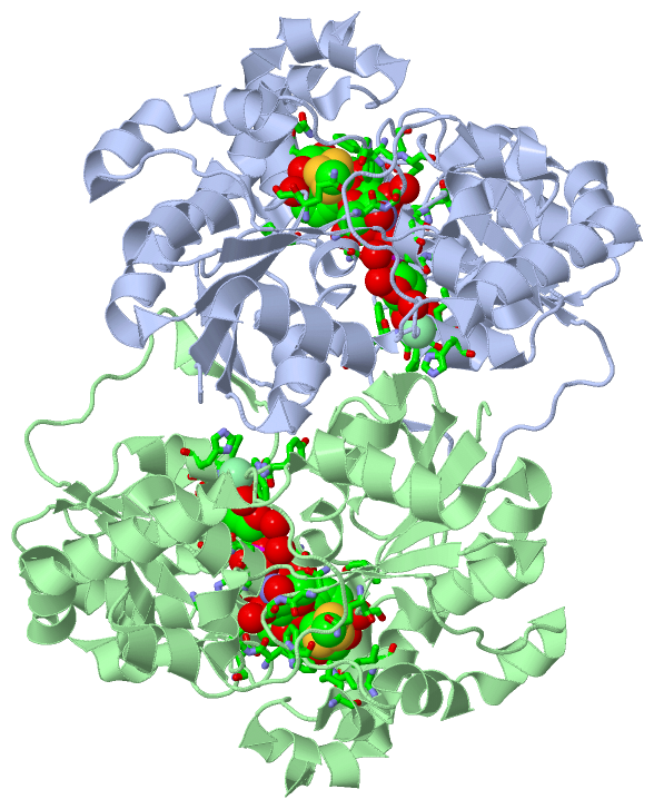
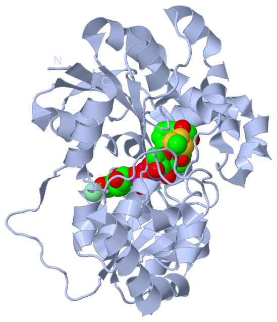
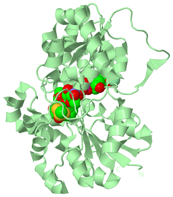
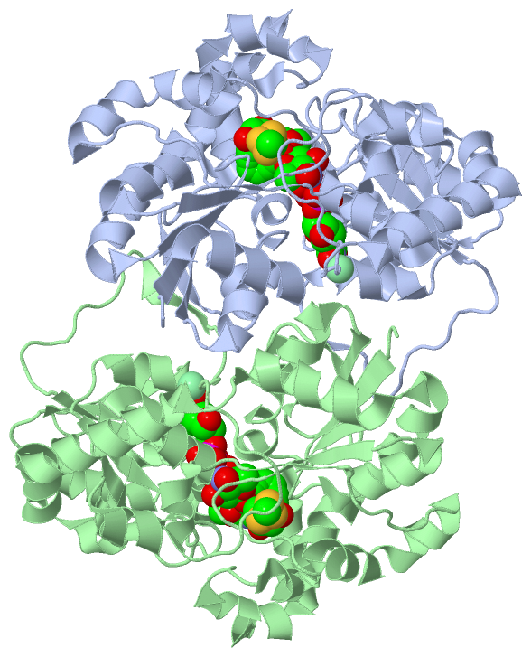
Asymmetric Unit (3, 6)
No.NameCountTypeFull Name
1C0T2Ligand/IonCALICHEAMICIN T0
2CL2Ligand/IonCHLORIDE ION
3TYD2Ligand/IonTHYMIDINE-5'-DIPHOSPHATE
Biological Unit 1 (2, 2)
No.NameCountTypeFull Name
1C0T1Ligand/IonCALICHEAMICIN T0
2CL-1Ligand/IonCHLORIDE ION
3TYD1Ligand/IonTHYMIDINE-5'-DIPHOSPHATE
Biological Unit 2 (2, 2)
No.NameCountTypeFull Name
1C0T1Ligand/IonCALICHEAMICIN T0
2CL-1Ligand/IonCHLORIDE ION
3TYD1Ligand/IonTHYMIDINE-5'-DIPHOSPHATE
Biological Unit 3 (2, 4)
No.NameCountTypeFull Name
1C0T2Ligand/IonCALICHEAMICIN T0
2CL-1Ligand/IonCHLORIDE ION
3TYD2Ligand/IonTHYMIDINE-5'-DIPHOSPHATE

(-) Sites  (6, 6)

Asymmetric Unit (6, 6)
No.NameEvidenceResiduesDescription
1AC1SOFTWAREGLY A:10 , GLY A:195 , GLY A:218 , THR A:219 , ILE A:220 , GLY A:249 , TRP A:268 , THR A:269 , PRO A:270 , LEU A:271 , HIS A:284 , GLY A:286 , GLY A:287 , GLY A:288 , THR A:289 , C0T A:378 , HOH A:380 , HOH A:388 , HOH A:416 , HOH A:472 , HOH A:496 , HOH A:498BINDING SITE FOR RESIDUE TYD A 377
2AC2SOFTWAREPRO A:7 , GLY A:10 , PHE A:60 , GLN A:88 , GLN A:116 , GLY A:117 , GLN A:137 , SER A:138 , TRP A:140 , GLY A:286 , GLY A:287 , ASP A:308 , PHE A:310 , GLN A:311 , TYD A:377 , HOH A:527 , HOH A:689BINDING SITE FOR RESIDUE C0T A 378
3AC3SOFTWARETYR A:194 , GLY A:195 , LEU A:271 , HIS A:272 , HOH A:498BINDING SITE FOR RESIDUE CL A 379
4AC4SOFTWAREGLY B:10 , GLY B:195 , GLY B:218 , THR B:219 , ILE B:220 , GLY B:249 , TRP B:268 , THR B:269 , PRO B:270 , LEU B:271 , HIS B:284 , GLY B:286 , GLY B:287 , GLY B:288 , THR B:289 , C0T B:378 , HOH B:384 , HOH B:391 , HOH B:413 , HOH B:453 , HOH B:512 , HOH B:547BINDING SITE FOR RESIDUE TYD B 377
5AC5SOFTWAREPRO B:7 , GLY B:10 , PHE B:60 , LEU B:82 , GLN B:88 , ILE B:89 , GLN B:116 , GLY B:117 , GLN B:137 , SER B:138 , TRP B:140 , ILE B:220 , GLY B:286 , GLY B:287 , ASP B:308 , PHE B:310 , GLN B:311 , TYD B:377 , HOH B:481 , HOH B:666BINDING SITE FOR RESIDUE C0T B 378
6AC6SOFTWARETYR B:194 , GLY B:195 , LEU B:271 , HIS B:272 , HOH B:547BINDING SITE FOR RESIDUE CL B 379

(-) SS Bonds  (0, 0)

(no "SS Bond" information available for 3OTI)

(-) Cis Peptide Bonds  (0, 0)

(no "Cis Peptide Bond" information available for 3OTI)

 Sequence-Structure Mapping

(-) SAPs(SNPs)/Variants  (0, 0)

(no "SAP(SNP)/Variant" information available for 3OTI)

(-) PROSITE Motifs  (0, 0)

(no "PROSITE Motif" information available for 3OTI)

(-) Exons   (0, 0)

(no "Exon" information available for 3OTI)

(-) Sequences/Alignments

Asymmetric Unit
   Reformat: Number of residues per line =  ('0' or empty: single-line sequence representation)
  Number of residues per labelling interval =   
  UniProt sequence: complete  aligned part    
   Show mapping: SCOP domains CATH domains Pfam domains Secondary structure (by author)
SAPs(SNPs) PROSITE motifs Exons
(details for a mapped element are shown in a popup box when the mouse pointer rests over it)
Chain A from PDB  Type:PROTEIN  Length:379
 aligned with Q8KND7_MICEC | Q8KND7 from UniProtKB/TrEMBL  Length:376

    Alignment length:379
                                1                                                                                                                                                                                                                                                                                                                                                                                      
                                |    6        16        26        36        46        56        66        76        86        96       106       116       126       136       146       156       166       176       186       196       206       216       226       236       246       256       266       276       286       296       306       316       326       336       346       356       366         
         Q8KND7_MICEC     - ----MLFVSSPGIGHLFPLIQLAWGFRTAGHDVLIAVAEHADRAAAAGLEVVDVAPDYSAVKVFEQVAKDNPRFAETVATRPAIDLEEWGVQIAAVNRPLVDGTMALVDDYRPDLVVYEQGATVGLLAADRAGVPAVQRNQSAWRTRGMHRSIASFLTDLMDKHQVSLPEPVATIESFPPSLLLEAEPEGWFMRWVPYGGGAVLGDRLPPVPARPEVAITMGTIELQAFGIGAVEPIIAAAGEVDADFVLALGDLDISPLGTLPRNVRAVGWTPLHTLLRTCTAVVHHGGGGTVMTAIDAGIPQLLAPDPRDQFQHTAREAVSRRGIGLVSTSDKVDADLLRRLIGDESLRTAAREVREEMVALPTPAETVRRIVERIS 375
               SCOP domains ------------------------------------------------------------------------------------------------------------------------------------------------------------------------------------------------------------------------------------------------------------------------------------------------------------------------------------------------------------------------------------------- SCOP domains
               CATH domains ------------------------------------------------------------------------------------------------------------------------------------------------------------------------------------------------------------------------------------------------------------------------------------------------------------------------------------------------------------------------------------------- CATH domains
               Pfam domains ------------------------------------------------------------------------------------------------------------------------------------------------------------------------------------------------------------------------------------------------------------------------------------------------------------------------------------------------------------------------------------------- Pfam domains
         Sec.struct. author ...eeeee...hhhhhh.hhhhhhhhhhh..eeeeee..hhhhhhh...eeee.....hhhhhhhhhhhhhhhhhhhhhhh...hhhhhhhhhhhhhh.hhhhhhhhhhhhh..eeeee..hhhhhhhhhhhh..eeee........hhhhhhhh.hhhhhhhh........eee...hhhhhh.................ee............eeee....hhhhhhh..hhhhhhhhhhh...eeeee.....hhhhh.....eeee...hhhhhhh...eeee..hhhhhhhhhhhh..eee...........hhhhhhhh..eee.hhhhhhhhhhhhhhhhhhhhhhhhhhhhhhhh..hhhhhhhhhhhhhh Sec.struct. author
                 SAPs(SNPs) ------------------------------------------------------------------------------------------------------------------------------------------------------------------------------------------------------------------------------------------------------------------------------------------------------------------------------------------------------------------------------------------- SAPs(SNPs)
                    PROSITE ------------------------------------------------------------------------------------------------------------------------------------------------------------------------------------------------------------------------------------------------------------------------------------------------------------------------------------------------------------------------------------------- PROSITE
                 Transcript ------------------------------------------------------------------------------------------------------------------------------------------------------------------------------------------------------------------------------------------------------------------------------------------------------------------------------------------------------------------------------------------- Transcript
                 3oti A  -3 RHMRVLFVSSPGIGHLFPLIQLAWGFRTAGHDVLIAVAEHADRAAAAGLEVVDVAPDYSAVKVFEQVAKDNPRFAETVATRPAIDLEEWGVQIAAVNRPLVDGTMALVDDYRPDLVVYEQGATVGLLAADRAGVPAVQRNQSAWRTRGMHRSIASFLTDLMDKHQVSLPEPVATIESFPPSLLLEAEPEGWFMRWVPYGGGAVLGDRLPPVPARPEVAITMGTIELQAFGIGAVEPIIAAAGEVDADFVLALGDLDISPLGTLPRNVRAVGWTPLHTLLRTCTAVVHHGGGGTVMTAIDAGIPQLLAPDPRDQFQHTAREAVSRRGIGLVSTSDKVDADLLRRLIGDESLRTAAREVREEMVALPTPAETVRRIVERIS 375
                                     6        16        26        36        46        56        66        76        86        96       106       116       126       136       146       156       166       176       186       196       206       216       226       236       246       256       266       276       286       296       306       316       326       336       346       356       366         

Chain B from PDB  Type:PROTEIN  Length:379
 aligned with Q8KND7_MICEC | Q8KND7 from UniProtKB/TrEMBL  Length:376

    Alignment length:379
                                1                                                                                                                                                                                                                                                                                                                                                                                      
                                |    6        16        26        36        46        56        66        76        86        96       106       116       126       136       146       156       166       176       186       196       206       216       226       236       246       256       266       276       286       296       306       316       326       336       346       356       366         
         Q8KND7_MICEC     - ----MLFVSSPGIGHLFPLIQLAWGFRTAGHDVLIAVAEHADRAAAAGLEVVDVAPDYSAVKVFEQVAKDNPRFAETVATRPAIDLEEWGVQIAAVNRPLVDGTMALVDDYRPDLVVYEQGATVGLLAADRAGVPAVQRNQSAWRTRGMHRSIASFLTDLMDKHQVSLPEPVATIESFPPSLLLEAEPEGWFMRWVPYGGGAVLGDRLPPVPARPEVAITMGTIELQAFGIGAVEPIIAAAGEVDADFVLALGDLDISPLGTLPRNVRAVGWTPLHTLLRTCTAVVHHGGGGTVMTAIDAGIPQLLAPDPRDQFQHTAREAVSRRGIGLVSTSDKVDADLLRRLIGDESLRTAAREVREEMVALPTPAETVRRIVERIS 375
               SCOP domains ------------------------------------------------------------------------------------------------------------------------------------------------------------------------------------------------------------------------------------------------------------------------------------------------------------------------------------------------------------------------------------------- SCOP domains
               CATH domains ------------------------------------------------------------------------------------------------------------------------------------------------------------------------------------------------------------------------------------------------------------------------------------------------------------------------------------------------------------------------------------------- CATH domains
           Pfam domains (1) -----Glyco_transf_28-3otiB03 B:2-146                                                                                                                  ------------------------DUF1205-3otiB01 B:171-264                                                                     --------------------------------------------------------------------------------------------------------------- Pfam domains (1)
           Pfam domains (2) -----Glyco_transf_28-3otiB04 B:2-146                                                                                                                  ------------------------DUF1205-3otiB02 B:171-264                                                                     --------------------------------------------------------------------------------------------------------------- Pfam domains (2)
         Sec.struct. author ...eeeee...hhhhhh.hhhhhhhhhhh..eeeeee..hhhhhhh...eeee.....hhhhhhhhhhhhhhhhhhhhhhh...hhhhhhhhhhhhhh.hhhhhhhhhhhhh..eeeee..hhhhhhhhhhhh..eeee........hhhhhhhh.hhhhhhhh........eee...hhhhhh.................eeee..........eeee....hhhhhhh..hhhhhhhhhhh...eeeee.....hhhhh.....eeee...hhhhhhh...eeee..hhhhhhhhhhhh..eee...........hhhhhhhh..eee.hhhhhhhhhhhhhhhhhhhhhhhhhhhhhhhh..hhhhhhhhhhhhhh Sec.struct. author
                 SAPs(SNPs) ------------------------------------------------------------------------------------------------------------------------------------------------------------------------------------------------------------------------------------------------------------------------------------------------------------------------------------------------------------------------------------------- SAPs(SNPs)
                    PROSITE ------------------------------------------------------------------------------------------------------------------------------------------------------------------------------------------------------------------------------------------------------------------------------------------------------------------------------------------------------------------------------------------- PROSITE
                 Transcript ------------------------------------------------------------------------------------------------------------------------------------------------------------------------------------------------------------------------------------------------------------------------------------------------------------------------------------------------------------------------------------------- Transcript
                 3oti B  -3 RHMRVLFVSSPGIGHLFPLIQLAWGFRTAGHDVLIAVAEHADRAAAAGLEVVDVAPDYSAVKVFEQVAKDNPRFAETVATRPAIDLEEWGVQIAAVNRPLVDGTMALVDDYRPDLVVYEQGATVGLLAADRAGVPAVQRNQSAWRTRGMHRSIASFLTDLMDKHQVSLPEPVATIESFPPSLLLEAEPEGWFMRWVPYGGGAVLGDRLPPVPARPEVAITMGTIELQAFGIGAVEPIIAAAGEVDADFVLALGDLDISPLGTLPRNVRAVGWTPLHTLLRTCTAVVHHGGGGTVMTAIDAGIPQLLAPDPRDQFQHTAREAVSRRGIGLVSTSDKVDADLLRRLIGDESLRTAAREVREEMVALPTPAETVRRIVERIS 375
                                     6        16        26        36        46        56        66        76        86        96       106       116       126       136       146       156       166       176       186       196       206       216       226       236       246       256       266       276       286       296       306       316       326       336       346       356       366         

   Legend:   → Mismatch (orange background)
  - → Gap (green background, '-', border residues have a numbering label)
    → Modified Residue (blue background, lower-case, 'x' indicates undefined single-letter code, labelled with number + name)
  x → Chemical Group (purple background, 'x', labelled with number + name, e.g. ACE or NH2)
  extra numbering lines below/above indicate numbering irregularities and modified residue names etc., number ends below/above '|'

 Classification and Annotation

(-) SCOP Domains  (0, 0)

(no "SCOP Domain" information available for 3OTI)

(-) CATH Domains  (0, 0)

(no "CATH Domain" information available for 3OTI)

(-) Pfam Domains  (2, 4)

Asymmetric Unit
(-)
Clan: GT-B (132)

(-) Gene Ontology  (1, 1)

Asymmetric Unit(hide GO term definitions)
Chain A,B   (Q8KND7_MICEC | Q8KND7)
molecular function
    GO:0046527    glucosyltransferase activity    Catalysis of the transfer of a glucosyl group to an acceptor molecule, typically another carbohydrate or a lipid.

 Visualization

(-) Interactive Views

Asymmetric Unit
  Complete Structure
    Jena3D(integrated viewing of ligand, site, SAP, PROSITE, SCOP information)
    WebMol | AstexViewer[tm]@PDBe
(Java Applets, require no local installation except for Java; loading may be slow)
    STRAP
(Java WebStart application, automatic local installation, requires Java; full application with system access!)
    RasMol
(require local installation)
    Molscript (VRML)
(requires installation of a VRML viewer; select preferred view via VRML and generate a mono or stereo PDF format file)
 
  Ligands, Modified Residues, Ions
    C0T  [ RasMol | Jena3D ]  +environment [ RasMol | Jena3D ]
    CL  [ RasMol | Jena3D ]  +environment [ RasMol | Jena3D ]
    TYD  [ RasMol | Jena3D ]  +environment [ RasMol | Jena3D ]
 
  Sites
    AC1  [ RasMol ]  +environment [ RasMol ]
    AC2  [ RasMol ]  +environment [ RasMol ]
    AC3  [ RasMol ]  +environment [ RasMol ]
    AC4  [ RasMol ]  +environment [ RasMol ]
    AC5  [ RasMol ]  +environment [ RasMol ]
    AC6  [ RasMol ]  +environment [ RasMol ]
 
  Cis Peptide Bonds
(no "Cis Peptide Bonds" information available for 3oti)
 
Biological Units
  Complete Structure
    Biological Unit 1  [ Jena3D ]
    Biological Unit 2  [ Jena3D ]
    Biological Unit 3  [ Jena3D ]

(-) Still Images

Jmol
  protein: cartoon or spacefill or dots and stick; nucleic acid: cartoon and stick; ligands: spacefill; active site: stick
Molscript
  protein, nucleic acid: cartoon; ligands: spacefill; active site: ball and stick

 Databases and Analysis Tools

(-) Databases

Access by PDB/NDB ID
  3oti
    Family and Domain InformationProDom | SYSTERS
    General Structural InformationGlycoscienceDB | MMDB | NDB | OCA | PDB | PDBe | PDBj | PDBsum | PDBWiki | PQS | PROTEOPEDIA
    Orientation in MembranesOPM
    Protein SurfaceSURFACE
    Secondary StructureDSSP (structure derived) | HSSP (homology derived)
    Structural GenomicsGeneCensus
    Structural NeighboursCE | VAST
    Structure ClassificationCATH | Dali | SCOP
    Validation and Original DataBMRB Data View | BMRB Restraints Grid | EDS | PROCHECK | RECOORD | WHAT_CHECK
 
Access by UniProt ID/Accession number
  Q8KND7_MICEC | Q8KND7
    Comparative Protein Structure ModelsModBase
    Genomic InformationEnsembl
    Protein-protein InteractionDIP
    Sequence, Family and Domain InformationInterPro | Pfam | SMART | UniProtKB/TrEMBL
 
Access by Enzyme Classificator   (EC Number)
  (no 'Enzyme Classificator' available)
    General Enzyme InformationBRENDA | EC-PDB | Enzyme | IntEnz
    PathwayKEGG | MetaCyc
 
Access by Disease Identifier   (MIM ID)
  (no 'MIM ID' available)
    Disease InformationOMIM
 
Access by GenAge ID
  (no 'GenAge ID' available)
    Age Related InformationGenAge

(-) Analysis Tools

Access by PDB/NDB ID
    Domain InformationXDom
    Interatomic Contacts of Structural UnitsCSU
    Ligand-protein ContactsLPC
    Protein CavitiescastP
    Sequence and Secondary StructurePDBCartoon
    Structure AlignmentSTRAP(Java WebStart application, automatic local installation, requires Java; full application with system access!)
    Structure and Sequence BrowserSTING
 
Access by UniProt ID/Accession number
  Q8KND7_MICEC | Q8KND7
    Protein Disorder PredictionDisEMBL | FoldIndex | GLOBPLOT (for more information see DisProt)

 Related Entries

(-) Entries Sharing at Least One Protein Chain (UniProt ID)

UniProtKB/TrEMBL
        Q8KND7_MICEC | Q8KND73d0q 3d0r

(-) Related Entries Specified in the PDB File

3d0q CRYSTAL STRUCTURE OF CALG3 FROM MICROMONOSPORA ECHINOSPORA DETERMINED IN SPACE GROUP I222
3d0r CRYSTAL STRUCTURE OF CALG3 FROM MICROMONOSPORA ECHINOSPORA DETERMINED IN SPACE GROUP P2(1)
3otg CRYSTAL STRUCTURE OF CALG1, CALICHEAMICIN GLYCOSTYLTRANSFERASE, TDP BOUND FORM
3oth CRYSTAL STRUCTURE OF CALG1, CALICHEAMICIN GLYCOSTYLTRANSFERASE, TDP AND CALICHEAMICIN ALPHA3I BOUND FORM RELATED ID: NATPRO-GO.119385 RELATED DB: TARGETTRACK