Show PDB file:   
         Plain Text   HTML   (compressed file size)
QuickSearch:   
by PDB,NDB,UniProt,PROSITE Code or Search Term(s)  
(-)Asymmetric Unit
(-)Asym. Unit - sites
(-)Biological Unit 1
(-)Biol. Unit 1 - sites
collapse expand < >
Image Asymmetric Unit
Asymmetric Unit  (Jmol Viewer)
Image Asym. Unit - sites
Asym. Unit - sites  (Jmol Viewer)
Image Biological Unit 1
Biological Unit 1  (Jmol Viewer)
Image Biol. Unit 1 - sites
Biol. Unit 1 - sites  (Jmol Viewer)

(-) Description

Title :  CRYSTAL STRUCTURE OF A HISTIDINE TRIAD FAMILY PROTEIN FROM ENTAMOEBA HISTOLYTICA, BOUND TO AMP
 
Authors :  Ssgcid, Seattle Structural Genomics Center For Infectious Dis (Ssgcid)
Date :  26 Aug 10  (Deposition) - 08 Sep 10  (Release) - 20 May 15  (Revision)
Method :  X-RAY DIFFRACTION
Resolution :  1.80
Chains :  Asym. Unit :  A
Biol. Unit 1:  A  (2x)
Keywords :  Ssgcid, Putative Hydrolase, Structural Genomics, Seattle Structural Genomics Center For Infectious Disease, Metal Binding Protein (Keyword Search: [Gene Ontology, PubMed, Web (Google))
 
Reference :  D. D. Lorimer, R. Choi, A. Abramov, S. Nakazawa Hewitt, A. S. Gardberg W. C. Van Voorhis, B. L. Staker, P. J. Myler, T. E. Edwards
Structures Of A Histidine Triad Family Protein From Entamoeba Histolytica Bound To Sulfate, Amp And Gmp.
Acta Crystallogr F Struct V. 71 572 2015 Biol Commun
PubMed-ID: 25945711  |  Reference-DOI: 10.1107/S2053230X1500237X

(-) Compounds

Molecule 1 - PUTATIVE HISTIDINE TRIAD FAMILY PROTEIN
    ChainsA
    EngineeredYES
    Expression SystemESCHERICHIA COLI
    Expression System PlasmidAVA0421
    Expression System StrainBL21(DE3)
    Expression System Taxid469008
    Expression System Vector TypePLASMID
    GeneEHI_093910
    Organism ScientificENTAMOEBA HISTOLYTICA
    Organism Taxid294381

 Structural Features

(-) Chains, Units

  1
Asymmetric Unit A
Biological Unit 1 (2x)A

Summary Information (see also Sequences/Alignments below)
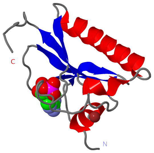
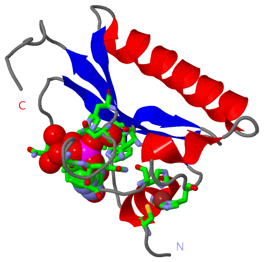
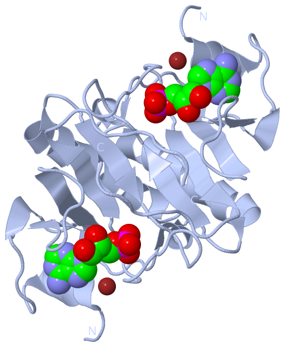
(-) Ligands, Modified Residues, Ions  (2, 2)

Asymmetric Unit (2, 2)
No.NameCountTypeFull Name
1AMP1Ligand/IonADENOSINE MONOPHOSPHATE
2ZN1Ligand/IonZINC ION
Biological Unit 1 (1, 2)
No.NameCountTypeFull Name
1AMP2Ligand/IonADENOSINE MONOPHOSPHATE
2ZN-1Ligand/IonZINC ION

(-) Sites  (2, 2)

Asymmetric Unit (2, 2)
No.NameEvidenceResiduesDescription
1AC1SOFTWARECYS A:5 , CYS A:8 , HIS A:47 , HIS A:97BINDING SITE FOR RESIDUE ZN A 114
2AC2SOFTWAREPHE A:29 , LYS A:30 , ASP A:31 , ILE A:32 , ASN A:33 , LEU A:41 , ASN A:86 , GLY A:92 , GLN A:93 , THR A:94 , HIS A:99 , HIS A:101 , HOH A:150 , HOH A:199 , HOH A:205 , HOH A:223 , HOH A:262 , HOH A:271BINDING SITE FOR RESIDUE AMP A 115

(-) SS Bonds  (0, 0)

(no "SS Bond" information available for 3OMF)

(-) Cis Peptide Bonds  (0, 0)

(no "Cis Peptide Bond" information available for 3OMF)

 Sequence-Structure Mapping

(-) SAPs(SNPs)/Variants  (0, 0)

(no "SAP(SNP)/Variant" information available for 3OMF)

(-) PROSITE Motifs  (2, 2)

Asymmetric Unit (2, 2)
 PROSITEUniProtKBPDB
No.IDACDescriptionIDLocationCountLocation
1HIT_2PS51084 HIT domain profile.HIT_ENTHI6-113  1A:6-113
2HIT_1PS00892 HIT domain signature.HIT_ENTHI86-104  1A:86-104
Biological Unit 1 (2, 4)
 PROSITEUniProtKBPDB
No.IDACDescriptionIDLocationCountLocation
1HIT_2PS51084 HIT domain profile.HIT_ENTHI6-113  2A:6-113
2HIT_1PS00892 HIT domain signature.HIT_ENTHI86-104  2A:86-104

(-) Exons   (0, 0)

(no "Exon" information available for 3OMF)

(-) Sequences/Alignments

Asymmetric Unit
   Reformat: Number of residues per line =  ('0' or empty: single-line sequence representation)
  Number of residues per labelling interval =   
  UniProt sequence: complete  aligned part    
   Show mapping: SCOP domains CATH domains Pfam domains Secondary structure (by author)
SAPs(SNPs) PROSITE motifs Exons
(details for a mapped element are shown in a popup box when the mouse pointer rests over it)
Chain A from PDB  Type:PROTEIN  Length:111
 aligned with HIT_ENTHI | C4LYI2 from UniProtKB/Swiss-Prot  Length:113

    Alignment length:111
                                    12        22        32        42        52        62        72        82        92       102       112 
            HIT_ENTHI     3 DSCIFCKIAQKQIPSTIVYEDDEIFAFKDINPIAPIHILVIPKQHIASLNEITEENEAFIGKVLYKVSLIGKKECPEGYRVVNNIGEDAGQTVKHIHFHILGGKKLAWDKL 113
               SCOP domains d3omfa_ A: automated matches                                                                                    SCOP domains
               CATH domains --------------------------------------------------------------------------------------------------------------- CATH domains
               Pfam domains --------------------------------------------------------------------------------------------------------------- Pfam domains
         Sec.struct. author ..hhhhhhhh......eeee...eeeee.......eeeeeee.....hhhhh...hhhhhhhhhhhhhhhhhhhh...eeee...............eeeee......... Sec.struct. author
                 SAPs(SNPs) --------------------------------------------------------------------------------------------------------------- SAPs(SNPs)
                PROSITE (1) ---HIT_2  PDB: A:6-113 UniProt: 6-113                                                                           PROSITE (1)
                PROSITE (2) -----------------------------------------------------------------------------------HIT_1  PDB: A:86-10--------- PROSITE (2)
                 Transcript --------------------------------------------------------------------------------------------------------------- Transcript
                 3omf A   3 DSCIFCKIAQKQIPSTIVYEDDEIFAFKDINPIAPIHILVIPKQHIASLNEITEENEAFIGKVLYKVSLIGKKECPEGYRVVNNIGEDAGQTVKHIHFHILGGKKLAWDKL 113
                                    12        22        32        42        52        62        72        82        92       102       112 

   Legend:   → Mismatch (orange background)
  - → Gap (green background, '-', border residues have a numbering label)
    → Modified Residue (blue background, lower-case, 'x' indicates undefined single-letter code, labelled with number + name)
  x → Chemical Group (purple background, 'x', labelled with number + name, e.g. ACE or NH2)
  extra numbering lines below/above indicate numbering irregularities and modified residue names etc., number ends below/above '|'

 Classification and Annotation

(-) SCOP Domains  (1, 1)

Asymmetric Unit

(-) CATH Domains  (0, 0)

(no "CATH Domain" information available for 3OMF)

(-) Pfam Domains  (0, 0)

(no "Pfam Domain" information available for 3OMF)

(-) Gene Ontology  (5, 5)

Asymmetric Unit(hide GO term definitions)
Chain A   (HIT_ENTHI | C4LYI2)
molecular function
    GO:0003824    catalytic activity    Catalysis of a biochemical reaction at physiological temperatures. In biologically catalyzed reactions, the reactants are known as substrates, and the catalysts are naturally occurring macromolecular substances known as enzymes. Enzymes possess specific binding sites for substrates, and are usually composed wholly or largely of protein, but RNA that has catalytic activity (ribozyme) is often also regarded as enzymatic.
    GO:0016787    hydrolase activity    Catalysis of the hydrolysis of various bonds, e.g. C-O, C-N, C-C, phosphoric anhydride bonds, etc. Hydrolase is the systematic name for any enzyme of EC class 3.
    GO:0000166    nucleotide binding    Interacting selectively and non-covalently with a nucleotide, any compound consisting of a nucleoside that is esterified with (ortho)phosphate or an oligophosphate at any hydroxyl group on the ribose or deoxyribose.
cellular component
    GO:0005737    cytoplasm    All of the contents of a cell excluding the plasma membrane and nucleus, but including other subcellular structures.
    GO:0005634    nucleus    A membrane-bounded organelle of eukaryotic cells in which chromosomes are housed and replicated. In most cells, the nucleus contains all of the cell's chromosomes except the organellar chromosomes, and is the site of RNA synthesis and processing. In some species, or in specialized cell types, RNA metabolism or DNA replication may be absent.

 Visualization

(-) Interactive Views

Asymmetric Unit
  Complete Structure
    Jena3D(integrated viewing of ligand, site, SAP, PROSITE, SCOP information)
    WebMol | AstexViewer[tm]@PDBe
(Java Applets, require no local installation except for Java; loading may be slow)
    STRAP
(Java WebStart application, automatic local installation, requires Java; full application with system access!)
    RasMol
(require local installation)
    Molscript (VRML)
(requires installation of a VRML viewer; select preferred view via VRML and generate a mono or stereo PDF format file)
 
  Ligands, Modified Residues, Ions
    AMP  [ RasMol | Jena3D ]  +environment [ RasMol | Jena3D ]
    ZN  [ RasMol | Jena3D ]  +environment [ RasMol | Jena3D ]
 
  Sites
    AC1  [ RasMol ]  +environment [ RasMol ]
    AC2  [ RasMol ]  +environment [ RasMol ]
 
  Cis Peptide Bonds
(no "Cis Peptide Bonds" information available for 3omf)
 
Biological Unit
  Complete Structure
    Biological Unit 1  [ Jena3D ]

(-) Still Images

Jmol
  protein: cartoon or spacefill or dots and stick; nucleic acid: cartoon and stick; ligands: spacefill; active site: stick
Molscript
  protein, nucleic acid: cartoon; ligands: spacefill; active site: ball and stick

 Databases and Analysis Tools

(-) Databases

Access by PDB/NDB ID
  3omf
    Family and Domain InformationProDom | SYSTERS
    General Structural InformationGlycoscienceDB | MMDB | NDB | OCA | PDB | PDBe | PDBj | PDBsum | PDBWiki | PQS | PROTEOPEDIA
    Orientation in MembranesOPM
    Protein SurfaceSURFACE
    Secondary StructureDSSP (structure derived) | HSSP (homology derived)
    Structural GenomicsGeneCensus
    Structural NeighboursCE | VAST
    Structure ClassificationCATH | Dali | SCOP
    Validation and Original DataBMRB Data View | BMRB Restraints Grid | EDS | PROCHECK | RECOORD | WHAT_CHECK
 
Access by UniProt ID/Accession number
  HIT_ENTHI | C4LYI2
    Comparative Protein Structure ModelsModBase
    Genomic InformationEnsembl
    Protein-protein InteractionDIP
    Sequence, Family and Domain InformationInterPro | Pfam | SMART | UniProtKB/SwissProt
 
Access by Enzyme Classificator   (EC Number)
  (no 'Enzyme Classificator' available)
    General Enzyme InformationBRENDA | EC-PDB | Enzyme | IntEnz
    PathwayKEGG | MetaCyc
 
Access by Disease Identifier   (MIM ID)
  (no 'MIM ID' available)
    Disease InformationOMIM
 
Access by GenAge ID
  (no 'GenAge ID' available)
    Age Related InformationGenAge

(-) Analysis Tools

Access by PDB/NDB ID
    Domain InformationXDom
    Interatomic Contacts of Structural UnitsCSU
    Ligand-protein ContactsLPC
    Protein CavitiescastP
    Sequence and Secondary StructurePDBCartoon
    Structure AlignmentSTRAP(Java WebStart application, automatic local installation, requires Java; full application with system access!)
    Structure and Sequence BrowserSTING
 
Access by UniProt ID/Accession number
  HIT_ENTHI | C4LYI2
    Protein Disorder PredictionDisEMBL | FoldIndex | GLOBPLOT (for more information see DisProt)

 Related Entries

(-) Entries Sharing at Least One Protein Chain (UniProt ID)

UniProtKB/Swiss-Prot
        HIT_ENTHI | C4LYI23oj7 3oxk

(-) Related Entries Specified in the PDB File

3oj7 STRUCTURE OF THE SAME HIT PROTEIN BOUND TO SULFATE RELATED ID: ENHIA.01296.A RELATED DB: TARGETDB
3oxk STRUCTURE OF THE SAME PROTEIN BOUND TO GMP