|
|
|
|
 Description
|
|
Compounds
|
||||||||||||||||||||||||||||||||||||||||||||||||||||
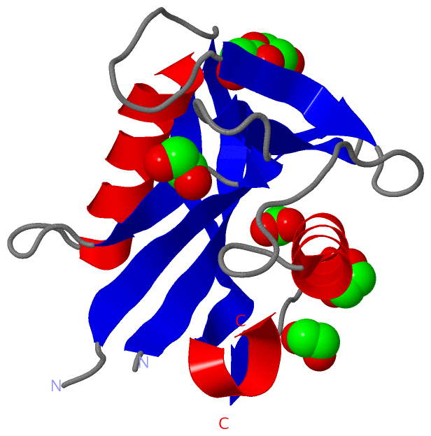
Chains, Units
Summary Information (see also Sequences/Alignments below) |
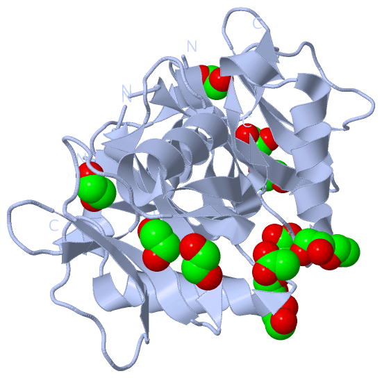
Ligands, Modified Residues, Ions (2, 6)| Asymmetric Unit (2, 6) Biological Unit 1 (2, 12) |
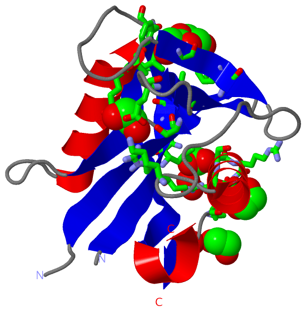
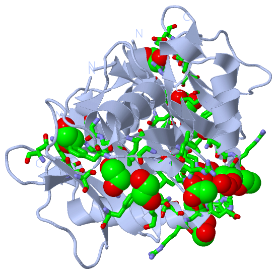
Sites (6, 6)
Asymmetric Unit (6, 6)
|
SS Bonds (0, 0)| (no "SS Bond" information available for 3O8S) |
Cis Peptide Bonds (0, 0)| (no "Cis Peptide Bond" information available for 3O8S) |
SAPs(SNPs)/Variants (0, 0)| (no "SAP(SNP)/Variant" information available for 3O8S) |
PROSITE Motifs (0, 0)| (no "PROSITE Motif" information available for 3O8S) |
Exons (0, 0)| (no "Exon" information available for 3O8S) |
Sequences/Alignments
Asymmetric Unit
Chain A from PDB Type:PROTEIN Length:134
SCOP domains -------------------------------------------------------------------------------------------------------------------------------------- SCOP domains
CATH domains -------------------------------------------------------------------------------------------------------------------------------------- CATH domains
Pfam domains -------------------------------------------------------------------------------------------------------------------------------------- Pfam domains
SAPs(SNPs) -------------------------------------------------------------------------------------------------------------------------------------- SAPs(SNPs)
PROSITE -------------------------------------------------------------------------------------------------------------------------------------- PROSITE
Transcript -------------------------------------------------------------------------------------------------------------------------------------- Transcript
3o8s A 67 TPKLDTRAAIFQEDKILLVQENDGLWSLPGGWCDVDQSVKDNVVKEVKEEAGLDVEAQRVVAILDKHKNNPRVTKVFILCRLLGGEFQPNSETVASGFFSLDDLPPLYLGKNTAEQLALCLEASRSEHWETRFD 205
76 86 96 106 116 126 136|| 151 161 171 181 191 201
137|
143
|
||||||||||||||||||||
SCOP Domains (0, 0)| (no "SCOP Domain" information available for 3O8S) |
CATH Domains (0, 0)| (no "CATH Domain" information available for 3O8S) |
Pfam Domains (0, 0)| (no "Pfam Domain" information available for 3O8S) |
Gene Ontology (0, 0)|
Asymmetric Unit(hide GO term definitions)
(no "Gene Ontology" information available for 3O8S)
|
Interactive Views
|
||||||||||||||||||||||||||||||||||||||||||||||||||||||||||||||||||||||||||||||||||||||||||||||||||||||||||||||||||||||||||||||||||||||||||||||||||||||||||||||||||||||||||||||||||
Still Images
|
||||||||||||||||
Databases
|
||||||||||||||||||||||||||||||||||||||||||||||||||||||||||||||||||||||||||||||||||||||||||||||||||||||||||||||||||||||||||||||||||||||||||||||||||||||||||||||||
Analysis Tools
|
|||||||||||||||||||||||||||||||||||||||||||||||||||||||||||||
Entries Sharing at Least One Protein Chain (UniProt ID)
Related Entries Specified in the PDB File
|
|