Show PDB file:   
         Plain Text   HTML   (compressed file size)
QuickSearch:   
by PDB,NDB,UniProt,PROSITE Code or Search Term(s)  
(-)Asymmetric Unit
(-)Asym. Unit - sites
(-)Biological Unit 1
(-)Biol. Unit 1 - sites
(-)Biological Unit 2
(-)Biological Unit 3
(-)Biological Unit 4
collapse expand < >
Image Asymmetric Unit
Asymmetric Unit  (Jmol Viewer)
Image Asym. Unit - sites
Asym. Unit - sites  (Jmol Viewer)
Image Biological Unit 1
Biological Unit 1  (Jmol Viewer)
Image Biol. Unit 1 - sites
Biol. Unit 1 - sites  (Jmol Viewer)
Image Biological Unit 2
Biological Unit 2  (Jmol Viewer)
Image Biological Unit 3
Biological Unit 3  (Jmol Viewer)
Image Biological Unit 4
Biological Unit 4  (Jmol Viewer)

(-) Description

Title :  THE CRYSTAL STRUCTURE OF THE PAS DOMAIN OF A GGDEF FAMILY PROTEIN FROM CHROMOBACTERIUM VIOLACEUM ATCC 12472.
 
Authors :  K. Tan, R. Wu, B. Feldmann, A. Joachimiak, Midwest Center For Struct Genomics (Mcsg)
Date :  17 Jun 10  (Deposition) - 18 Aug 10  (Release) - 18 Aug 10  (Revision)
Method :  X-RAY DIFFRACTION
Resolution :  2.37
Chains :  Asym. Unit :  A,B,C,D
Biol. Unit 1:  A,B  (2x)
Biol. Unit 2:  A,B  (1x)
Biol. Unit 3:  C,D  (2x)
Biol. Unit 4:  C,D  (1x)
Keywords :  Structural Genomics, Psi-2, Protein Structure Initiative, Midwest Center For Structural Genomics, Mcsg, Unknown Function (Keyword Search: [Gene Ontology, PubMed, Web (Google))
 
Reference :  K. Tan, R. Wu, B. Feldmann, A. Joachimiak
The Crystal Structure Of The Pas Domain Of A Ggdef Family Protein From Chromobacterium Violaceum Atcc 12472.
To Be Published
PubMed: search

(-) Compounds

Molecule 1 - PROBABLE GGDEF FAMILY PROTEIN
    ChainsA, B, C, D
    EngineeredYES
    Expression SystemESCHERICHIA COLI
    Expression System PlasmidPMCSG7
    Expression System StrainBL21
    Expression System Taxid511693
    Expression System Vector TypePLASMID
    GeneCV_1582
    Organism ScientificCHROMOBACTERIUM VIOLACEUM
    Organism Taxid243365
    StrainATCC 12472

 Structural Features

(-) Chains, Units

  1234
Asymmetric Unit ABCD
Biological Unit 1 (2x)AB  
Biological Unit 2 (1x)AB  
Biological Unit 3 (2x)  CD
Biological Unit 4 (1x)  CD

Summary Information (see also Sequences/Alignments below)

(-) Ligands, Modified Residues, Ions  (4, 16)

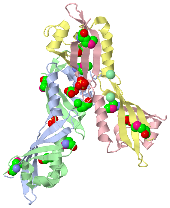
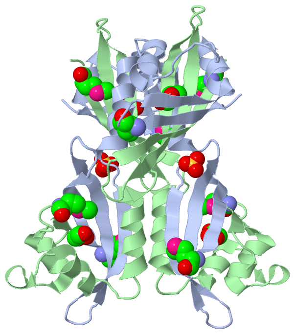
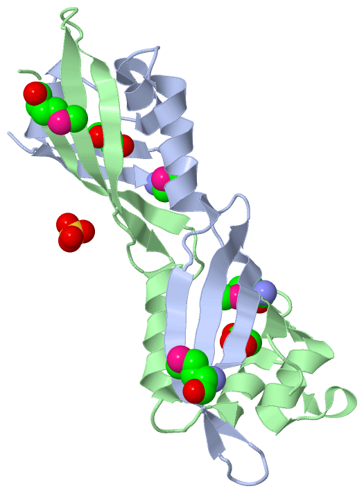
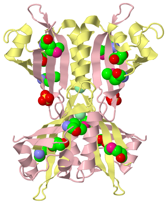
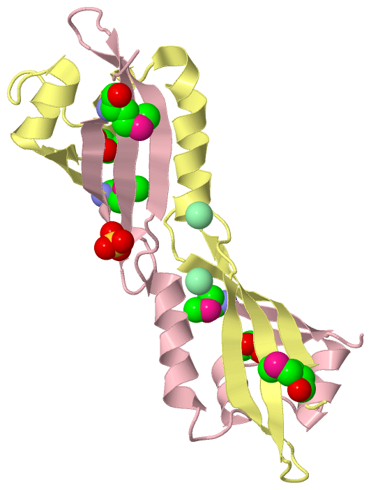
Asymmetric Unit (4, 16)
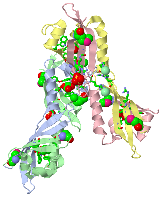
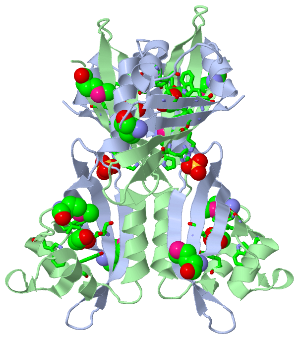
No.NameCountTypeFull Name
1CL2Ligand/IonCHLORIDE ION
2GOL4Ligand/IonGLYCEROL
3MSE8Mod. Amino AcidSELENOMETHIONINE
4SO42Ligand/IonSULFATE ION
Biological Unit 1 (3, 14)
No.NameCountTypeFull Name
1CL-1Ligand/IonCHLORIDE ION
2GOL4Ligand/IonGLYCEROL
3MSE8Mod. Amino AcidSELENOMETHIONINE
4SO42Ligand/IonSULFATE ION
Biological Unit 2 (3, 7)
No.NameCountTypeFull Name
1CL-1Ligand/IonCHLORIDE ION
2GOL2Ligand/IonGLYCEROL
3MSE4Mod. Amino AcidSELENOMETHIONINE
4SO41Ligand/IonSULFATE ION
Biological Unit 3 (3, 14)
No.NameCountTypeFull Name
1CL-1Ligand/IonCHLORIDE ION
2GOL4Ligand/IonGLYCEROL
3MSE8Mod. Amino AcidSELENOMETHIONINE
4SO42Ligand/IonSULFATE ION
Biological Unit 4 (3, 7)
No.NameCountTypeFull Name
1CL-1Ligand/IonCHLORIDE ION
2GOL2Ligand/IonGLYCEROL
3MSE4Mod. Amino AcidSELENOMETHIONINE
4SO41Ligand/IonSULFATE ION

(-) Sites  (8, 8)

Asymmetric Unit (8, 8)
No.NameEvidenceResiduesDescription
1AC1SOFTWAREGLU A:95 , TRP B:18 , LEU B:20 , TRP B:29 , TYR B:51BINDING SITE FOR RESIDUE GOL A 123
2AC2SOFTWARETRP A:18 , LEU A:27 , TRP A:29 , TYR A:51 , TYR B:81 , GLU B:95 , VAL B:114BINDING SITE FOR RESIDUE GOL B 123
3AC3SOFTWAREPRO B:76 , ARG B:96 , HIS B:98BINDING SITE FOR RESIDUE SO4 B 124
4AC4SOFTWAREARG C:96 , HIS C:98 , ARG D:96BINDING SITE FOR RESIDUE SO4 C 123
5AC5SOFTWAREARG C:75BINDING SITE FOR RESIDUE CL C 124
6AC6SOFTWAREGLU C:95 , TRP D:18 , LEU D:27 , TRP D:29 , TYR D:51BINDING SITE FOR RESIDUE GOL D 123
7AC7SOFTWARETRP C:18 , LEU C:27 , TRP C:29 , TYR D:81 , GLU D:95BINDING SITE FOR RESIDUE GOL D 124
8AC8SOFTWAREHIS D:70 , ARG D:75BINDING SITE FOR RESIDUE CL D 125

(-) SS Bonds  (0, 0)

(no "SS Bond" information available for 3NJA)

(-) Cis Peptide Bonds  (0, 0)

(no "Cis Peptide Bond" information available for 3NJA)

 Sequence-Structure Mapping

(-) SAPs(SNPs)/Variants  (0, 0)

(no "SAP(SNP)/Variant" information available for 3NJA)

(-) PROSITE Motifs  (0, 0)

(no "PROSITE Motif" information available for 3NJA)

(-) Exons   (0, 0)

(no "Exon" information available for 3NJA)

(-) Sequences/Alignments

Asymmetric Unit
   Reformat: Number of residues per line =  ('0' or empty: single-line sequence representation)
  Number of residues per labelling interval =   
  UniProt sequence: complete  aligned part    
   Show mapping: SCOP domains CATH domains Pfam domains Secondary structure (by author)
SAPs(SNPs) PROSITE motifs Exons
(details for a mapped element are shown in a popup box when the mouse pointer rests over it)
Chain A from PDB  Type:PROTEIN  Length:106
 aligned with Q7NXP4_CHRVO | Q7NXP4 from UniProtKB/TrEMBL  Length:427

    Alignment length:106
                                    23        33        43        53        63        73        83        93       103       113      
         Q7NXP4_CHRVO    14 GIGSWVLHMESGRLEWSQAVHDIFGTDSATFDATEDAYFQRVHPDDRARVRRELDRHVLGDRPFDVEYRIVRPDGQVRELLERNHIQRQASGQVDHLWGTVIDMTE 119
               SCOP domains ---------------------------------------------------------------------------------------------------------- SCOP domains
               CATH domains ---------------------------------------------------------------------------------------------------------- CATH domains
               Pfam domains ---------------------------------------------------------------------------------------------------------- Pfam domains
         Sec.struct. author ..eeeeee....eeeehhhhhhhhh........hhhhhhhhh...hhhhhhhhhhhhhhh...eeeeeeee.....eeeeeeeeeeee.....eeeeeeeeee... Sec.struct. author
                 SAPs(SNPs) ---------------------------------------------------------------------------------------------------------- SAPs(SNPs)
                    PROSITE ---------------------------------------------------------------------------------------------------------- PROSITE
                 Transcript ---------------------------------------------------------------------------------------------------------- Transcript
                 3nja A  14 GIGSWVLHmESGRLEWSQAVHDIFGTDSATFDATEDAYFQRVHPDDRARVRRELDRHVLGDRPFDVEYRIVRPDGQVRELLERNHIQRQASGQVDHLWGTVIDmTE 119
                                    23        33        43        53        63        73        83        93       103       113   |  
                                   22-MSE                                                                                        117-MSE

Chain B from PDB  Type:PROTEIN  Length:108
 aligned with Q7NXP4_CHRVO | Q7NXP4 from UniProtKB/TrEMBL  Length:427

    Alignment length:108
                                    21        31        41        51        61        71        81        91       101       111        
         Q7NXP4_CHRVO    12 DAGIGSWVLHMESGRLEWSQAVHDIFGTDSATFDATEDAYFQRVHPDDRARVRRELDRHVLGDRPFDVEYRIVRPDGQVRELLERNHIQRQASGQVDHLWGTVIDMTE 119
               SCOP domains ------------------------------------------------------------------------------------------------------------ SCOP domains
               CATH domains ------------------------------------------------------------------------------------------------------------ CATH domains
               Pfam domains ------------------------------------------------------------------------------------------------------------ Pfam domains
         Sec.struct. author ....eeeeee.....eeehhhhhhhhh.hhhhh..hhhhhhhh....hhhhhhhhhhhhhh....eeeeeeee.....eeeeeeeeeeee.....eeeeeeeeee... Sec.struct. author
                 SAPs(SNPs) ------------------------------------------------------------------------------------------------------------ SAPs(SNPs)
                    PROSITE ------------------------------------------------------------------------------------------------------------ PROSITE
                 Transcript ------------------------------------------------------------------------------------------------------------ Transcript
                 3nja B  12 DAGIGSWVLHmESGRLEWSQAVHDIFGTDSATFDATEDAYFQRVHPDDRARVRRELDRHVLGDRPFDVEYRIVRPDGQVRELLERNHIQRQASGQVDHLWGTVIDmTE 119
                                    21|       31        41        51        61        71        81        91       101       111     |  
                                     22-MSE                                                                                        117-MSE

Chain C from PDB  Type:PROTEIN  Length:108
 aligned with Q7NXP4_CHRVO | Q7NXP4 from UniProtKB/TrEMBL  Length:427

    Alignment length:108
                                    22        32        42        52        62        72        82        92       102       112        
         Q7NXP4_CHRVO    13 AGIGSWVLHMESGRLEWSQAVHDIFGTDSATFDATEDAYFQRVHPDDRARVRRELDRHVLGDRPFDVEYRIVRPDGQVRELLERNHIQRQASGQVDHLWGTVIDMTEH 120
               SCOP domains ------------------------------------------------------------------------------------------------------------ SCOP domains
               CATH domains ------------------------------------------------------------------------------------------------------------ CATH domains
               Pfam domains ------------------------------------------------------------------------------------------------------------ Pfam domains
         Sec.struct. author ...eeeeee.....eeehhhhhhhhh........hhhhhhh.....hhhhhhhhhhhhhh....eeeeeeee.....eeeeeeeeeeee.....eeeeeeeeee.... Sec.struct. author
                 SAPs(SNPs) ------------------------------------------------------------------------------------------------------------ SAPs(SNPs)
                    PROSITE ------------------------------------------------------------------------------------------------------------ PROSITE
                 Transcript ------------------------------------------------------------------------------------------------------------ Transcript
                 3nja C  13 AGIGSWVLHmESGRLEWSQAVHDIFGTDSATFDATEDAYFQRVHPDDRARVRRELDRHVLGDRPFDVEYRIVRPDGQVRELLERNHIQRQASGQVDHLWGTVIDmTEH 120
                                    22        32        42        52        62        72        82        92       102       112    |   
                                    22-MSE                                                                                        117-MSE

Chain D from PDB  Type:PROTEIN  Length:105
 aligned with Q7NXP4_CHRVO | Q7NXP4 from UniProtKB/TrEMBL  Length:427

    Alignment length:105
                                    24        34        44        54        64        74        84        94       104       114     
         Q7NXP4_CHRVO    15 IGSWVLHMESGRLEWSQAVHDIFGTDSATFDATEDAYFQRVHPDDRARVRRELDRHVLGDRPFDVEYRIVRPDGQVRELLERNHIQRQASGQVDHLWGTVIDMTE 119
               SCOP domains --------------------------------------------------------------------------------------------------------- SCOP domains
               CATH domains --------------------------------------------------------------------------------------------------------- CATH domains
           Pfam domains (1) -----------PAS_3-3njaD01 D:26-114                                                                   ----- Pfam domains (1)
           Pfam domains (2) -----------PAS_3-3njaD02 D:26-114                                                                   ----- Pfam domains (2)
           Pfam domains (3) -----------PAS_3-3njaD03 D:26-114                                                                   ----- Pfam domains (3)
           Pfam domains (4) -----------PAS_3-3njaD04 D:26-114                                                                   ----- Pfam domains (4)
         Sec.struct. author .eeeeee.....eeehhhhhhhhh........hhhhhhh..hhhhhhhhhhhhhhhhh....eeeeeeee.....eeeeeeeeeeee.....eeeeeeeeee... Sec.struct. author
                 SAPs(SNPs) --------------------------------------------------------------------------------------------------------- SAPs(SNPs)
                    PROSITE --------------------------------------------------------------------------------------------------------- PROSITE
                 Transcript --------------------------------------------------------------------------------------------------------- Transcript
                 3nja D  15 IGSWVLHmESGRLEWSQAVHDIFGTDSATFDATEDAYFQRVHPDDRARVRRELDRHVLGDRPFDVEYRIVRPDGQVRELLERNHIQRQASGQVDHLWGTVIDmTE 119
                                   |24        34        44        54        64        74        84        94       104       114  |  
                                  22-MSE                                                                                        117-MSE

   Legend:   → Mismatch (orange background)
  - → Gap (green background, '-', border residues have a numbering label)
    → Modified Residue (blue background, lower-case, 'x' indicates undefined single-letter code, labelled with number + name)
  x → Chemical Group (purple background, 'x', labelled with number + name, e.g. ACE or NH2)
  extra numbering lines below/above indicate numbering irregularities and modified residue names etc., number ends below/above '|'

 Classification and Annotation

(-) SCOP Domains  (0, 0)

(no "SCOP Domain" information available for 3NJA)

(-) CATH Domains  (0, 0)

(no "CATH Domain" information available for 3NJA)

(-) Pfam Domains  (1, 4)

Asymmetric Unit
(-)
Clan: PAS_Fold (84)

(-) Gene Ontology  (4, 4)

Asymmetric Unit(hide GO term definitions)
Chain A,B,C,D   (Q7NXP4_CHRVO | Q7NXP4)
molecular function
    GO:0000155    phosphorelay sensor kinase activity    Catalysis of the phosphorylation of a histidine residue in response to detection of an extracellular signal such as a chemical ligand or change in environment, to initiate a change in cell state or activity. The two-component sensor is a histidine kinase that autophosphorylates a histidine residue in its active site. The phosphate is then transferred to an aspartate residue in a downstream response regulator, to trigger a response.
biological process
    GO:0000160    phosphorelay signal transduction system    A conserved series of molecular signals found in prokaryotes and eukaryotes; involves autophosphorylation of a histidine kinase and the transfer of the phosphate group to an aspartate that then acts as a phospho-donor to response regulator proteins.
    GO:0023014    signal transduction by protein phosphorylation    A process in which the transfer of one or more phosphate groups to a substrate transmits a signal to the phosphorylated substrate.
cellular component
    GO:0005622    intracellular    The living contents of a cell; the matter contained within (but not including) the plasma membrane, usually taken to exclude large vacuoles and masses of secretory or ingested material. In eukaryotes it includes the nucleus and cytoplasm.

 Visualization

(-) Interactive Views

Asymmetric Unit
  Complete Structure
    Jena3D(integrated viewing of ligand, site, SAP, PROSITE, SCOP information)
    WebMol | AstexViewer[tm]@PDBe
(Java Applets, require no local installation except for Java; loading may be slow)
    STRAP
(Java WebStart application, automatic local installation, requires Java; full application with system access!)
    RasMol
(require local installation)
    Molscript (VRML)
(requires installation of a VRML viewer; select preferred view via VRML and generate a mono or stereo PDF format file)
 
  Ligands, Modified Residues, Ions
    CL  [ RasMol | Jena3D ]  +environment [ RasMol | Jena3D ]
    GOL  [ RasMol | Jena3D ]  +environment [ RasMol | Jena3D ]
    MSE  [ RasMol | Jena3D ]  +environment [ RasMol | Jena3D ]
    SO4  [ RasMol | Jena3D ]  +environment [ RasMol | Jena3D ]
 
  Sites
    AC1  [ RasMol ]  +environment [ RasMol ]
    AC2  [ RasMol ]  +environment [ RasMol ]
    AC3  [ RasMol ]  +environment [ RasMol ]
    AC4  [ RasMol ]  +environment [ RasMol ]
    AC5  [ RasMol ]  +environment [ RasMol ]
    AC6  [ RasMol ]  +environment [ RasMol ]
    AC7  [ RasMol ]  +environment [ RasMol ]
    AC8  [ RasMol ]  +environment [ RasMol ]
 
  Cis Peptide Bonds
(no "Cis Peptide Bonds" information available for 3nja)
 
Biological Units
  Complete Structure
    Biological Unit 1  [ Jena3D ]
    Biological Unit 2  [ Jena3D ]
    Biological Unit 3  [ Jena3D ]
    Biological Unit 4  [ Jena3D ]

(-) Still Images

Jmol
  protein: cartoon or spacefill or dots and stick; nucleic acid: cartoon and stick; ligands: spacefill; active site: stick
Molscript
  protein, nucleic acid: cartoon; ligands: spacefill; active site: ball and stick

 Databases and Analysis Tools

(-) Databases

Access by PDB/NDB ID
  3nja
    Family and Domain InformationProDom | SYSTERS
    General Structural InformationGlycoscienceDB | MMDB | NDB | OCA | PDB | PDBe | PDBj | PDBsum | PDBWiki | PQS | PROTEOPEDIA
    Orientation in MembranesOPM
    Protein SurfaceSURFACE
    Secondary StructureDSSP (structure derived) | HSSP (homology derived)
    Structural GenomicsGeneCensus
    Structural NeighboursCE | VAST
    Structure ClassificationCATH | Dali | SCOP
    Validation and Original DataBMRB Data View | BMRB Restraints Grid | EDS | PROCHECK | RECOORD | WHAT_CHECK
 
Access by UniProt ID/Accession number
  Q7NXP4_CHRVO | Q7NXP4
    Comparative Protein Structure ModelsModBase
    Genomic InformationEnsembl
    Protein-protein InteractionDIP
    Sequence, Family and Domain InformationInterPro | Pfam | SMART | UniProtKB/TrEMBL
 
Access by Enzyme Classificator   (EC Number)
  (no 'Enzyme Classificator' available)
    General Enzyme InformationBRENDA | EC-PDB | Enzyme | IntEnz
    PathwayKEGG | MetaCyc
 
Access by Disease Identifier   (MIM ID)
  (no 'MIM ID' available)
    Disease InformationOMIM
 
Access by GenAge ID
  (no 'GenAge ID' available)
    Age Related InformationGenAge

(-) Analysis Tools

Access by PDB/NDB ID
    Domain InformationXDom
    Interatomic Contacts of Structural UnitsCSU
    Ligand-protein ContactsLPC
    Protein CavitiescastP
    Sequence and Secondary StructurePDBCartoon
    Structure AlignmentSTRAP(Java WebStart application, automatic local installation, requires Java; full application with system access!)
    Structure and Sequence BrowserSTING
 
Access by UniProt ID/Accession number
  Q7NXP4_CHRVO | Q7NXP4
    Protein Disorder PredictionDisEMBL | FoldIndex | GLOBPLOT (for more information see DisProt)

 Related Entries

(-) Entries Sharing at Least One Protein Chain (UniProt ID)

(no "Entries Sharing at Least One Protein Chain" available for 3NJA)

(-) Related Entries Specified in the PDB File

(no "Related Entries Specified in the PDB File" available for 3NJA)