|
|
|
|
 Description
|
|
Compounds
|
||||||||||||||||||||||||||||||||||||||||||||||||||||||||||||||||||||||||||||||
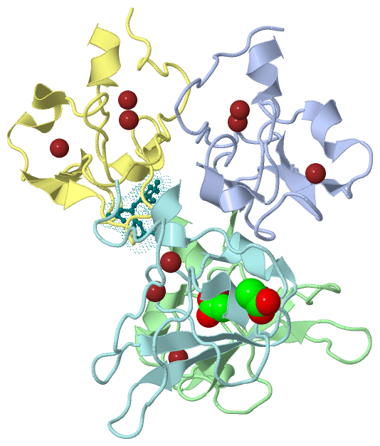
Chains, Units
Summary Information (see also Sequences/Alignments below) |
Ligands, Modified Residues, Ions (2, 14)| Asymmetric Unit (2, 14) Biological Unit 1 (0, 0) Biological Unit 2 (0, 0) Biological Unit 3 (0, 0) Biological Unit 4 (1, 2) |
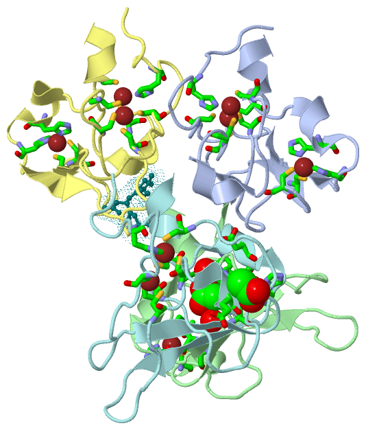
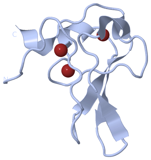
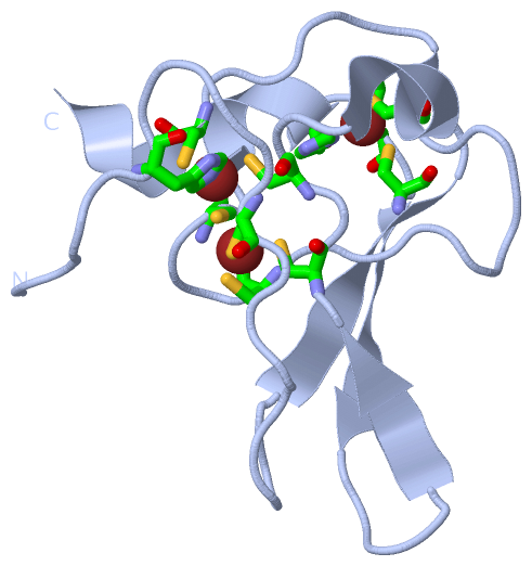
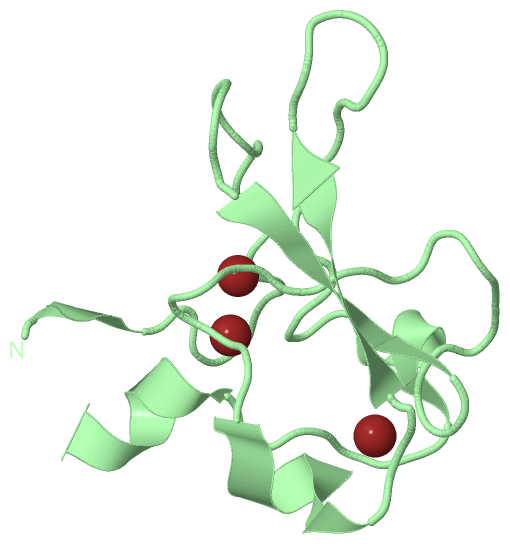
Sites (14, 14)
Asymmetric Unit (14, 14)
|
SS Bonds (0, 0)| (no "SS Bond" information available for 3NIL) |
Cis Peptide Bonds (0, 0)| (no "Cis Peptide Bond" information available for 3NIL) |
SAPs(SNPs)/Variants (0, 0)| (no "SAP(SNP)/Variant" information available for 3NIL) |
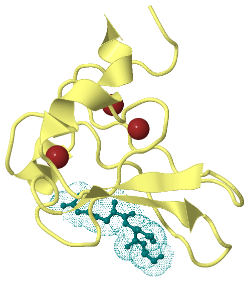
PROSITE Motifs (1, 4)
Asymmetric Unit (1, 4)
|
||||||||||||||||||||||||||||||||||||||||||||||||||||||||||||||||||||||||||||||||||||||||||||||||||||||||||||||||||||||||
Exons (0, 0)| (no "Exon" information available for 3NIL) |
Sequences/Alignments
Asymmetric UnitChain A from PDB Type:PROTEIN Length:81 aligned with UBR1_YEAST | P19812 from UniProtKB/Swiss-Prot Length:1950 Alignment length:81 122 132 142 152 162 172 182 192 UBR1_YEAST 113 GDVHKHTGRNCGRKFKIGEPLYRCHECGCDDTCVLCIHCFNPKDHVNHHVCTDICTEFTSGICDCGDEEAWNSPLHCKAEE 193 SCOP domains --------------------------------------------------------------------------------- SCOP domains CATH domains --------------------------------------------------------------------------------- CATH domains Pfam domains --------------------------------------------------------------------------------- Pfam domains SAPs(SNPs) --------------------------------------------------------------------------------- SAPs(SNPs) PROSITE --------ZF_UBR PDB: A:121-193 UniProt: 121-194 PROSITE Transcript --------------------------------------------------------------------------------- Transcript 3nil A 113 GSVHKHTGRNCGRKFKIGEPLYRCHECGCDDTCVLCIHCFNPKDHVNHHVCTDICTEFTSGICDCGDEEAWNSPLHCKAEE 193 122 132 142 152 162 172 182 192 Chain B from PDB Type:PROTEIN Length:82 aligned with UBR1_YEAST | P19812 from UniProtKB/Swiss-Prot Length:1950 Alignment length:82 122 132 142 152 162 172 182 192 UBR1_YEAST 113 GDVHKHTGRNCGRKFKIGEPLYRCHECGCDDTCVLCIHCFNPKDHVNHHVCTDICTEFTSGICDCGDEEAWNSPLHCKAEEQ 194 SCOP domains ---------------------------------------------------------------------------------- SCOP domains CATH domains ---------------------------------------------------------------------------------- CATH domains Pfam domains ---------------------------------------------------------------------------------- Pfam domains SAPs(SNPs) ---------------------------------------------------------------------------------- SAPs(SNPs) PROSITE --------ZF_UBR PDB: B:121-194 UniProt: 121-194 PROSITE Transcript ---------------------------------------------------------------------------------- Transcript 3nil B 113 GSVHKHTGRNCGRKFKIGEPLYRCHECGCDDTCVLCIHCFNPKDHVNHHVCTDICTEFTSGICDCGDEEAWNSPLHCKAEEQ 194 122 132 142 152 162 172 182 192 Chain D from PDB Type:PROTEIN Length:81 aligned with UBR1_YEAST | P19812 from UniProtKB/Swiss-Prot Length:1950 Alignment length:81 122 132 142 152 162 172 182 192 UBR1_YEAST 113 GDVHKHTGRNCGRKFKIGEPLYRCHECGCDDTCVLCIHCFNPKDHVNHHVCTDICTEFTSGICDCGDEEAWNSPLHCKAEE 193 SCOP domains --------------------------------------------------------------------------------- SCOP domains CATH domains --------------------------------------------------------------------------------- CATH domains Pfam domains --------------------------------------------------------------------------------- Pfam domains SAPs(SNPs) --------------------------------------------------------------------------------- SAPs(SNPs) PROSITE --------ZF_UBR PDB: D:121-193 UniProt: 121-194 PROSITE Transcript --------------------------------------------------------------------------------- Transcript 3nil D 113 GSVHKHTGRNCGRKFKIGEPLYRCHECGCDDTCVLCIHCFNPKDHVNHHVCTDICTEFTSGICDCGDEEAWNSPLHCKAEE 193 122 132 142 152 162 172 182 192 Chain F from PDB Type:PROTEIN Length:81 aligned with UBR1_YEAST | P19812 from UniProtKB/Swiss-Prot Length:1950 Alignment length:81 122 132 142 152 162 172 182 192 UBR1_YEAST 113 GDVHKHTGRNCGRKFKIGEPLYRCHECGCDDTCVLCIHCFNPKDHVNHHVCTDICTEFTSGICDCGDEEAWNSPLHCKAEE 193 SCOP domains --------------------------------------------------------------------------------- SCOP domains CATH domains --------------------------------------------------------------------------------- CATH domains Pfam domains (1) --------zf-UBR-3nilF01 F:121-193 Pfam domains (1) Pfam domains (2) --------zf-UBR-3nilF02 F:121-193 Pfam domains (2) Pfam domains (3) --------zf-UBR-3nilF03 F:121-193 Pfam domains (3) Pfam domains (4) --------zf-UBR-3nilF04 F:121-193 Pfam domains (4) SAPs(SNPs) --------------------------------------------------------------------------------- SAPs(SNPs) PROSITE --------ZF_UBR PDB: F:121-193 UniProt: 121-194 PROSITE Transcript --------------------------------------------------------------------------------- Transcript 3nil F 113 GSVHKHTGRNCGRKFKIGEPLYRCHECGCDDTCVLCIHCFNPKDHVNHHVCTDICTEFTSGICDCGDEEAWNSPLHCKAEE 193 122 132 142 152 162 172 182 192
Chain X from PDB Type:PROTEIN Length:3
SCOP domains --- SCOP domains
CATH domains --- CATH domains
Pfam domains --- Pfam domains
SAPs(SNPs) --- SAPs(SNPs)
PROSITE --- PROSITE
Transcript --- Transcript
3nil X 1 RDA 3
|
||||||||||||||||||||
SCOP Domains (0, 0)| (no "SCOP Domain" information available for 3NIL) |
CATH Domains (0, 0)| (no "CATH Domain" information available for 3NIL) |
Pfam Domains (1, 4)
Asymmetric Unit
|
Gene Ontology (17, 17)|
Asymmetric Unit(hide GO term definitions) Chain A,B,D,F (UBR1_YEAST | P19812)
|
||||||||||||||||||||||||||||||||||||||||||||||||||||||||||||||||||||||||||||||||||||||||||||||||||||||||||||||||||||||||
Interactive Views
|
|||||||||||||||||||||||||||||||||||||||||||||||||||||||||||||||||||||||||||||||||||||||||||||||||||||||||||||||||||||||||||||||||||||||||||||||||||||||||||||||||||||||||||||||||||||||||||||||||||||||||||||||||||||||||||||||||||||||||||||||||||||||||
Still Images
|
||||||||||||||||
Databases
|
||||||||||||||||||||||||||||||||||||||||||||||||||||||||||||||||||||||||||||||||||||||||||||||||||||||||||||||||||||||||||||||||||||||||||||||||||||||||||||||||
Analysis Tools
|
|||||||||||||||||||||||||||||||||||||||||||||||||||||||||||||
Entries Sharing at Least One Protein Chain (UniProt ID)
Related Entries Specified in the PDB File
|
|