Show PDB file:   
         Plain Text   HTML   (compressed file size)
QuickSearch:   
by PDB,NDB,UniProt,PROSITE Code or Search Term(s)  
(-)Asymmetric Unit
(-)Biological Unit 1
(-)Biological Unit 2
(-)Biological Unit 3
collapse expand < >
Image Asymmetric Unit
Asymmetric Unit  (Jmol Viewer)
Image Biological Unit 1
Biological Unit 1  (Jmol Viewer)
Image Biological Unit 2
Biological Unit 2  (Jmol Viewer)
Image Biological Unit 3
Biological Unit 3  (Jmol Viewer)

(-) Description

Title :  CRYSTAL STRUCTURE OF A RESPONSE REGULATOR FROM MYXOCOCCUS XANTHUS
 
Authors :  K. Palani, S. K. Burley, S. Swaminathan, New York Sgx Research Cent Structural Genomics (Nysgxrc)
Date :  14 Jun 10  (Deposition) - 21 Jul 10  (Release) - 21 Jul 10  (Revision)
Method :  X-RAY DIFFRACTION
Resolution :  2.19
Chains :  Asym. Unit :  A,B
Biol. Unit 1:  A  (1x)
Biol. Unit 2:  B  (1x)
Biol. Unit 3:  A,B  (1x)
Keywords :  Protein Structure Initiative Ii(Psi Ii), Nysgxrc, Structural Genomics, New York Sgx Research Center For Structural Genomics, Signaling Protein (Keyword Search: [Gene Ontology, PubMed, Web (Google))
 
Reference :  K. Palani, S. K. Burley, S. Swaminathan
Crystal Structure Of A Response Regulator From Myxococcus Xanthus
To Be Published
PubMed: search

(-) Compounds

Molecule 1 - RESPONSE REGULATOR
    ChainsA, B
    EngineeredYES
    Expression SystemESCHERICHIA COLI
    Expression System PlasmidBC - PSGX3 (BC)
    Expression System StrainBL21(DE3)
    Expression System Taxid469008
    Expression System Vector TypePLASMID
    GeneMXAN_5889
    Organism ScientificMYXOCOCCUS XANTHUS
    Organism Taxid246197
    StrainDK 1622

 Structural Features

(-) Chains, Units

  12
Asymmetric Unit AB
Biological Unit 1 (1x)A 
Biological Unit 2 (1x) B
Biological Unit 3 (1x)AB

Summary Information (see also Sequences/Alignments below)

(-) Ligands, Modified Residues, Ions  (1, 6)

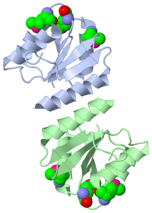
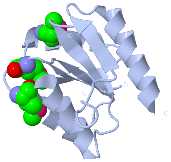
Asymmetric Unit (1, 6)
No.NameCountTypeFull Name
1MSE6Mod. Amino AcidSELENOMETHIONINE
Biological Unit 1 (1, 3)
No.NameCountTypeFull Name
1MSE3Mod. Amino AcidSELENOMETHIONINE
Biological Unit 2 (1, 3)
No.NameCountTypeFull Name
1MSE3Mod. Amino AcidSELENOMETHIONINE
Biological Unit 3 (1, 6)
No.NameCountTypeFull Name
1MSE6Mod. Amino AcidSELENOMETHIONINE

(-) Sites  (0, 0)

(no "Site" information available for 3NHM)

(-) SS Bonds  (0, 0)

(no "SS Bond" information available for 3NHM)

(-) Cis Peptide Bonds  (2, 2)

Asymmetric Unit
No.Residues
1Lys A:119 -Pro A:120
2Lys B:119 -Pro B:120

 Sequence-Structure Mapping

(-) SAPs(SNPs)/Variants  (0, 0)

(no "SAP(SNP)/Variant" information available for 3NHM)

(-) PROSITE Motifs  (0, 0)

(no "PROSITE Motif" information available for 3NHM)

(-) Exons   (0, 0)

(no "Exon" information available for 3NHM)

(-) Sequences/Alignments

Asymmetric Unit
   Reformat: Number of residues per line =  ('0' or empty: single-line sequence representation)
  Number of residues per labelling interval =   
  UniProt sequence: complete  aligned part    
   Show mapping: SCOP domains CATH domains Pfam domains Secondary structure (by author)
SAPs(SNPs) PROSITE motifs Exons
(details for a mapped element are shown in a popup box when the mouse pointer rests over it)
Chain A from PDB  Type:PROTEIN  Length:115
 aligned with Q1CZZ7_MYXXD | Q1CZZ7 from UniProtKB/TrEMBL  Length:153

    Alignment length:120
                                    28        38        48        58        68        78        88        98       108       118       128       138
         Q1CZZ7_MYXXD    19 PKVLIVENSWTMRETLRLLLSGEFDCTTAADGASGLQQALAHPPDVLISDVNMDGMDGYALCGHFRSEPTLKHIPVIFVSGYAPRTEGPADQPVPDAYLVKPVKPPVLIAQLHALLARAE 138
               SCOP domains d3nhma_ A: automated matches                                                                                             SCOP domains
               CATH domains ------------------------------------------------------------------------------------------------------------------------ CATH domains
               Pfam domains ------------------------------------------------------------------------------------------------------------------------ Pfam domains
         Sec.struct. author .eeeee..hhhhhhhhhhhhh...eeeee.hhhhhhhhhhhh...eeee.......hhhhhhhhhhhh.......eeeee...-----........eeee...hhhhhhhhhhhhhhhhh Sec.struct. author
                 SAPs(SNPs) ------------------------------------------------------------------------------------------------------------------------ SAPs(SNPs)
                    PROSITE ------------------------------------------------------------------------------------------------------------------------ PROSITE
                 Transcript ------------------------------------------------------------------------------------------------------------------------ Transcript
                 3nhm A  19 PKVLIVENSWTmRETLRLLLSGEFDCTTAADGASGLQQALAHPPDVLISDVNmDGmDGYALCGHFRSEPTLKHIPVIFVSGYA-----PADQPVPDAYLVKPVKPPVLIAQLHALLARAE 138
                                    28 |      38        48        58        68  |  |  78        88        98  |    108       118       128       138
                                      30-MSE                                   71-MSE                       101   107                               
                                                                                  74-MSE                                                            

Chain B from PDB  Type:PROTEIN  Length:115
 aligned with Q1CZZ7_MYXXD | Q1CZZ7 from UniProtKB/TrEMBL  Length:153

    Alignment length:120
                                    28        38        48        58        68        78        88        98       108       118       128       138
         Q1CZZ7_MYXXD    19 PKVLIVENSWTMRETLRLLLSGEFDCTTAADGASGLQQALAHPPDVLISDVNMDGMDGYALCGHFRSEPTLKHIPVIFVSGYAPRTEGPADQPVPDAYLVKPVKPPVLIAQLHALLARAE 138
               SCOP domains d3nhmb_ B: automated matches                                                                                             SCOP domains
               CATH domains ------------------------------------------------------------------------------------------------------------------------ CATH domains
           Pfam domains (1) --Response_reg-3nhmB01 B:21-131                                                                                  ------- Pfam domains (1)
           Pfam domains (2) --Response_reg-3nhmB02 B:21-131                                                                                  ------- Pfam domains (2)
         Sec.struct. author .eeeee..hhhhhhhhhhhhh...eeeee.hhhhhhhhhhhh...eeeee......hhhhhhhhhhhh.......eeeee...-----........eeee...hhhhhhhhhhhhhhhhh Sec.struct. author
                 SAPs(SNPs) ------------------------------------------------------------------------------------------------------------------------ SAPs(SNPs)
                    PROSITE ------------------------------------------------------------------------------------------------------------------------ PROSITE
                 Transcript ------------------------------------------------------------------------------------------------------------------------ Transcript
                 3nhm B  19 PKVLIVENSWTmRETLRLLLSGEFDCTTAADGASGLQQALAHPPDVLISDVNmDGmDGYALCGHFRSEPTLKHIPVIFVSGYA-----PADQPVPDAYLVKPVKPPVLIAQLHALLARAE 138
                                    28 |      38        48        58        68  |  |  78        88        98  |    108       118       128       138
                                      30-MSE                                   71-MSE                       101   107                               
                                                                                  74-MSE                                                            

   Legend:   → Mismatch (orange background)
  - → Gap (green background, '-', border residues have a numbering label)
    → Modified Residue (blue background, lower-case, 'x' indicates undefined single-letter code, labelled with number + name)
  x → Chemical Group (purple background, 'x', labelled with number + name, e.g. ACE or NH2)
  extra numbering lines below/above indicate numbering irregularities and modified residue names etc., number ends below/above '|'

 Classification and Annotation

(-) SCOP Domains  (1, 2)

Asymmetric Unit

(-) CATH Domains  (0, 0)

(no "CATH Domain" information available for 3NHM)

(-) Pfam Domains  (1, 2)

Asymmetric Unit
(-)
Clan: CheY (97)

(-) Gene Ontology  (2, 2)

Asymmetric Unit(hide GO term definitions)
Chain A,B   (Q1CZZ7_MYXXD | Q1CZZ7)
biological process
    GO:0000160    phosphorelay signal transduction system    A conserved series of molecular signals found in prokaryotes and eukaryotes; involves autophosphorylation of a histidine kinase and the transfer of the phosphate group to an aspartate that then acts as a phospho-donor to response regulator proteins.
cellular component
    GO:0005622    intracellular    The living contents of a cell; the matter contained within (but not including) the plasma membrane, usually taken to exclude large vacuoles and masses of secretory or ingested material. In eukaryotes it includes the nucleus and cytoplasm.

 Visualization

(-) Interactive Views

Asymmetric Unit
  Complete Structure
    Jena3D(integrated viewing of ligand, site, SAP, PROSITE, SCOP information)
    WebMol | AstexViewer[tm]@PDBe
(Java Applets, require no local installation except for Java; loading may be slow)
    STRAP
(Java WebStart application, automatic local installation, requires Java; full application with system access!)
    RasMol
(require local installation)
    Molscript (VRML)
(requires installation of a VRML viewer; select preferred view via VRML and generate a mono or stereo PDF format file)
 
  Ligands, Modified Residues, Ions
    MSE  [ RasMol | Jena3D ]  +environment [ RasMol | Jena3D ]
 
  Sites
(no "Sites" information available for 3nhm)
 
  Cis Peptide Bonds
    Lys A:119 - Pro A:120   [ RasMol ]  
    Lys B:119 - Pro B:120   [ RasMol ]  
 
Biological Units
  Complete Structure
    Biological Unit 1  [ Jena3D ]
    Biological Unit 2  [ Jena3D ]
    Biological Unit 3  [ Jena3D ]

(-) Still Images

Jmol
  protein: cartoon or spacefill or dots and stick; nucleic acid: cartoon and stick; ligands: spacefill; active site: stick
Molscript
  protein, nucleic acid: cartoon; ligands: spacefill; active site: ball and stick

 Databases and Analysis Tools

(-) Databases

Access by PDB/NDB ID
  3nhm
    Family and Domain InformationProDom | SYSTERS
    General Structural InformationGlycoscienceDB | MMDB | NDB | OCA | PDB | PDBe | PDBj | PDBsum | PDBWiki | PQS | PROTEOPEDIA
    Orientation in MembranesOPM
    Protein SurfaceSURFACE
    Secondary StructureDSSP (structure derived) | HSSP (homology derived)
    Structural GenomicsGeneCensus
    Structural NeighboursCE | VAST
    Structure ClassificationCATH | Dali | SCOP
    Validation and Original DataBMRB Data View | BMRB Restraints Grid | EDS | PROCHECK | RECOORD | WHAT_CHECK
 
Access by UniProt ID/Accession number
  Q1CZZ7_MYXXD | Q1CZZ7
    Comparative Protein Structure ModelsModBase
    Genomic InformationEnsembl
    Protein-protein InteractionDIP
    Sequence, Family and Domain InformationInterPro | Pfam | SMART | UniProtKB/TrEMBL
 
Access by Enzyme Classificator   (EC Number)
  (no 'Enzyme Classificator' available)
    General Enzyme InformationBRENDA | EC-PDB | Enzyme | IntEnz
    PathwayKEGG | MetaCyc
 
Access by Disease Identifier   (MIM ID)
  (no 'MIM ID' available)
    Disease InformationOMIM
 
Access by GenAge ID
  (no 'GenAge ID' available)
    Age Related InformationGenAge

(-) Analysis Tools

Access by PDB/NDB ID
    Domain InformationXDom
    Interatomic Contacts of Structural UnitsCSU
    Ligand-protein ContactsLPC
    Protein CavitiescastP
    Sequence and Secondary StructurePDBCartoon
    Structure AlignmentSTRAP(Java WebStart application, automatic local installation, requires Java; full application with system access!)
    Structure and Sequence BrowserSTING
 
Access by UniProt ID/Accession number
  Q1CZZ7_MYXXD | Q1CZZ7
    Protein Disorder PredictionDisEMBL | FoldIndex | GLOBPLOT (for more information see DisProt)

 Related Entries

(-) Entries Sharing at Least One Protein Chain (UniProt ID)

(no "Entries Sharing at Least One Protein Chain" available for 3NHM)

(-) Related Entries Specified in the PDB File

(no "Related Entries Specified in the PDB File" available for 3NHM)