Show PDB file:   
         Plain Text   HTML   (compressed file size)
QuickSearch:   
by PDB,NDB,UniProt,PROSITE Code or Search Term(s)  
(-)Asym./Biol. Unit
(-)Asym./Biol. Unit - sites
collapse expand < >
Image Asym./Biol. Unit
Asym./Biol. Unit  (Jmol Viewer)
Image Asym./Biol. Unit - sites
Asym./Biol. Unit - sites  (Jmol Viewer)

(-) Description

Title :  THE LACTOSE-SPECIFIC IIB COMPONENT DOMAIN STRUCTURE OF THE PHOSPHOENOLPYRUVATE:CARBOHYDRATE PHOSPHOTRANSFERASE SYSTEM (PTS) FROM STREPTOCOCCUS PNEUMONIAE.
 
Authors :  M. E. Cuff, G. Chhor, S. Clancy, A. Joachimiak, Midwest Center For St Genomics (Mcsg)
Date :  03 Jun 10  (Deposition) - 08 Sep 10  (Release) - 08 Sep 10  (Revision)
Method :  X-RAY DIFFRACTION
Resolution :  1.30
Chains :  Asym./Biol. Unit :  A
Keywords :  Pts_iib_lactose, Phosphoenolpyruvate:Carbohydrate System, P-Loop, Phosphorylation, Structural Genomics, Psi-2, Protein Structure Initiative, Midwest Center For Structural Genomics, Mcsg, Transferase (Keyword Search: [Gene Ontology, PubMed, Web (Google))
 
Reference :  M. E. Cuff, G. Chhor, S. Clancy, A. Joachimiak
Structure Of The Lactose-Specific Iib Component Domain Of The Phosphoenolpyruvate:Carbohydrate Phosphotransferase System (Pts) From Streptococcus Pneumoniae.
To Be Published
PubMed: search

(-) Compounds

Molecule 1 - PTS SYSTEM, LACTOSE-SPECIFIC IIBC COMPONENTS
    ChainsA
    EngineeredYES
    Expression SystemESCHERICHIA COLI
    Expression System PlasmidPMCSG19
    Expression System StrainMODIFIED BL21
    Expression System Taxid562
    Expression System Vector TypePLASMID
    FragmentRESIDUES 453-557
    GeneLACE-1, SP_0478
    Organism ScientificSTREPTOCOCCUS PNEUMONIAE
    Organism Taxid170187
    StrainTIGR4

 Structural Features

(-) Chains, Units

  1
Asymmetric/Biological Unit A

Summary Information (see also Sequences/Alignments below)
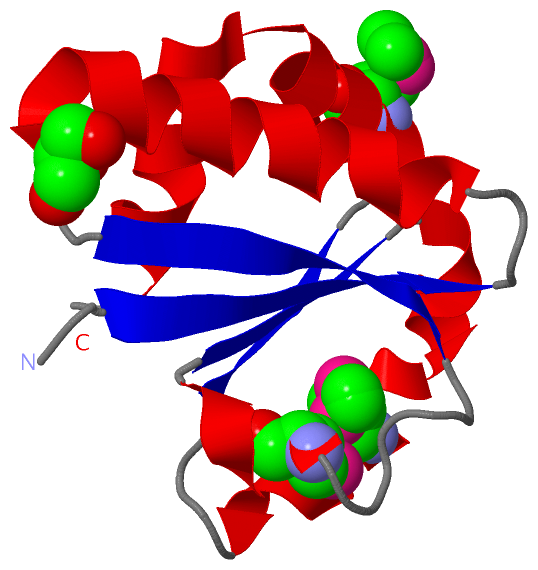
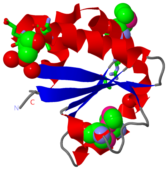
(-) Ligands, Modified Residues, Ions  (2, 4)

Asymmetric/Biological Unit (2, 4)
No.NameCountTypeFull Name
1GOL1Ligand/IonGLYCEROL
2MSE3Mod. Amino AcidSELENOMETHIONINE

(-) Sites  (1, 1)

Asymmetric Unit (1, 1)
No.NameEvidenceResiduesDescription
1AC1SOFTWAREHOH A:58 , HOH A:99 , HOH A:122 , ALA A:479 , ASN A:480 , GLU A:483 , VAL A:484 , ALA A:532 , ARG A:534BINDING SITE FOR RESIDUE GOL A 1

(-) SS Bonds  (0, 0)

(no "SS Bond" information available for 3NBM)

(-) Cis Peptide Bonds  (0, 0)

(no "Cis Peptide Bond" information available for 3NBM)

 Sequence-Structure Mapping

(-) SAPs(SNPs)/Variants  (0, 0)

(no "SAP(SNP)/Variant" information available for 3NBM)

(-) PROSITE Motifs  (0, 0)

(no "PROSITE Motif" information available for 3NBM)

(-) Exons   (0, 0)

(no "Exon" information available for 3NBM)

(-) Sequences/Alignments

Asymmetric/Biological Unit
   Reformat: Number of residues per line =  ('0' or empty: single-line sequence representation)
  Number of residues per labelling interval =   
  UniProt sequence: complete  aligned part    
   Show mapping: SCOP domains CATH domains Pfam domains Secondary structure (by author)
SAPs(SNPs) PROSITE motifs Exons
(details for a mapped element are shown in a popup box when the mouse pointer rests over it)
Chain A from PDB  Type:PROTEIN  Length:104
 aligned with A0A0H2UNS1_S | A0A0H2UNS1 from UniProtKB/TrEMBL  Length:559

    Alignment length:104
                                   463       473       483       493       503       513       523       533       543       553    
         A0A0H2UNS1_S   454 KELKVLVLCAGSGTSAQLANAINEGANLTEVRVIANSGAYGAHYDIMGVYDLIILAPQVRSYYREMKVDAERLGIQIVATRGMEYIHLTKSPSKALQFVLEHYQ 557
               SCOP domains -------------------------------------------------------------------------------------------------------- SCOP domains
               CATH domains -------------------------------------------------------------------------------------------------------- CATH domains
               Pfam domains -------------------------------------------------------------------------------------------------------- Pfam domains
         Sec.struct. author ..eeeeeee...hhhhhhhhhhhhhhhhhh..eeeeeee......hhhhh.eeee.hhhhhhhhhhhhhhh....eeee.hhhhhhhhhhhhhhhhhhhhhh.. Sec.struct. author
                 SAPs(SNPs) -------------------------------------------------------------------------------------------------------- SAPs(SNPs)
                    PROSITE -------------------------------------------------------------------------------------------------------- PROSITE
                 Transcript -------------------------------------------------------------------------------------------------------- Transcript
                 3nbm A 454 KELKVLVLCAGSGTSAQLANAINEGANLTEVRVIANSGAYGAHYDImGVYDLIILAPQVRSYYREmKVDAERLGIQIVATRGmEYIHLTKSPSKALQFVLEHYQ 557
                                   463       473       483       493      |503       513     | 523       533  |    543       553    
                                                                        500-MSE            519-MSE          536-MSE                 

   Legend:   → Mismatch (orange background)
  - → Gap (green background, '-', border residues have a numbering label)
    → Modified Residue (blue background, lower-case, 'x' indicates undefined single-letter code, labelled with number + name)
  x → Chemical Group (purple background, 'x', labelled with number + name, e.g. ACE or NH2)
  extra numbering lines below/above indicate numbering irregularities and modified residue names etc., number ends below/above '|'

 Classification and Annotation

(-) SCOP Domains  (0, 0)

(no "SCOP Domain" information available for 3NBM)

(-) CATH Domains  (0, 0)

(no "CATH Domain" information available for 3NBM)

(-) Pfam Domains  (0, 0)

(no "Pfam Domain" information available for 3NBM)

(-) Gene Ontology  (0, 0)

Asymmetric/Biological Unit(hide GO term definitions)
    (no "Gene Ontology" information available for 3NBM)

 Visualization

(-) Interactive Views

Asymmetric/Biological Unit
  Complete Structure
    Jena3D(integrated viewing of ligand, site, SAP, PROSITE, SCOP information)
    WebMol | AstexViewer[tm]@PDBe
(Java Applets, require no local installation except for Java; loading may be slow)
    STRAP
(Java WebStart application, automatic local installation, requires Java; full application with system access!)
    RasMol
(require local installation)
    Molscript (VRML)
(requires installation of a VRML viewer; select preferred view via VRML and generate a mono or stereo PDF format file)
 
  Ligands, Modified Residues, Ions
    GOL  [ RasMol | Jena3D ]  +environment [ RasMol | Jena3D ]
    MSE  [ RasMol | Jena3D ]  +environment [ RasMol | Jena3D ]
 
  Sites
    AC1  [ RasMol ]  +environment [ RasMol ]
 
  Cis Peptide Bonds
(no "Cis Peptide Bonds" information available for 3nbm)
 

(-) Still Images

Jmol
  protein: cartoon or spacefill or dots and stick; nucleic acid: cartoon and stick; ligands: spacefill; active site: stick
Molscript
  protein, nucleic acid: cartoon; ligands: spacefill; active site: ball and stick

 Databases and Analysis Tools

(-) Databases

Access by PDB/NDB ID
  3nbm
    Family and Domain InformationProDom | SYSTERS
    General Structural InformationGlycoscienceDB | MMDB | NDB | OCA | PDB | PDBe | PDBj | PDBsum | PDBWiki | PQS | PROTEOPEDIA
    Orientation in MembranesOPM
    Protein SurfaceSURFACE
    Secondary StructureDSSP (structure derived) | HSSP (homology derived)
    Structural GenomicsGeneCensus
    Structural NeighboursCE | VAST
    Structure ClassificationCATH | Dali | SCOP
    Validation and Original DataBMRB Data View | BMRB Restraints Grid | EDS | PROCHECK | RECOORD | WHAT_CHECK
 
Access by UniProt ID/Accession number
  A0A0H2UNS1_S | A0A0H2UNS1
    Comparative Protein Structure ModelsModBase
    Genomic InformationEnsembl
    Protein-protein InteractionDIP
    Sequence, Family and Domain InformationInterPro | Pfam | SMART | UniProtKB/TrEMBL
 
Access by Enzyme Classificator   (EC Number)
  (no 'Enzyme Classificator' available)
    General Enzyme InformationBRENDA | EC-PDB | Enzyme | IntEnz
    PathwayKEGG | MetaCyc
 
Access by Disease Identifier   (MIM ID)
  (no 'MIM ID' available)
    Disease InformationOMIM
 
Access by GenAge ID
  (no 'GenAge ID' available)
    Age Related InformationGenAge

(-) Analysis Tools

Access by PDB/NDB ID
    Domain InformationXDom
    Interatomic Contacts of Structural UnitsCSU
    Ligand-protein ContactsLPC
    Protein CavitiescastP
    Sequence and Secondary StructurePDBCartoon
    Structure AlignmentSTRAP(Java WebStart application, automatic local installation, requires Java; full application with system access!)
    Structure and Sequence BrowserSTING
 
Access by UniProt ID/Accession number
  A0A0H2UNS1_S | A0A0H2UNS1
    Protein Disorder PredictionDisEMBL | FoldIndex | GLOBPLOT (for more information see DisProt)

 Related Entries

(-) Entries Sharing at Least One Protein Chain (UniProt ID)

(no "Entries Sharing at Least One Protein Chain" available for 3NBM)

(-) Related Entries Specified in the PDB File

(no "Related Entries Specified in the PDB File" available for 3NBM)