|
|
|
|
 Description
|
|
Compounds
|
||||||||||||||||||||||||||||||||||||||||||||
Chains, Units
Summary Information (see also Sequences/Alignments below) |
Ligands, Modified Residues, Ions (3, 4)
Asymmetric Unit (3, 4)
|
Sites (4, 4)
Asymmetric Unit (4, 4)
|
SS Bonds (0, 0)| (no "SS Bond" information available for 3MXU) |
Cis Peptide Bonds (0, 0)| (no "Cis Peptide Bond" information available for 3MXU) |
SAPs(SNPs)/Variants (0, 0)| (no "SAP(SNP)/Variant" information available for 3MXU) |
PROSITE Motifs (2, 2)
Asymmetric Unit (2, 2)
|
||||||||||||||||||||||||||||||||||||||||||||||||||||||||||||||||||||||||||||||||||||||||||||||||
Exons (0, 0)| (no "Exon" information available for 3MXU) |
Sequences/Alignments
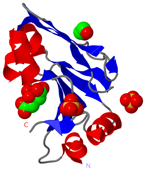
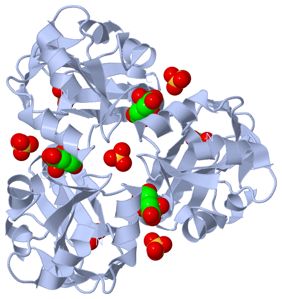
Asymmetric UnitChain A from PDB Type:PROTEIN Length:134 aligned with GCSH_BARHE | Q6G2F0 from UniProtKB/Swiss-Prot Length:122 Alignment length:134 1 - | 7 17 27 37 47 57 67 77 87 97 107 117 GCSH_BARHE - -------------MSKTYFTQDHEWLSVEGQVVTVGITDYAQEQLGDLVFIDLPQNGTKLSKGDAAAVVESVKAASDVYAPLDGEVVEINAALAESPELVNQKAETEGWLWKMTVQDETQLERLLDEAAYKELI 121 SCOP domains d3mxua_ A: automated matches SCOP domains CATH domains -------------------------------------------------------------------------------------------------------------------------------------- CATH domains Pfam domains ----------------GCV_H-3mxuA01 A:4-121 Pfam domains SAPs(SNPs) -------------------------------------------------------------------------------------------------------------------------------------- SAPs(SNPs) PROSITE (1) -------------------------------BIOTINYL_LIPOYL PDB: A:19-101 UniProt: 19-101 -------------------- PROSITE (1) PROSITE (2) --------------------------------------------------------LIPOYL PDB: A:44-73 ------------------------------------------------ PROSITE (2) Transcript -------------------------------------------------------------------------------------------------------------------------------------- Transcript 3mxu A -12 MGTLEAQTQGPGSMSKTYFTQDHEWLSVEGQVVTVGITDYAQEQLGDLVFIDLPQNGTKLSKGDAAAVVESVKAASDVYAPLDGEVVEINAALAESPELVNQKAETEGWLWKMTVQDETQLERLLDEAAYKELI 121 -3 7 17 27 37 47 57 67 77 87 97 107 117
|
||||||||||||||||||||
SCOP Domains (1, 1)
Asymmetric Unit
 |
CATH Domains (0, 0)| (no "CATH Domain" information available for 3MXU) |
Pfam Domains (1, 1)
Asymmetric Unit
|
Gene Ontology (2, 2)|
Asymmetric Unit(hide GO term definitions) Chain A (GCSH_BARHE | Q6G2F0)
|
||||||||||||||||||||||||
Interactive Views
|
||||||||||||||||||||||||||||||||||||||||||||||||||||||||||||||||||||||||||||||||||||||||||||||||||||||||||||||||||||||||||||||||||||||||||||||||||||||||||||||||||||||||||||||||
Still Images
|
||||||||||||||||
Databases
|
||||||||||||||||||||||||||||||||||||||||||||||||||||||||||||||||||||||||||||||||||||||||||||||||||||||||||||||||||||||||||||||||||||||||||||||||||||||||||||||||
Analysis Tools
|
|||||||||||||||||||||||||||||||||||||||||||||||||||||||||||||
Entries Sharing at Least One Protein Chain (UniProt ID)
Related Entries Specified in the PDB File
|
|