Show PDB file:   
         Plain Text   HTML   (compressed file size)
QuickSearch:   
by PDB,NDB,UniProt,PROSITE Code or Search Term(s)  
(-)Asym./Biol. Unit
collapse expand < >
Image Asym./Biol. Unit
Asym./Biol. Unit  (Jmol Viewer)

(-) Description

Title :  CRYSTAL STRUCTURE OF A DOMAIN FROM A PUTATIVE UDP-N-ACETYLMURAMATE:L-ALANYL-GAMMA-D-GLUTAMAYL-MEDO-DIAMINOPIMELATE LIGASE FROM HAEMOPHILUS DUCREYI 35000HP
 
Authors :  E. V. Filippova, G. Minasov, L. Shuvalova, O. Kiryukhina, S. Clancy, A. Joachimiak, F. W. Anderson, Midwest Center For Structural Gen (Mcsg)
Date :  04 May 10  (Deposition) - 16 Jun 10  (Release) - 16 Jun 10  (Revision)
Method :  X-RAY DIFFRACTION
Resolution :  1.90
Chains :  Asym./Biol. Unit :  A
Keywords :  Structural Genomics, Mcsg, Psi-2, Protein Structure Initiative, Midwest Center For Structural Genomics, Ligase (Keyword Search: [Gene Ontology, PubMed, Web (Google))
 
Reference :  E. V. Filippova, G. Minasov, L. Shuvalova, O. Kiryukhina, S. Clancy, A. Joachimiak, F. W. Anderson
Crystal Structure Of A Domain From A Putative Udp-N-Acetylmuramate:L-Alanyl-Gamma-D-Glutamayl-Medo- Diaminopimelate Ligase From Haemophilus Ducreyi 35000Hp
To Be Published
PubMed: search

(-) Compounds

Molecule 1
    ChainsA
    EngineeredYES
    Expression SystemESCHERICHIA COLI
    Expression System PlasmidPMCSG19
    Expression System StrainBL21MAGIC
    Expression System Taxid511693
    Expression System Vector TypePLASMID
    GeneHD_0703, MPL
    Organism ScientificHAEMOPHILUS DUCREYI
    Organism Taxid233412
    Strain35000HP

 Structural Features

(-) Chains, Units

  1
Asymmetric/Biological Unit A

Summary Information (see also Sequences/Alignments below)

(-) Ligands, Modified Residues, Ions  (1, 2)

Asymmetric/Biological Unit (1, 2)
No.NameCountTypeFull Name
1MSE2Mod. Amino AcidSELENOMETHIONINE

(-) Sites  (0, 0)

(no "Site" information available for 3MVN)

(-) SS Bonds  (0, 0)

(no "SS Bond" information available for 3MVN)

(-) Cis Peptide Bonds  (0, 0)

(no "Cis Peptide Bond" information available for 3MVN)

 Sequence-Structure Mapping

(-) SAPs(SNPs)/Variants  (0, 0)

(no "SAP(SNP)/Variant" information available for 3MVN)

(-) PROSITE Motifs  (0, 0)

(no "PROSITE Motif" information available for 3MVN)

(-) Exons   (0, 0)

(no "Exon" information available for 3MVN)

(-) Sequences/Alignments

Asymmetric/Biological Unit
   Reformat: Number of residues per line =  ('0' or empty: single-line sequence representation)
  Number of residues per labelling interval =   
  UniProt sequence: complete  aligned part    
   Show mapping: SCOP domains CATH domains Pfam domains Secondary structure (by author)
SAPs(SNPs) PROSITE motifs Exons
(details for a mapped element are shown in a popup box when the mouse pointer rests over it)
Chain A from PDB  Type:PROTEIN  Length:123
 aligned with Q7VN73_HAEDU | Q7VN73 from UniProtKB/TrEMBL  Length:459

    Alignment length:136
                                   326       336       346       356       366       376       386       396       406       416       426       436       446      
         Q7VN73_HAEDU   317 RLEVKGVVNNITVYDDFAHHPTAITATIDALRAKVGQQRILAVLEPRSNTMKMGVHKHELATSLQDADSVFIYQPPTIEWQVSEVLANLAQPAISADDVDELVMRIVQQAKPNDHILIMSNGAFGGIHQKLLTALA 452
               SCOP domains ---------------------------------------------------------------------------------------------------------------------------------------- SCOP domains
               CATH domains ---------------------------------------------------------------------------------------------------------------------------------------- CATH domains
               Pfam domains Mur_ligase_C-3mvnA01 A:317-391                                                 --------------------------------------------------------- Pfam domains
         Sec.struct. author ..eeeeee..eeeeee...hhhhhhhhhhhhhhhhh...eeeee...---------hhhhhhhhh...eeeee..----.hhhhhhh.....eeee.hhhhhhhhhhhhh....eeeee...hhhhhhhhhhhhh. Sec.struct. author
                 SAPs(SNPs) ---------------------------------------------------------------------------------------------------------------------------------------- SAPs(SNPs)
                    PROSITE ---------------------------------------------------------------------------------------------------------------------------------------- PROSITE
                 Transcript ---------------------------------------------------------------------------------------------------------------------------------------- Transcript
                 3mvn A 317 RLEVKGVVNNITVYDDFAHHPTAITATIDALRAKVGQQRILAVLEPR---------KHELATSLQDADSVFIYQP----WQVSEVLANLAQPAISADDVDELVmRIVQQAKPNDHILImSNGAFGGIHQKLLTALA 452
                                   326       336       346       356      |  -      |376       386    |  396       406       416   |   426       436       446      
                                                                        363       373               391  396                     420-MSE        435-MSE             

   Legend:   → Mismatch (orange background)
  - → Gap (green background, '-', border residues have a numbering label)
    → Modified Residue (blue background, lower-case, 'x' indicates undefined single-letter code, labelled with number + name)
  x → Chemical Group (purple background, 'x', labelled with number + name, e.g. ACE or NH2)
  extra numbering lines below/above indicate numbering irregularities and modified residue names etc., number ends below/above '|'

 Classification and Annotation

(-) SCOP Domains  (0, 0)

(no "SCOP Domain" information available for 3MVN)

(-) CATH Domains  (0, 0)

(no "CATH Domain" information available for 3MVN)

(-) Pfam Domains  (1, 1)

Asymmetric/Biological Unit

(-) Gene Ontology  (13, 13)

Asymmetric/Biological Unit(hide GO term definitions)
Chain A   (Q7VN73_HAEDU | Q7VN73)
molecular function
    GO:0005524    ATP binding    Interacting selectively and non-covalently with ATP, adenosine 5'-triphosphate, a universally important coenzyme and enzyme regulator.
    GO:0016881    acid-amino acid ligase activity    Catalysis of the ligation of an acid to an amino acid via a carbon-nitrogen bond, with the concomitant hydrolysis of the diphosphate bond in ATP or a similar triphosphate.
    GO:0016874    ligase activity    Catalysis of the joining of two substances, or two groups within a single molecule, with the concomitant hydrolysis of the diphosphate bond in ATP or a similar triphosphate.
    GO:0000166    nucleotide binding    Interacting selectively and non-covalently with a nucleotide, any compound consisting of a nucleoside that is esterified with (ortho)phosphate or an oligophosphate at any hydroxyl group on the ribose or deoxyribose.
biological process
    GO:0009058    biosynthetic process    The chemical reactions and pathways resulting in the formation of substances; typically the energy-requiring part of metabolism in which simpler substances are transformed into more complex ones.
    GO:0007049    cell cycle    The progression of biochemical and morphological phases and events that occur in a cell during successive cell replication or nuclear replication events. Canonically, the cell cycle comprises the replication and segregation of genetic material followed by the division of the cell, but in endocycles or syncytial cells nuclear replication or nuclear division may not be followed by cell division.
    GO:0051301    cell division    The process resulting in division and partitioning of components of a cell to form more cells; may or may not be accompanied by the physical separation of a cell into distinct, individually membrane-bounded daughter cells.
    GO:0071555    cell wall organization    A process that results in the assembly, arrangement of constituent parts, or disassembly of the cell wall, the rigid or semi-rigid envelope lying outside the cell membrane of plant, fungal and most prokaryotic cells, maintaining their shape and protecting them from osmotic lysis.
    GO:0009252    peptidoglycan biosynthetic process    The chemical reactions and pathways resulting in the formation of peptidoglycans, any of a class of glycoconjugates found in bacterial cell walls.
    GO:0009254    peptidoglycan turnover    The continual breakdown and regeneration of peptidoglycan required to maintain the cell wall.
    GO:0008360    regulation of cell shape    Any process that modulates the surface configuration of a cell.
cellular component
    GO:0016021    integral component of membrane    The component of a membrane consisting of the gene products and protein complexes having at least some part of their peptide sequence embedded in the hydrophobic region of the membrane.
    GO:0016020    membrane    A lipid bilayer along with all the proteins and protein complexes embedded in it an attached to it.
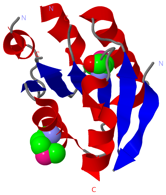
 Visualization

(-) Interactive Views

Asymmetric/Biological Unit
  Complete Structure
    Jena3D(integrated viewing of ligand, site, SAP, PROSITE, SCOP information)
    WebMol | AstexViewer[tm]@PDBe
(Java Applets, require no local installation except for Java; loading may be slow)
    STRAP
(Java WebStart application, automatic local installation, requires Java; full application with system access!)
    RasMol
(require local installation)
    Molscript (VRML)
(requires installation of a VRML viewer; select preferred view via VRML and generate a mono or stereo PDF format file)
 
  Ligands, Modified Residues, Ions
    MSE  [ RasMol | Jena3D ]  +environment [ RasMol | Jena3D ]
 
  Sites
(no "Sites" information available for 3mvn)
 
  Cis Peptide Bonds
(no "Cis Peptide Bonds" information available for 3mvn)
 

(-) Still Images

Jmol
  protein: cartoon or spacefill or dots and stick; nucleic acid: cartoon and stick; ligands: spacefill; active site: stick
Molscript
  protein, nucleic acid: cartoon; ligands: spacefill; active site: ball and stick

 Databases and Analysis Tools

(-) Databases

Access by PDB/NDB ID
  3mvn
    Family and Domain InformationProDom | SYSTERS
    General Structural InformationGlycoscienceDB | MMDB | NDB | OCA | PDB | PDBe | PDBj | PDBsum | PDBWiki | PQS | PROTEOPEDIA
    Orientation in MembranesOPM
    Protein SurfaceSURFACE
    Secondary StructureDSSP (structure derived) | HSSP (homology derived)
    Structural GenomicsGeneCensus
    Structural NeighboursCE | VAST
    Structure ClassificationCATH | Dali | SCOP
    Validation and Original DataBMRB Data View | BMRB Restraints Grid | EDS | PROCHECK | RECOORD | WHAT_CHECK
 
Access by UniProt ID/Accession number
  Q7VN73_HAEDU | Q7VN73
    Comparative Protein Structure ModelsModBase
    Genomic InformationEnsembl
    Protein-protein InteractionDIP
    Sequence, Family and Domain InformationInterPro | Pfam | SMART | UniProtKB/TrEMBL
 
Access by Enzyme Classificator   (EC Number)
  (no 'Enzyme Classificator' available)
    General Enzyme InformationBRENDA | EC-PDB | Enzyme | IntEnz
    PathwayKEGG | MetaCyc
 
Access by Disease Identifier   (MIM ID)
  (no 'MIM ID' available)
    Disease InformationOMIM
 
Access by GenAge ID
  (no 'GenAge ID' available)
    Age Related InformationGenAge

(-) Analysis Tools

Access by PDB/NDB ID
    Domain InformationXDom
    Interatomic Contacts of Structural UnitsCSU
    Ligand-protein ContactsLPC
    Protein CavitiescastP
    Sequence and Secondary StructurePDBCartoon
    Structure AlignmentSTRAP(Java WebStart application, automatic local installation, requires Java; full application with system access!)
    Structure and Sequence BrowserSTING
 
Access by UniProt ID/Accession number
  Q7VN73_HAEDU | Q7VN73
    Protein Disorder PredictionDisEMBL | FoldIndex | GLOBPLOT (for more information see DisProt)

 Related Entries

(-) Entries Sharing at Least One Protein Chain (UniProt ID)

(no "Entries Sharing at Least One Protein Chain" available for 3MVN)

(-) Related Entries Specified in the PDB File

(no "Related Entries Specified in the PDB File" available for 3MVN)