Show PDB file:   
         Plain Text   HTML   (compressed file size)
QuickSearch:   
by PDB,NDB,UniProt,PROSITE Code or Search Term(s)  
(-)Asymmetric Unit
(-)Asym. Unit - sites
(-)Biological Unit 1
(-)Biol. Unit 1 - sites
(-)Biological Unit 2
collapse expand < >
Image Asymmetric Unit
Asymmetric Unit  (Jmol Viewer)
Image Asym. Unit - sites
Asym. Unit - sites  (Jmol Viewer)
Image Biological Unit 1
Biological Unit 1  (Jmol Viewer)
Image Biol. Unit 1 - sites
Biol. Unit 1 - sites  (Jmol Viewer)
Image Biological Unit 2
Biological Unit 2  (Jmol Viewer)

(-) Description

Title :  STRUCTURE OF PLASMODIUM FALCIPARUM ARGINASE IN COMPLEX WITH ABH
 
Authors :  D. P. Dowling, M. Ilies, K. L. Olszewski, S. Portugal, M. M. Mota, M. Llin D. W. Christianson
Date :  20 Apr 10  (Deposition) - 23 Jun 10  (Release) - 14 Jul 10  (Revision)
Method :  X-RAY DIFFRACTION
Resolution :  2.14
Chains :  Asym. Unit :  A
Biol. Unit 1:  A  (3x)
Biol. Unit 2:  A  (6x)
Keywords :  Malaria, Abh, Lcr, Parasite, L-Arginine, Boronic Acid, Metallohydrolase, Binuclear, Manganese, Hydrolase (Keyword Search: [Gene Ontology, PubMed, Web (Google))
 
Reference :  D. P. Dowling, M. Ilies, K. L. Olszewski, S. Portugal, M. M. Mota, M. Llinas, D. W. Christianson
Crystal Structure Of Arginase From Plasmodium Falciparum An Implications For L-Arginine Depletion In Malarial Infection .
Biochemistry V. 49 5600 2010
PubMed-ID: 20527960  |  Reference-DOI: 10.1021/BI100390Z

(-) Compounds

Molecule 1 - ARGINASE
    ChainsA
    EC Number3.5.3.1
    EngineeredYES
    Expression SystemESCHERICHIA COLI
    Expression System PlasmidPET28A
    Expression System StrainBL21-CODONPLUSTM(DE3)-RIL
    Expression System Taxid469008
    Expression System Vector TypePLASMID
    FragmentUNP RESIDUES 22-411
    GenePFI0320W
    Organism ScientificPLASMODIUM FALCIPARUM 3D7
    Organism Taxid36329
    StrainISOLATE 3D7

 Structural Features

(-) Chains, Units

  1
Asymmetric Unit A
Biological Unit 1 (3x)A
Biological Unit 2 (6x)A

Summary Information (see also Sequences/Alignments below)

(-) Ligands, Modified Residues, Ions  (3, 4)

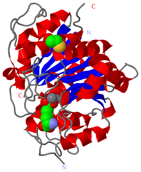
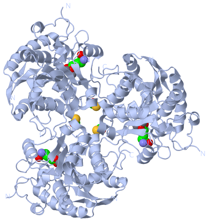
Asymmetric Unit (3, 4)
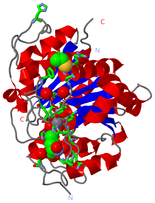
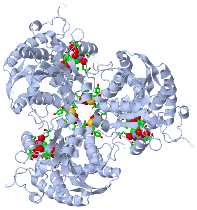
No.NameCountTypeFull Name
1ABH1Ligand/Ion2(S)-AMINO-6-BORONOHEXANOIC ACID
2BME1Ligand/IonBETA-MERCAPTOETHANOL
3MN2Ligand/IonMANGANESE (II) ION
Biological Unit 1 (2, 6)
No.NameCountTypeFull Name
1ABH3Ligand/Ion2(S)-AMINO-6-BORONOHEXANOIC ACID
2BME3Ligand/IonBETA-MERCAPTOETHANOL
3MN-1Ligand/IonMANGANESE (II) ION
Biological Unit 2 (2, 12)
No.NameCountTypeFull Name
1ABH6Ligand/Ion2(S)-AMINO-6-BORONOHEXANOIC ACID
2BME6Ligand/IonBETA-MERCAPTOETHANOL
3MN-1Ligand/IonMANGANESE (II) ION

(-) Sites  (4, 4)

Asymmetric Unit (4, 4)
No.NameEvidenceResiduesDescription
1AC1SOFTWAREHIS A:193 , ASP A:216 , ASP A:220 , ASP A:323 , MN A:413 , ABH A:551BINDING SITE FOR RESIDUE MN A 412
2AC2SOFTWAREASP A:216 , HIS A:218 , ASP A:323 , ASP A:325 , MN A:412 , ABH A:551BINDING SITE FOR RESIDUE MN A 413
3AC3SOFTWAREHIS A:193 , ASP A:216 , HIS A:218 , ASP A:220 , ASN A:222 , SER A:229 , HIS A:233 , ASP A:274 , GLU A:277 , ASP A:323 , ASP A:325 , GLU A:368 , HIS A:381 , MN A:412 , MN A:413 , HOH A:424 , HOH A:445 , HOH A:475 , HOH A:525BINDING SITE FOR RESIDUE ABH A 551
4AC4SOFTWAREASP A:328 , PHE A:331 , GLY A:341 , ASN A:344 , TYR A:345 , ARG A:346 , HOH A:435 , HOH A:545BINDING SITE FOR RESIDUE BME A 414

(-) SS Bonds  (0, 0)

(no "SS Bond" information available for 3MMR)

(-) Cis Peptide Bonds  (2, 2)

Asymmetric Unit
No.Residues
1Gly A:190 -Gly A:191
2Asp A:377 -Lys A:378

 Sequence-Structure Mapping

(-) SAPs(SNPs)/Variants  (0, 0)

(no "SAP(SNP)/Variant" information available for 3MMR)

(-) PROSITE Motifs  (0, 0)

(no "PROSITE Motif" information available for 3MMR)

(-) Exons   (0, 0)

(no "Exon" information available for 3MMR)

(-) Sequences/Alignments

Asymmetric Unit
   Reformat: Number of residues per line =  ('0' or empty: single-line sequence representation)
  Number of residues per labelling interval =   
  UniProt sequence: complete  aligned part    
   Show mapping: SCOP domains CATH domains Pfam domains Secondary structure (by author)
SAPs(SNPs) PROSITE motifs Exons
(details for a mapped element are shown in a popup box when the mouse pointer rests over it)
Chain A from PDB  Type:PROTEIN  Length:308
 aligned with Q8I384_PLAF7 | Q8I384 from UniProtKB/TrEMBL  Length:411

    Alignment length:390
                                    31        41        51        61        71        81        91       101       111       121       131       141       151       161       171       181       191       201       211       221       231       241       251       261       271       281       291       301       311       321       331       341       351       361       371       381       391       401       411
         Q8I384_PLAF7    22 KNVSIIGSPLAAGQPLGGVQLACDDLRKLGLHNVIDVLGWKYEDIGNIDNGDNEMKQEKKTNNYINNNDNNNDNNNDNNNDNNNNCYIPNGVIKEKKHDLSNNKMNGYVNHNFYGNYEENNVISTNDKYKNNCYYDNIRNIKEIGIFSKNLFDTMSNELRKKNFVLNIGGDHGVAFSSILSSLQMYQNLRVIWIDAHGDINIPETSPSGNYHGMTLAHTLGLFKKKVPYFEWSENLTYLKPENTAIIGIRDIDAYEKIILKKCNINYYTIFDIEKNGIYNTICTALEKIDPNSNCPIHISLDIDSVDNVFAPGTGTVAKGGLNYREINLLMKILAETKRVVSMDLVEYNPSLDEVDKKVHGDSLPILDNATKTGKLCLELIARVLGYDIV 411
               SCOP domains ------------------------------------------------------------------------------------------------------------------------------------------------------------------------------------------------------------------------------------------------------------------------------------------------------------------------------------------------------------------------------------------------------ SCOP domains
               CATH domains ------------------------------------------------------------------------------------------------------------------------------------------------------------------------------------------------------------------------------------------------------------------------------------------------------------------------------------------------------------------------------------------------------ CATH domains
               Pfam domains Arginase-3mmrA01 A:22-407                                                                                                                                                                                                                                                                                                                                                                         ---- Pfam domains
         Sec.struct. author .eeeeeee.........hhhhhhhhhhhhhhhhhhhhh..eeeeeee...----------------------------------------------------------------------------------.......hhhhhhhhhhhhhhhhhhhhh...eeeeee.hhhhhhhhhhhhhhhh...eeeee...............hhhhhhhhhhh.........hhhhh.....hhh.eeeeee...hhhhhhhhhhh..eeeehhhhhhhhhhhhhhhhhhhhh......eeeeee.hhh................hhhhhhhhhhhhhhh..eeeeeee..hhhhh.................hhhhhhhhhhhhhhh..... Sec.struct. author
                 SAPs(SNPs) ------------------------------------------------------------------------------------------------------------------------------------------------------------------------------------------------------------------------------------------------------------------------------------------------------------------------------------------------------------------------------------------------------ SAPs(SNPs)
                    PROSITE ------------------------------------------------------------------------------------------------------------------------------------------------------------------------------------------------------------------------------------------------------------------------------------------------------------------------------------------------------------------------------------------------------ PROSITE
                 Transcript ------------------------------------------------------------------------------------------------------------------------------------------------------------------------------------------------------------------------------------------------------------------------------------------------------------------------------------------------------------------------------------------------------ Transcript
                 3mmr A  22 KNVSIIGSPLAAGQPLGGVQLACDDLRKLGLHNVIDVLGWKYEDIGNIDN----------------------------------------------------------------------------------CYYDNIRNIKEIGIFSKNLFDTMSNELRKKNFVLNIGGDHGVAFSSILSSLQMYQNLRVIWIDAHGDINIPETSPSGNYHGMTLAHTLGLFKKKVPYFEWSENLTYLKPENTAIIGIRDIDAYEKIILKKCNINYYTIFDIEKNGIYNTICTALEKIDPNSNCPIHISLDIDSVDNVFAPGTGTVAKGGLNYREINLLMKILAETKRVVSMDLVEYNPSLDEVDKKVHGDSLPILDNATKTGKLCLELIARVLGYDIV 411
                                    31        41        51        61        71         -         -         -         -         -         -         -         -  |    161       171       181       191       201       211       221       231       241       251       261       271       281       291       301       311       321       331       341       351       361       371       381       391       401       411
                                                                            71                                                                                154                                                                                                                                                                                                                                                                 

   Legend:   → Mismatch (orange background)
  - → Gap (green background, '-', border residues have a numbering label)
    → Modified Residue (blue background, lower-case, 'x' indicates undefined single-letter code, labelled with number + name)
  x → Chemical Group (purple background, 'x', labelled with number + name, e.g. ACE or NH2)
  extra numbering lines below/above indicate numbering irregularities and modified residue names etc., number ends below/above '|'

 Classification and Annotation

(-) SCOP Domains  (0, 0)

(no "SCOP Domain" information available for 3MMR)

(-) CATH Domains  (0, 0)

(no "CATH Domain" information available for 3MMR)

(-) Pfam Domains  (1, 1)

Asymmetric Unit
(-)
Clan: Arginase (56)

(-) Gene Ontology  (3, 3)

Asymmetric Unit(hide GO term definitions)
Chain A   (Q8I384_PLAF7 | Q8I384)
molecular function
    GO:0004053    arginase activity    Catalysis of the reaction: L-arginine + H2O = L-ornithine + urea.
    GO:0016787    hydrolase activity    Catalysis of the hydrolysis of various bonds, e.g. C-O, C-N, C-C, phosphoric anhydride bonds, etc. Hydrolase is the systematic name for any enzyme of EC class 3.
    GO:0046872    metal ion binding    Interacting selectively and non-covalently with any metal ion.

 Visualization

(-) Interactive Views

Asymmetric Unit
  Complete Structure
    Jena3D(integrated viewing of ligand, site, SAP, PROSITE, SCOP information)
    WebMol | AstexViewer[tm]@PDBe
(Java Applets, require no local installation except for Java; loading may be slow)
    STRAP
(Java WebStart application, automatic local installation, requires Java; full application with system access!)
    RasMol
(require local installation)
    Molscript (VRML)
(requires installation of a VRML viewer; select preferred view via VRML and generate a mono or stereo PDF format file)
 
  Ligands, Modified Residues, Ions
    ABH  [ RasMol | Jena3D ]  +environment [ RasMol | Jena3D ]
    BME  [ RasMol | Jena3D ]  +environment [ RasMol | Jena3D ]
    MN  [ RasMol | Jena3D ]  +environment [ RasMol | Jena3D ]
 
  Sites
    AC1  [ RasMol ]  +environment [ RasMol ]
    AC2  [ RasMol ]  +environment [ RasMol ]
    AC3  [ RasMol ]  +environment [ RasMol ]
    AC4  [ RasMol ]  +environment [ RasMol ]
 
  Cis Peptide Bonds
    Asp A:377 - Lys A:378   [ RasMol ]  
    Gly A:190 - Gly A:191   [ RasMol ]  
 
Biological Units
  Complete Structure
    Biological Unit 1  [ Jena3D ]
    Biological Unit 2  [ Jena3D ]

(-) Still Images

Jmol
  protein: cartoon or spacefill or dots and stick; nucleic acid: cartoon and stick; ligands: spacefill; active site: stick
Molscript
  protein, nucleic acid: cartoon; ligands: spacefill; active site: ball and stick

 Databases and Analysis Tools

(-) Databases

Access by PDB/NDB ID
  3mmr
    Family and Domain InformationProDom | SYSTERS
    General Structural InformationGlycoscienceDB | MMDB | NDB | OCA | PDB | PDBe | PDBj | PDBsum | PDBWiki | PQS | PROTEOPEDIA
    Orientation in MembranesOPM
    Protein SurfaceSURFACE
    Secondary StructureDSSP (structure derived) | HSSP (homology derived)
    Structural GenomicsGeneCensus
    Structural NeighboursCE | VAST
    Structure ClassificationCATH | Dali | SCOP
    Validation and Original DataBMRB Data View | BMRB Restraints Grid | EDS | PROCHECK | RECOORD | WHAT_CHECK
 
Access by UniProt ID/Accession number
  Q8I384_PLAF7 | Q8I384
    Comparative Protein Structure ModelsModBase
    Genomic InformationEnsembl
    Protein-protein InteractionDIP
    Sequence, Family and Domain InformationInterPro | Pfam | SMART | UniProtKB/TrEMBL
 
Access by Enzyme Classificator   (EC Number)
  3.5.3.1
    General Enzyme InformationBRENDA | EC-PDB | Enzyme | IntEnz
    PathwayKEGG | MetaCyc
 
Access by Disease Identifier   (MIM ID)
  (no 'MIM ID' available)
    Disease InformationOMIM
 
Access by GenAge ID
  (no 'GenAge ID' available)
    Age Related InformationGenAge

(-) Analysis Tools

Access by PDB/NDB ID
    Domain InformationXDom
    Interatomic Contacts of Structural UnitsCSU
    Ligand-protein ContactsLPC
    Protein CavitiescastP
    Sequence and Secondary StructurePDBCartoon
    Structure AlignmentSTRAP(Java WebStart application, automatic local installation, requires Java; full application with system access!)
    Structure and Sequence BrowserSTING
 
Access by UniProt ID/Accession number
  Q8I384_PLAF7 | Q8I384
    Protein Disorder PredictionDisEMBL | FoldIndex | GLOBPLOT (for more information see DisProt)

 Related Entries

(-) Entries Sharing at Least One Protein Chain (UniProt ID)

UniProtKB/TrEMBL
        Q8I384_PLAF7 | Q8I3843sl0 3sl1

(-) Related Entries Specified in the PDB File

(no "Related Entries Specified in the PDB File" available for 3MMR)