|
|
|
|
 Description
|
|
Compounds
|
||||||||||||||||||||||||||||||||||||||||||||||||||||||||
Chains, Units
Summary Information (see also Sequences/Alignments below) |
Ligands, Modified Residues, Ions (3, 11)
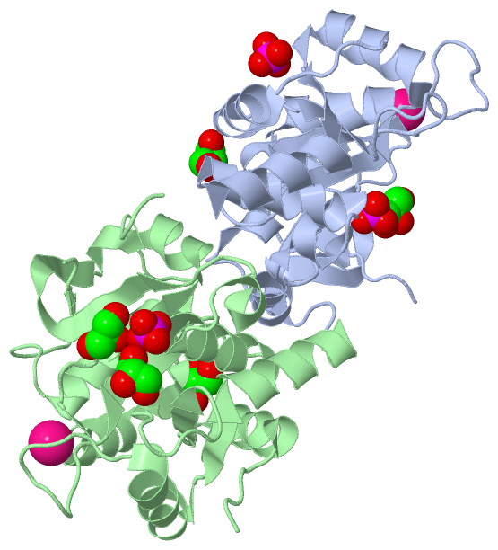
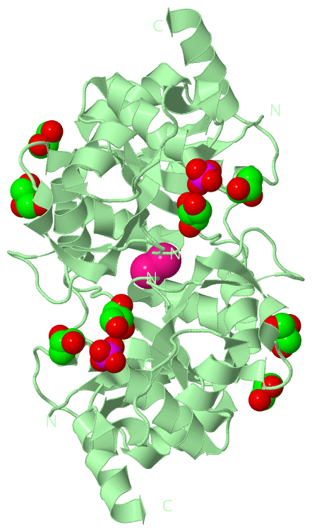
Asymmetric Unit (3, 11)
|
Sites (11, 11)
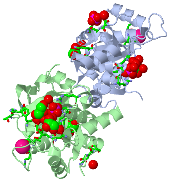
Asymmetric Unit (11, 11)
|
SS Bonds (0, 0)| (no "SS Bond" information available for 3MIO) |
Cis Peptide Bonds (2, 2)
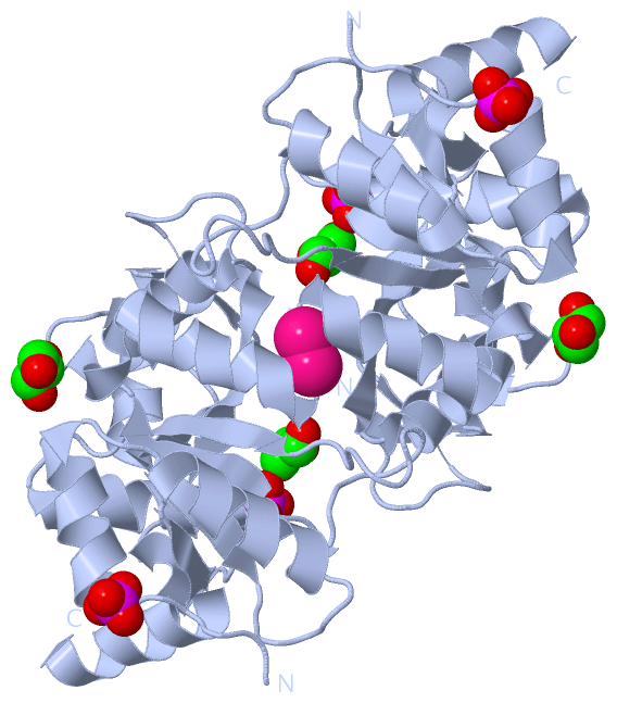
Asymmetric Unit
|
||||||||||||
SAPs(SNPs)/Variants (0, 0)| (no "SAP(SNP)/Variant" information available for 3MIO) |
PROSITE Motifs (0, 0)| (no "PROSITE Motif" information available for 3MIO) |
Exons (0, 0)| (no "Exon" information available for 3MIO) |
Sequences/Alignments
Asymmetric UnitChain A from PDB Type:PROTEIN Length:195 aligned with RIBBA_MYCTA | A5U2B7 from UniProtKB/Swiss-Prot Length:425 Alignment length:206 10 20 30 40 50 60 70 80 90 100 110 120 130 140 150 160 170 180 190 200 RIBBA_MYCTA 1 MTRLDSVERAVADIAAGKAVIVIDDEDRENEGDLIFAAEKATPEMVAFMVRYTSGYLCVPLDGAICDRLGLLPMYAVNQDKHGTAYTVTVDARNGIGTGISASDRATTMRLLADPTSVADDFTRPGHVVPLRAKDGGVLRRPGHTEAAVDLARMAGLQPAGAICEIVSQKDEGSMAHTDELRVFADEHGLALITIADLIEWRRKHE 206 SCOP domains d3mioa_ A: automated matches SCOP domains CATH domains -------------------------------------------------------------------------------------------------------------------------------------------------------------------------------------------------------------- CATH domains Pfam domains -------------------------------------------------------------------------------------------------------------------------------------------------------------------------------------------------------------- Pfam domains SAPs(SNPs) -------------------------------------------------------------------------------------------------------------------------------------------------------------------------------------------------------------- SAPs(SNPs) PROSITE -------------------------------------------------------------------------------------------------------------------------------------------------------------------------------------------------------------- PROSITE Transcript -------------------------------------------------------------------------------------------------------------------------------------------------------------------------------------------------------------- Transcript 3mio A 1 MTRLDSVERAVADIAAGKAVIVIDDEDRENEGDLIFAAEKATPEMVAFMVRYTSGYLCVPLDGAICDRLGLLPM-----------YTVTVDARNGIGTGISASDRATTMRLLADPTSVADDFTRPGHVVPLRAKDGGVLRRPGHTEAAVDLARMAGLQPAGAICEIVSQKDEGSMAHTDELRVFADEHGLALITIADLIEWRRKHE 206 10 20 30 40 50 60 70 | - | 90 100 110 120 130 140 150 160 170 180 190 200 74 86 Chain B from PDB Type:PROTEIN Length:194 aligned with RIBBA_MYCTA | A5U2B7 from UniProtKB/Swiss-Prot Length:425 Alignment length:206 10 20 30 40 50 60 70 80 90 100 110 120 130 140 150 160 170 180 190 200 RIBBA_MYCTA 1 MTRLDSVERAVADIAAGKAVIVIDDEDRENEGDLIFAAEKATPEMVAFMVRYTSGYLCVPLDGAICDRLGLLPMYAVNQDKHGTAYTVTVDARNGIGTGISASDRATTMRLLADPTSVADDFTRPGHVVPLRAKDGGVLRRPGHTEAAVDLARMAGLQPAGAICEIVSQKDEGSMAHTDELRVFADEHGLALITIADLIEWRRKHE 206 SCOP domains d3miob_ B: automated matches SCOP domains CATH domains -------------------------------------------------------------------------------------------------------------------------------------------------------------------------------------------------------------- CATH domains Pfam domains (1) ------DHBP_synthase-3mioB01 B:7-202 ---- Pfam domains (1) Pfam domains (2) ------DHBP_synthase-3mioB02 B:7-202 ---- Pfam domains (2) SAPs(SNPs) -------------------------------------------------------------------------------------------------------------------------------------------------------------------------------------------------------------- SAPs(SNPs) PROSITE -------------------------------------------------------------------------------------------------------------------------------------------------------------------------------------------------------------- PROSITE Transcript -------------------------------------------------------------------------------------------------------------------------------------------------------------------------------------------------------------- Transcript 3mio B 1 MTRLDSVERAVADIAAGKAVIVIDDEDRENEGDLIFAAEKATPEMVAFMVRYTSGYLCVPLDGAICDRLGLLPM------------TVTVDARNGIGTGISASDRATTMRLLADPTSVADDFTRPGHVVPLRAKDGGVLRRPGHTEAAVDLARMAGLQPAGAICEIVSQKDEGSMAHTDELRVFADEHGLALITIADLIEWRRKHE 206 10 20 30 40 50 60 70 | - | 90 100 110 120 130 140 150 160 170 180 190 200 74 87
|
||||||||||||||||||||
SCOP Domains (1, 2)
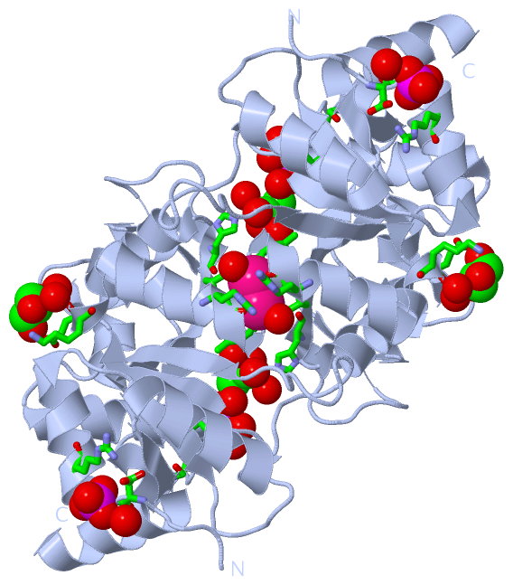
Asymmetric Unit
 |
CATH Domains (0, 0)| (no "CATH Domain" information available for 3MIO) |
Pfam Domains (1, 2)
Asymmetric Unit
|
Gene Ontology (13, 13)|
Asymmetric Unit(hide GO term definitions) Chain A,B (RIBBA_MYCTA | A5U2B7)
|
||||||||||||||||||||||||||||||||||||||||||||||||||||||||||||||||||||||||||||||||||||||||||
Interactive Views
|
|||||||||||||||||||||||||||||||||||||||||||||||||||||||||||||||||||||||||||||||||||||||||||||||||||||||||||||||||||||||||||||||||||||||||||||||||||||||||||||||||||||||||||||||||||||||||||||||||||||||||||||||||||||||||||||||||||||||||
Still Images
|
||||||||||||||||
Databases
|
||||||||||||||||||||||||||||||||||||||||||||||||||||||||||||||||||||||||||||||||||||||||||||||||||||||||||||||||||||||||||||||||||||||||||||||||||||||||||||||||
Analysis Tools
|
|||||||||||||||||||||||||||||||||||||||||||||||||||||||||||||
Entries Sharing at Least One Protein Chain (UniProt ID)
Related Entries Specified in the PDB File
|
|