Show PDB file:   
         Plain Text   HTML   (compressed file size)
QuickSearch:   
by PDB,NDB,UniProt,PROSITE Code or Search Term(s)  
(-)Asymmetric Unit
(-)Asym. Unit - sites
(-)Biological Unit 1
(-)Biol. Unit 1 - sites
(-)Biological Unit 2
collapse expand < >
Image Asymmetric Unit
Asymmetric Unit  (Jmol Viewer)
Image Asym. Unit - sites
Asym. Unit - sites  (Jmol Viewer)
Image Biological Unit 1
Biological Unit 1  (Jmol Viewer)
Image Biol. Unit 1 - sites
Biol. Unit 1 - sites  (Jmol Viewer)
Image Biological Unit 2
Biological Unit 2  (Jmol Viewer)

(-) Description

Title :  CRYSTAL STRUCTURE OF PUTATIVE ELECTRON TRANSPORT PROTEIN AQ_2194 FROM AQUIFEX AEOLICUS VF5
 
Authors :  V. N. Malashkevich, R. Toro, J. M. Sauder, S. K. Burley, S. C. Almo, New Y Research Center For Structural Genomics (Nysgxrc)
Date :  31 Mar 10  (Deposition) - 14 Apr 10  (Release) - 13 Jul 11  (Revision)
Method :  X-RAY DIFFRACTION
Resolution :  1.50
Chains :  Asym. Unit :  A,B
Biol. Unit 1:  A  (1x)
Biol. Unit 2:  B  (1x)
Keywords :  Electron Transfer Protein, Electron Transport, Structural Genomics, Psi-2, Protein Structure Initiative, New York Sgx Research Center For Structural Genomics, Nysgxrc (Keyword Search: [Gene Ontology, PubMed, Web (Google))
 
Reference :  V. N. Malashkevich, R. Toro, J. M. Sauder, S. K. Burley, S. C. Almo
Crystal Structure Of Putative Electron Transport Protein Aq_2194 From Aquifex Aeolicus Vf5
To Be Published
PubMed: search

(-) Compounds

Molecule 1 - PUTATIVE UNCHARACTERIZED PROTEIN
    ChainsA, B
    EngineeredYES
    Expression SystemESCHERICHIA COLI
    Expression System PlasmidBC-PSGX3(BC)
    Expression System StrainBL21(DE3)CODON+RIL
    Expression System Taxid562
    Expression System Vector TypePLASMID
    GeneAQ_2194, NP_214507.1
    Organism ScientificAQUIFEX AEOLICUS
    Organism Taxid63363

 Structural Features

(-) Chains, Units

  12
Asymmetric Unit AB
Biological Unit 1 (1x)A 
Biological Unit 2 (1x) B

Summary Information (see also Sequences/Alignments below)

(-) Ligands, Modified Residues, Ions  (1, 2)

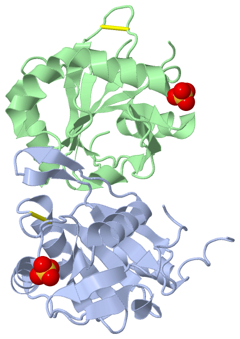
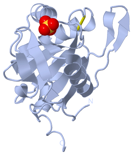
Asymmetric Unit (1, 2)
No.NameCountTypeFull Name
1SO42Ligand/IonSULFATE ION
Biological Unit 1 (1, 1)
No.NameCountTypeFull Name
1SO41Ligand/IonSULFATE ION
Biological Unit 2 (1, 1)
No.NameCountTypeFull Name
1SO41Ligand/IonSULFATE ION

(-) Sites  (2, 2)

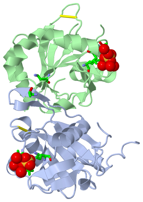
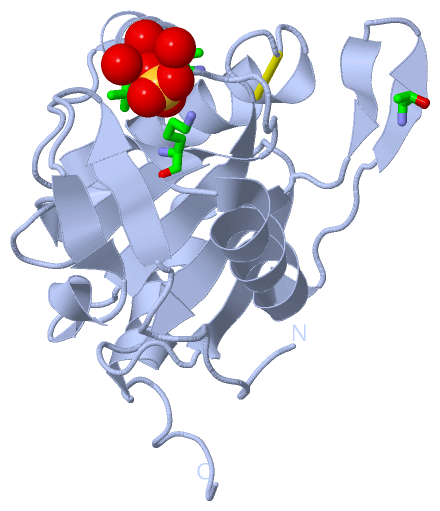
Asymmetric Unit (2, 2)
No.NameEvidenceResiduesDescription
1AC1SOFTWARETHR A:77 , LEU A:78 , LYS A:99 , THR A:119 , HOH A:231 , HOH A:325 , HOH A:370 , LYS B:93BINDING SITE FOR RESIDUE SO4 A 501
2AC2SOFTWARETHR B:77 , LEU B:78 , LYS B:99 , HOH B:201 , HOH B:207 , HOH B:285 , HOH B:304BINDING SITE FOR RESIDUE SO4 B 501

(-) SS Bonds  (2, 2)

Asymmetric Unit
No.Residues
1A:40 -A:44
2B:40 -B:44

(-) Cis Peptide Bonds  (0, 0)

(no "Cis Peptide Bond" information available for 3ME7)

 Sequence-Structure Mapping

(-) SAPs(SNPs)/Variants  (0, 0)

(no "SAP(SNP)/Variant" information available for 3ME7)

(-) PROSITE Motifs  (0, 0)

(no "PROSITE Motif" information available for 3ME7)

(-) Exons   (0, 0)

(no "Exon" information available for 3ME7)

(-) Sequences/Alignments

Asymmetric Unit
   Reformat: Number of residues per line =  ('0' or empty: single-line sequence representation)
  Number of residues per labelling interval =   
  UniProt sequence: complete  aligned part    
   Show mapping: SCOP domains CATH domains Pfam domains Secondary structure (by author)
SAPs(SNPs) PROSITE motifs Exons
(details for a mapped element are shown in a popup box when the mouse pointer rests over it)
Chain A from PDB  Type:PROTEIN  Length:169
 aligned with O67938_AQUAE | O67938 from UniProtKB/TrEMBL  Length:232

    Alignment length:174
                                    39        49        59        69        79        89        99       109       119       129       139       149       159       169       179       189       199    
         O67938_AQUAE    30 TLGTYVPGDITLVDSYGNEFQLKNLKGKPIILSPIYTHCRAACPLITKSLLKVIPKLGTPGKDFWVITFTFDPKDTLEDIKRFQKEYGIDGKGWKVVKAKTSEDLFKLLDAIDFRFMTAGNDFIHPNVVVVLSPELQIKDYIYGVNYNYLEFVNALRLARGETALPEGFRSYLF 203
               SCOP domains ------------------------------------------------------------------------------------------------------------------------------------------------------------------------------ SCOP domains
               CATH domains ------------------------------------------------------------------------------------------------------------------------------------------------------------------------------ CATH domains
               Pfam domains ------------------------------------------------------------------------------------------------------------------------------------------------------------------------------ Pfam domains
         Sec.struct. author ..........eeee....eee.hhhh...eeeeee.....hhhhhhhhhhhhhhhhhh......eeeeee.....hhhhhhhhhhhh......eeeeee.hhhhhhhhhhhh....eee..eee...eeeee.....eeeeee....hhhhhhhhhhhhh..-----....... Sec.struct. author
                 SAPs(SNPs) ------------------------------------------------------------------------------------------------------------------------------------------------------------------------------ SAPs(SNPs)
                    PROSITE ------------------------------------------------------------------------------------------------------------------------------------------------------------------------------ PROSITE
                 Transcript ------------------------------------------------------------------------------------------------------------------------------------------------------------------------------ Transcript
                 3me7 A   2 SLGTYVPGDITLVDSYGNEFQLKNLKGKPIILSPIYTHCRAACPLITKSLLKVIPKLGTPGKDFWVITFTFDPKDTLEDIKRFQKEYGIDGKGWKVVKAKTSEDLFKLLDAIDFRFMTAGNDFIHPNVVVVLSPELQIKDYIYGVNYNYLEFVNALRLARGE-----GHHHHHH 170
                                    11        21        31        41        51        61        71        81        91       101       111       121       131       141       151       161 |     166    
                                                                                                                                                                                           163   164      

Chain B from PDB  Type:PROTEIN  Length:158
 aligned with O67938_AQUAE | O67938 from UniProtKB/TrEMBL  Length:232

    Alignment length:162
                                    39        49        59        69        79        89        99       109       119       129       139       149       159       169       179       189  
         O67938_AQUAE    30 TLGTYVPGDITLVDSYGNEFQLKNLKGKPIILSPIYTHCRAACPLITKSLLKVIPKLGTPGKDFWVITFTFDPKDTLEDIKRFQKEYGIDGKGWKVVKAKTSEDLFKLLDAIDFRFMTAGNDFIHPNVVVVLSPELQIKDYIYGVNYNYLEFVNALRLARGE 191
               SCOP domains ------------------------------------------------------------------------------------------------------------------------------------------------------------------ SCOP domains
               CATH domains ------------------------------------------------------------------------------------------------------------------------------------------------------------------ CATH domains
           Pfam domains (1) -SCO1-SenC-3me7B01 B:3-146                                                                                                                       ----------------- Pfam domains (1)
           Pfam domains (2) -SCO1-SenC-3me7B02 B:3-146                                                                                                                       ----------------- Pfam domains (2)
         Sec.struct. author ..........eeee....eee.hhhh...eeeeee.........hhhhhhhhhhhhhh......eeeeee.....hhhhhhhhhhhhh.....eeeeee.hhhhhhhhhhhhh......----....eeeee.....eeeeee....hhhhhhhhhhhhhh. Sec.struct. author
                 SAPs(SNPs) ------------------------------------------------------------------------------------------------------------------------------------------------------------------ SAPs(SNPs)
                    PROSITE ------------------------------------------------------------------------------------------------------------------------------------------------------------------ PROSITE
                 Transcript ------------------------------------------------------------------------------------------------------------------------------------------------------------------ Transcript
                 3me7 B   2 SLGTYVPGDITLVDSYGNEFQLKNLKGKPIILSPIYTHCRAACPLITKSLLKVIPKLGTPGKDFWVITFTFDPKDTLEDIKRFQKEYGIDGKGWKVVKAKTSEDLFKLLDAIDFRFMTA----IHPNVVVVLSPELQIKDYIYGVNYNYLEFVNALRLARGE 163
                                    11        21        31        41        51        61        71        81        91       101       111        |-   |   131       141       151       161  
                                                                                                                                                120  125                                      

   Legend:   → Mismatch (orange background)
  - → Gap (green background, '-', border residues have a numbering label)
    → Modified Residue (blue background, lower-case, 'x' indicates undefined single-letter code, labelled with number + name)
  x → Chemical Group (purple background, 'x', labelled with number + name, e.g. ACE or NH2)
  extra numbering lines below/above indicate numbering irregularities and modified residue names etc., number ends below/above '|'

 Classification and Annotation

(-) SCOP Domains  (0, 0)

(no "SCOP Domain" information available for 3ME7)

(-) CATH Domains  (0, 0)

(no "CATH Domain" information available for 3ME7)

(-) Pfam Domains  (1, 2)

Asymmetric Unit

(-) Gene Ontology  (2, 2)

Asymmetric Unit(hide GO term definitions)
Chain A,B   (O67938_AQUAE | O67938)
cellular component
    GO:0016021    integral component of membrane    The component of a membrane consisting of the gene products and protein complexes having at least some part of their peptide sequence embedded in the hydrophobic region of the membrane.
    GO:0016020    membrane    A lipid bilayer along with all the proteins and protein complexes embedded in it an attached to it.

 Visualization

(-) Interactive Views

Asymmetric Unit
  Complete Structure
    Jena3D(integrated viewing of ligand, site, SAP, PROSITE, SCOP information)
    WebMol | AstexViewer[tm]@PDBe
(Java Applets, require no local installation except for Java; loading may be slow)
    STRAP
(Java WebStart application, automatic local installation, requires Java; full application with system access!)
    RasMol
(require local installation)
    Molscript (VRML)
(requires installation of a VRML viewer; select preferred view via VRML and generate a mono or stereo PDF format file)
 
  Ligands, Modified Residues, Ions
    SO4  [ RasMol | Jena3D ]  +environment [ RasMol | Jena3D ]
 
  Sites
    AC1  [ RasMol ]  +environment [ RasMol ]
    AC2  [ RasMol ]  +environment [ RasMol ]
 
  Cis Peptide Bonds
(no "Cis Peptide Bonds" information available for 3me7)
 
Biological Units
  Complete Structure
    Biological Unit 1  [ Jena3D ]
    Biological Unit 2  [ Jena3D ]

(-) Still Images

Jmol
  protein: cartoon or spacefill or dots and stick; nucleic acid: cartoon and stick; ligands: spacefill; active site: stick
Molscript
  protein, nucleic acid: cartoon; ligands: spacefill; active site: ball and stick

 Databases and Analysis Tools

(-) Databases

Access by PDB/NDB ID
  3me7
    Family and Domain InformationProDom | SYSTERS
    General Structural InformationGlycoscienceDB | MMDB | NDB | OCA | PDB | PDBe | PDBj | PDBsum | PDBWiki | PQS | PROTEOPEDIA
    Orientation in MembranesOPM
    Protein SurfaceSURFACE
    Secondary StructureDSSP (structure derived) | HSSP (homology derived)
    Structural GenomicsGeneCensus
    Structural NeighboursCE | VAST
    Structure ClassificationCATH | Dali | SCOP
    Validation and Original DataBMRB Data View | BMRB Restraints Grid | EDS | PROCHECK | RECOORD | WHAT_CHECK
 
Access by UniProt ID/Accession number
  O67938_AQUAE | O67938
    Comparative Protein Structure ModelsModBase
    Genomic InformationEnsembl
    Protein-protein InteractionDIP
    Sequence, Family and Domain InformationInterPro | Pfam | SMART | UniProtKB/TrEMBL
 
Access by Enzyme Classificator   (EC Number)
  (no 'Enzyme Classificator' available)
    General Enzyme InformationBRENDA | EC-PDB | Enzyme | IntEnz
    PathwayKEGG | MetaCyc
 
Access by Disease Identifier   (MIM ID)
  (no 'MIM ID' available)
    Disease InformationOMIM
 
Access by GenAge ID
  (no 'GenAge ID' available)
    Age Related InformationGenAge

(-) Analysis Tools

Access by PDB/NDB ID
    Domain InformationXDom
    Interatomic Contacts of Structural UnitsCSU
    Ligand-protein ContactsLPC
    Protein CavitiescastP
    Sequence and Secondary StructurePDBCartoon
    Structure AlignmentSTRAP(Java WebStart application, automatic local installation, requires Java; full application with system access!)
    Structure and Sequence BrowserSTING
 
Access by UniProt ID/Accession number
  O67938_AQUAE | O67938
    Protein Disorder PredictionDisEMBL | FoldIndex | GLOBPLOT (for more information see DisProt)

 Related Entries

(-) Entries Sharing at Least One Protein Chain (UniProt ID)

UniProtKB/TrEMBL
        O67938_AQUAE | O679383me8

(-) Related Entries Specified in the PDB File

3me8 RELATED ID: NYSGXRC-11213H RELATED DB: TARGETDB