|
|
|
|
 Description
|
|
Compounds
|
||||||||||||||||||||||||||||||||||||||||||||||||||||||||||||
Chains, Units
Summary Information (see also Sequences/Alignments below) |
Ligands, Modified Residues, Ions (2, 3)| Asymmetric Unit (2, 3) Biological Unit 1 (1, 1) Biological Unit 2 (2, 2) |
Sites (3, 3)
Asymmetric Unit (3, 3)
|
SS Bonds (0, 0)| (no "SS Bond" information available for 3MCF) |
Cis Peptide Bonds (0, 0)| (no "Cis Peptide Bond" information available for 3MCF) |
SAPs(SNPs)/Variants (0, 0)| (no "SAP(SNP)/Variant" information available for 3MCF) |
PROSITE Motifs (2, 4)
Asymmetric Unit (2, 4)
|
||||||||||||||||||||||||||||||||||||||||||||||||||||||||||||||||||||||||||||||||||||||||||||||||
Exons (0, 0)| (no "Exon" information available for 3MCF) |
Sequences/Alignments
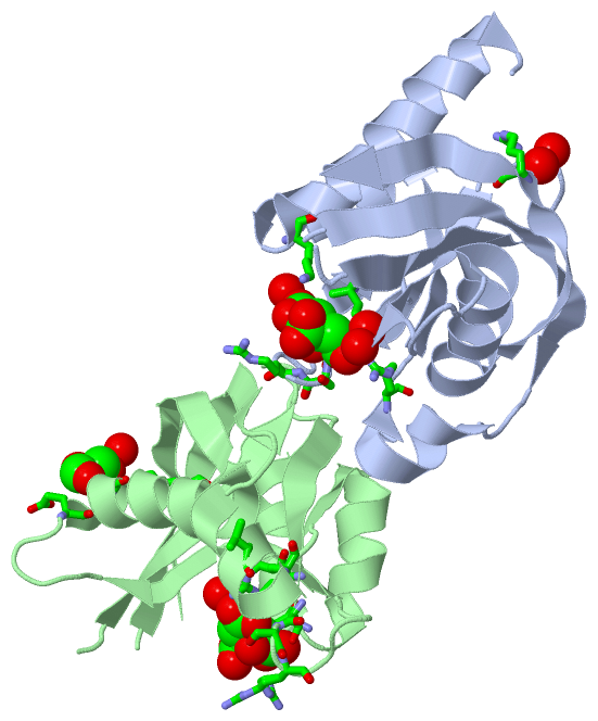
Asymmetric UnitChain A from PDB Type:PROTEIN Length:130 aligned with NUD10_HUMAN | Q8NFP7 from UniProtKB/Swiss-Prot Length:164 Alignment length:136 25 35 45 55 65 75 85 95 105 115 125 135 145 NUD10_HUMAN 16 FKKRAACLCFRSEREDEVLLVSSSRYPDRWIVPGGGMEPEEEPGGAAVREVYEEAGVKGKLGRLLGVFEQNQDPKHRTYVYVLTVTELLEDWEDSVSIGRKREWFKVEDAIKVLQCHKPVHAEYLEKLKLGGSPTN 151 SCOP domains d3mcfa_ A: automated matches SCOP domains CATH domains ---------------------------------------------------------------------------------------------------------------------------------------- CATH domains Pfam domains ---------------------------------------------------------------------------------------------------------------------------------------- Pfam domains SAPs(SNPs) ---------------------------------------------------------------------------------------------------------------------------------------- SAPs(SNPs) PROSITE (1) -NUDIX PDB: A:17-144 UniProt: 17-144 ------- PROSITE (1) PROSITE (2) ----------------------------------NUDIX_BOX PDB: A:50-7-------------------------------------------------------------------------------- PROSITE (2) Transcript ---------------------------------------------------------------------------------------------------------------------------------------- Transcript 3mcf A 16 MKKRAACLCFRSEREDEVLLVSSSRYPDRWIVPGGGMEPEEEPGGAAVREVYEEAGVKGKLGRLLGVFE------HRTYVYVLTVTELLEDWEDSVSIGRKREWFKVEDAIKVLQCHKPVHAEYLEKLKAHHHHHH 151 25 35 45 55 65 75 |- | 95 105 115 125 135 145 84 91 Chain B from PDB Type:PROTEIN Length:123 aligned with NUD10_HUMAN | Q8NFP7 from UniProtKB/Swiss-Prot Length:164 Alignment length:129 25 35 45 55 65 75 85 95 105 115 125 135 NUD10_HUMAN 16 FKKRAACLCFRSEREDEVLLVSSSRYPDRWIVPGGGMEPEEEPGGAAVREVYEEAGVKGKLGRLLGVFEQNQDPKHRTYVYVLTVTELLEDWEDSVSIGRKREWFKVEDAIKVLQCHKPVHAEYLEKLK 144 SCOP domains d3mcfb_ B: automated matches SCOP domains CATH domains --------------------------------------------------------------------------------------------------------------------------------- CATH domains Pfam domains (1) -NUDIX-3mcfB01 B:17-143 - Pfam domains (1) Pfam domains (2) -NUDIX-3mcfB02 B:17-143 - Pfam domains (2) SAPs(SNPs) --------------------------------------------------------------------------------------------------------------------------------- SAPs(SNPs) PROSITE (1) -NUDIX PDB: B:17-144 UniProt: 17-144 PROSITE (1) PROSITE (2) ----------------------------------NUDIX_BOX PDB: B:50-7------------------------------------------------------------------------- PROSITE (2) Transcript --------------------------------------------------------------------------------------------------------------------------------- Transcript 3mcf B 16 MKKRAACLCFRSEREDEVLLVSSSRYPDRWIVPGGGMEPEEEPGGAAVREVYEEAGVKGKLGRLLGVFE------HRTYVYVLTVTELLEDWEDSVSIGRKREWFKVEDAIKVLQCHKPVHAEYLEKLK 144 25 35 45 55 65 75 |- | 95 105 115 125 135 84 91
|
||||||||||||||||||||
SCOP Domains (1, 2)
Asymmetric Unit
 |
CATH Domains (0, 0)| (no "CATH Domain" information available for 3MCF) |
Pfam Domains (1, 2)
Asymmetric Unit
|
Gene Ontology (16, 16)|
Asymmetric Unit(hide GO term definitions) Chain A,B (NUD10_HUMAN | Q8NFP7)
|
||||||||||||||||||||||||||||||||||||||||||||||||||||||||||||||||||||||||||||||||||||||||||||||||||||||||||||||||||
Interactive Views
|
||||||||||||||||||||||||||||||||||||||||||||||||||||||||||||||||||||||||||||||||||||||||||||||||||||||||||||||||||||||||||||||||||||||||||||||||||||||||||||||||||
Still Images
|
||||||||||||||||
Databases
|
||||||||||||||||||||||||||||||||||||||||||||||||||||||||||||||||||||||||||||||||||||||||||||||||||||||||||||||||||||||||||||||||||||||||||||||||||||||||||||||||||||||||||||||||
Analysis Tools
|
|||||||||||||||||||||||||||||||||||||||||||||||||||||||||||||
Entries Sharing at Least One Protein Chain (UniProt ID)
Related Entries Specified in the PDB File
|
|