| molecular function |
|---|
| | GO:0005524 | | ATP binding | | Interacting selectively and non-covalently with ATP, adenosine 5'-triphosphate, a universally important coenzyme and enzyme regulator. |
| | GO:0016301 | | kinase activity | | Catalysis of the transfer of a phosphate group, usually from ATP, to a substrate molecule. |
| | GO:0000166 | | nucleotide binding | | Interacting selectively and non-covalently with a nucleotide, any compound consisting of a nucleoside that is esterified with (ortho)phosphate or an oligophosphate at any hydroxyl group on the ribose or deoxyribose. |
| | GO:0000155 | | phosphorelay sensor kinase activity | | Catalysis of the phosphorylation of a histidine residue in response to detection of an extracellular signal such as a chemical ligand or change in environment, to initiate a change in cell state or activity. The two-component sensor is a histidine kinase that autophosphorylates a histidine residue in its active site. The phosphate is then transferred to an aspartate residue in a downstream response regulator, to trigger a response. |
| | GO:0004673 | | protein histidine kinase activity | | Catalysis of the reaction: ATP + protein L-histidine = ADP + protein phospho-L-histidine. |
| | GO:0016740 | | transferase activity | | Catalysis of the transfer of a group, e.g. a methyl group, glycosyl group, acyl group, phosphorus-containing, or other groups, from one compound (generally regarded as the donor) to another compound (generally regarded as the acceptor). Transferase is the systematic name for any enzyme of EC class 2. |
| | GO:0016772 | | transferase activity, transferring phosphorus-containing groups | | Catalysis of the transfer of a phosphorus-containing group from one compound (donor) to another (acceptor). |
| biological process |
|---|
| | GO:0018106 | | peptidyl-histidine phosphorylation | | The phosphorylation of peptidyl-histidine to form peptidyl-1'-phospho-L-histidine (otherwise known as tau-phosphohistidine, tele-phosphohistidine) or peptidyl-3'-phospho-L-histidine (otherwise known as pi-phosphohistidine, pros-phosphohistidine). |
| | GO:0000160 | | phosphorelay signal transduction system | | A conserved series of molecular signals found in prokaryotes and eukaryotes; involves autophosphorylation of a histidine kinase and the transfer of the phosphate group to an aspartate that then acts as a phospho-donor to response regulator proteins. |
| | GO:0016310 | | phosphorylation | | The process of introducing a phosphate group into a molecule, usually with the formation of a phosphoric ester, a phosphoric anhydride or a phosphoric amide. |
| | GO:0006355 | | regulation of transcription, DNA-templated | | Any process that modulates the frequency, rate or extent of cellular DNA-templated transcription. |
| | GO:0007165 | | signal transduction | | The cellular process in which a signal is conveyed to trigger a change in the activity or state of a cell. Signal transduction begins with reception of a signal (e.g. a ligand binding to a receptor or receptor activation by a stimulus such as light), or for signal transduction in the absence of ligand, signal-withdrawal or the activity of a constitutively active receptor. Signal transduction ends with regulation of a downstream cellular process, e.g. regulation of transcription or regulation of a metabolic process. Signal transduction covers signaling from receptors located on the surface of the cell and signaling via molecules located within the cell. For signaling between cells, signal transduction is restricted to events at and within the receiving cell. |
| | GO:0023014 | | signal transduction by protein phosphorylation | | A process in which the transfer of one or more phosphate groups to a substrate transmits a signal to the phosphorylated substrate. |
| cellular component |
|---|
| | GO:0005622 | | intracellular | | The living contents of a cell; the matter contained within (but not including) the plasma membrane, usually taken to exclude large vacuoles and masses of secretory or ingested material. In eukaryotes it includes the nucleus and cytoplasm. |
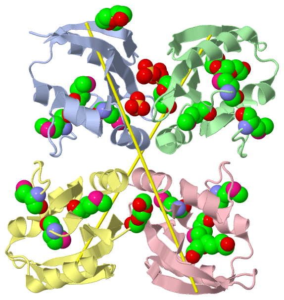
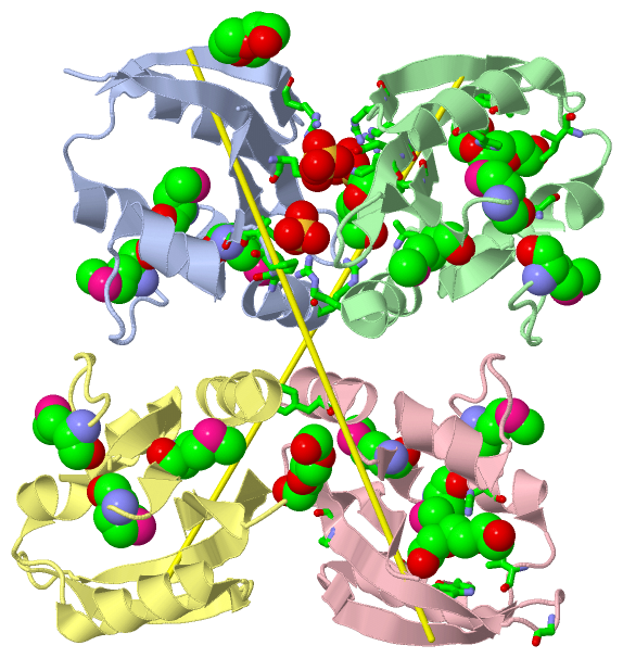
 Description
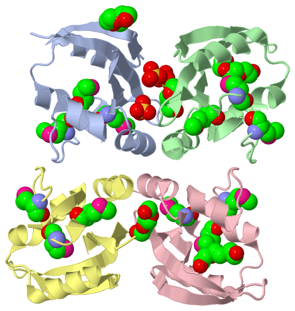
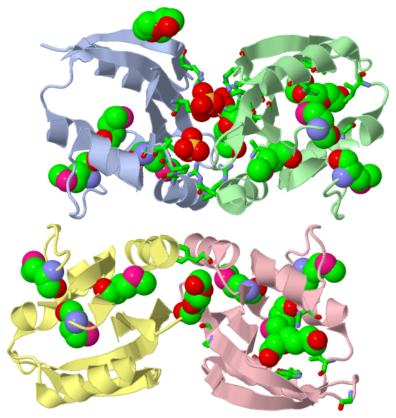