Show PDB file:   
         Plain Text   HTML   (compressed file size)
QuickSearch:   
by PDB,NDB,UniProt,PROSITE Code or Search Term(s)  
(-)Asymmetric Unit
(-)Asym. Unit - sites
(-)Biological Unit 1
(-)Biol. Unit 1 - sites
(-)Biological Unit 2
collapse expand < >
Image Asymmetric Unit
Asymmetric Unit  (Jmol Viewer)
Image Asym. Unit - sites
Asym. Unit - sites  (Jmol Viewer)
Image Biological Unit 1
Biological Unit 1  (Jmol Viewer)
Image Biol. Unit 1 - sites
Biol. Unit 1 - sites  (Jmol Viewer)
Image Biological Unit 2
Biological Unit 2  (Jmol Viewer)

(-) Description

Title :  CRYSTAL STRUCTURE OF CYANOBACTERIAL PSBQ FROM SYNECHOCYSTIS SP. PCC 6803 COMPLEXED WITH ZN2+
 
Authors :  S. A. Jackson, R. D. Fagerlund, S. M. Wilbanks, J. J. Eaton-Rye
Date :  12 Feb 10  (Deposition) - 31 Mar 10  (Release) - 07 Apr 10  (Revision)
Method :  X-RAY DIFFRACTION
Resolution :  1.85
Chains :  Asym. Unit :  A,B
Biol. Unit 1:  A  (1x)
Biol. Unit 2:  B  (1x)
Keywords :  Photosynthesis, Four Helix Bundle (Keyword Search: [Gene Ontology, PubMed, Web (Google))
 
Reference :  S. A. Jackson, R. D. Fagerlund, S. M. Wilbanks, J. J. Eaton-Rye
Crystal Structure Of Psbq From Synechocystis Sp. Pcc 6803 At 1. 8 A: Implications For Binding And Function In Cyanobacterial Photosystem Ii
Biochemistry V. 49 2765 2010
PubMed-ID: 20210304  |  Reference-DOI: 10.1021/BI100217H

(-) Compounds

Molecule 1 - SLL1638 PROTEIN
    ChainsA, B
    EngineeredYES
    Expression SystemESCHERICHIA COLI
    Expression System PlasmidPGEX-6-P-3
    Expression System StrainBL21(DE3)
    Expression System Taxid469008
    Expression System Vector TypePLASMID
    FragmentRESIDUES 21-149
    GeneSLL1638
    Organism ScientificSYNECHOCYSTIS SP.
    Organism Taxid1148
    StrainPCC 6803
    SynonymPSBQ

 Structural Features

(-) Chains, Units

  12
Asymmetric Unit AB
Biological Unit 1 (1x)A 
Biological Unit 2 (1x) B

Summary Information (see also Sequences/Alignments below)

(-) Ligands, Modified Residues, Ions  (1, 4)

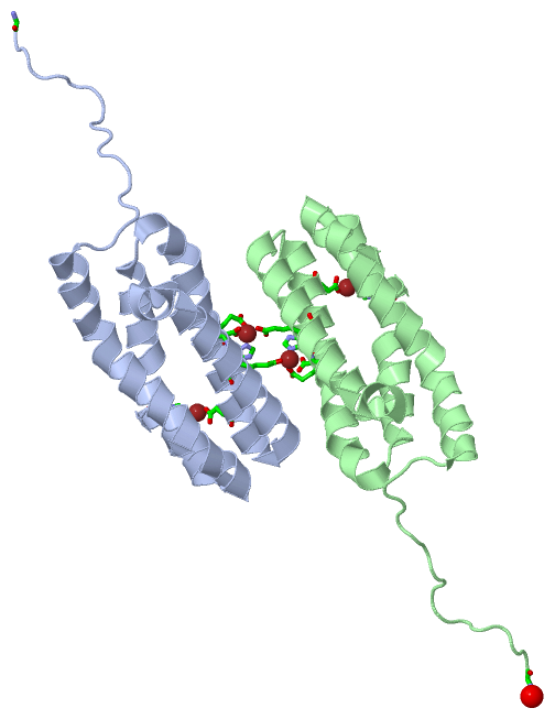
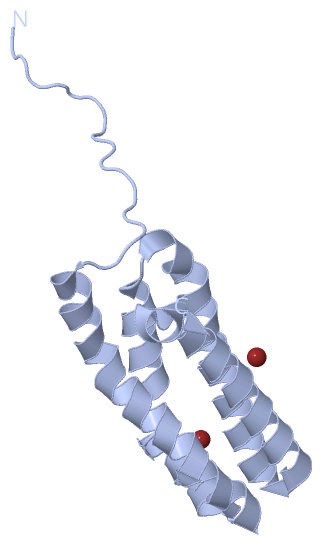
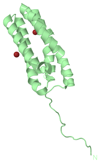
Asymmetric Unit (1, 4)
No.NameCountTypeFull Name
1ZN4Ligand/IonZINC ION
Biological Unit 1 (0, 0)
No.NameCountTypeFull Name
1ZN-1Ligand/IonZINC ION
Biological Unit 2 (0, 0)
No.NameCountTypeFull Name
1ZN-1Ligand/IonZINC ION

(-) Sites  (4, 4)

Asymmetric Unit (4, 4)
No.NameEvidenceResiduesDescription
1AC1SOFTWAREGLU A:107 , HIS A:111 , ASP A:137 , GLU B:133BINDING SITE FOR RESIDUE ZN A 1
2AC2SOFTWAREHIS A:76 , ASP A:116 , GLY B:17 , HOH B:239BINDING SITE FOR RESIDUE ZN A 3
3AC3SOFTWAREGLY A:17 , HIS B:76 , ASP B:116 , HOH B:240BINDING SITE FOR RESIDUE ZN B 2
4AC4SOFTWAREGLU A:133 , GLU B:107 , HIS B:111 , ASP B:137BINDING SITE FOR RESIDUE ZN B 4

(-) SS Bonds  (0, 0)

(no "SS Bond" information available for 3LS1)

(-) Cis Peptide Bonds  (0, 0)

(no "Cis Peptide Bond" information available for 3LS1)

 Sequence-Structure Mapping

(-) SAPs(SNPs)/Variants  (0, 0)

(no "SAP(SNP)/Variant" information available for 3LS1)

(-) PROSITE Motifs  (0, 0)

(no "PROSITE Motif" information available for 3LS1)

(-) Exons   (0, 0)

(no "Exon" information available for 3LS1)

(-) Sequences/Alignments

Asymmetric Unit
   Reformat: Number of residues per line =  ('0' or empty: single-line sequence representation)
  Number of residues per labelling interval =   
  UniProt sequence: complete  aligned part    
   Show mapping: SCOP domains CATH domains Pfam domains Secondary structure (by author)
SAPs(SNPs) PROSITE motifs Exons
(details for a mapped element are shown in a popup box when the mouse pointer rests over it)
Chain A from PDB  Type:PROTEIN  Length:133
 aligned with P73048_SYNY3 | P73048 from UniProtKB/TrEMBL  Length:149

    Alignment length:133
                                    26        36        46        56        66        76        86        96       106       116       126       136       146   
         P73048_SYNY3    17 TVLVSCSSPQVEIPTTYSPEKIAQLQVYVNPIAVARDGMEKRLQGLIADQNWVDTQTYIHGPLGQLRRDMLGLASSLLPKDQDKAKTLAKEVFGHLERLDAAAKDRNGSQAKIQYQEALADFDSFLNLLPQAS 149
               SCOP domains ------------------------------------------------------------------------------------------------------------------------------------- SCOP domains
               CATH domains ------------------------------------------------------------------------------------------------------------------------------------- CATH domains
               Pfam domains ------------------------------------------------------------------------------------------------------------------------------------- Pfam domains
         Sec.struct. author .................hhhhhhhhhhhhhhhhhhhhhhhhhhhhhhhh.hhhhhhhhhhh...hhhhhhhhhhh..hhhhhhhhhhhhhhhhhhhhhhhhhhhhhhhhhhhhhhhhhhhhhhhhhhhhhhhh Sec.struct. author
                 SAPs(SNPs) ------------------------------------------------------------------------------------------------------------------------------------- SAPs(SNPs)
                    PROSITE ------------------------------------------------------------------------------------------------------------------------------------- PROSITE
                 Transcript ------------------------------------------------------------------------------------------------------------------------------------- Transcript
                 3ls1 A  17 GPLGSCSSPQVEIPTTYSPEKIAQLQVYVNPIAVARDGMEKRLQGLIADQNWVDTQTYIHGPLGQLRRDMLGLASSLLPKDQDKAKTLAKEVFGHLERLDAAAKDRNGSQAKIQYQEALADFDSFLNLLPQAS 149
                                    26        36        46        56        66        76        86        96       106       116       126       136       146   

Chain B from PDB  Type:PROTEIN  Length:133
 aligned with P73048_SYNY3 | P73048 from UniProtKB/TrEMBL  Length:149

    Alignment length:133
                                    26        36        46        56        66        76        86        96       106       116       126       136       146   
         P73048_SYNY3    17 TVLVSCSSPQVEIPTTYSPEKIAQLQVYVNPIAVARDGMEKRLQGLIADQNWVDTQTYIHGPLGQLRRDMLGLASSLLPKDQDKAKTLAKEVFGHLERLDAAAKDRNGSQAKIQYQEALADFDSFLNLLPQAS 149
               SCOP domains ------------------------------------------------------------------------------------------------------------------------------------- SCOP domains
               CATH domains ------------------------------------------------------------------------------------------------------------------------------------- CATH domains
           Pfam domains (1) -------------------------------PsbQ-3ls1B01 B:48-146                                                                              --- Pfam domains (1)
           Pfam domains (2) -------------------------------PsbQ-3ls1B02 B:48-146                                                                              --- Pfam domains (2)
         Sec.struct. author .................hhhhhhhhhhhhhhhhhhhhhhhhhhhhhhhh.hhhhhhhhhhh...hhhhhhhhhhh..hhhhhhhhhhhhhhhhhhhhhhhhhhhh.hhhhhhhhhhhhhhhhhhhhhhhhhhh Sec.struct. author
                 SAPs(SNPs) ------------------------------------------------------------------------------------------------------------------------------------- SAPs(SNPs)
                    PROSITE ------------------------------------------------------------------------------------------------------------------------------------- PROSITE
                 Transcript ------------------------------------------------------------------------------------------------------------------------------------- Transcript
                 3ls1 B  17 GPLGSCSSPQVEIPTTYSPEKIAQLQVYVNPIAVARDGMEKRLQGLIADQNWVDTQTYIHGPLGQLRRDMLGLASSLLPKDQDKAKTLAKEVFGHLERLDAAAKDRNGSQAKIQYQEALADFDSFLNLLPQAS 149
                                    26        36        46        56        66        76        86        96       106       116       126       136       146   

   Legend:   → Mismatch (orange background)
  - → Gap (green background, '-', border residues have a numbering label)
    → Modified Residue (blue background, lower-case, 'x' indicates undefined single-letter code, labelled with number + name)
  x → Chemical Group (purple background, 'x', labelled with number + name, e.g. ACE or NH2)
  extra numbering lines below/above indicate numbering irregularities and modified residue names etc., number ends below/above '|'

 Classification and Annotation

(-) SCOP Domains  (0, 0)

(no "SCOP Domain" information available for 3LS1)

(-) CATH Domains  (0, 0)

(no "CATH Domain" information available for 3LS1)

(-) Pfam Domains  (1, 2)

Asymmetric Unit

(-) Gene Ontology  (7, 7)

Asymmetric Unit(hide GO term definitions)
Chain A,B   (P73048_SYNY3 | P73048)
molecular function
    GO:0005509    calcium ion binding    Interacting selectively and non-covalently with calcium ions (Ca2+).
    GO:0046872    metal ion binding    Interacting selectively and non-covalently with any metal ion.
biological process
    GO:0015979    photosynthesis    The synthesis by organisms of organic chemical compounds, especially carbohydrates, from carbon dioxide (CO2) using energy obtained from light rather than from the oxidation of chemical compounds.
cellular component
    GO:0019898    extrinsic component of membrane    The component of a membrane consisting of gene products and protein complexes that are loosely bound to one of its surfaces, but not integrated into the hydrophobic region.
    GO:0009523    photosystem II    A photosystem that contains a pheophytin-quinone reaction center with associated accessory pigments and electron carriers. In cyanobacteria and chloroplasts, in the presence of light, PSII functions as a water-plastoquinone oxidoreductase, transferring electrons from water to plastoquinone, whereas other photosynthetic bacteria carry out anoxygenic photosynthesis and oxidize other compounds to re-reduce the photoreaction center.
    GO:0009654    photosystem II oxygen evolving complex    A complex, composed of a cluster of manganese, calcium and chloride ions bound to extrinsic proteins, that catalyzes the splitting of water to O2 and 4 H+. In cyanobacteria there are five extrinsic proteins in OEC (PsbO, PsbP-like, PsbQ-like, PsbU and PsbV), while in plants there are only three (PsbO, PsbP and PsbQ).
    GO:0030096    plasma membrane-derived thylakoid photosystem II    A protein complex, located in the membrane-derived thylakoid, containing the P680 reaction center. In the light, PSII functions as a water-plastoquinone oxidoreductase, transferring electrons from water to plastoquinone.

 Visualization

(-) Interactive Views

Asymmetric Unit
  Complete Structure
    Jena3D(integrated viewing of ligand, site, SAP, PROSITE, SCOP information)
    WebMol | AstexViewer[tm]@PDBe
(Java Applets, require no local installation except for Java; loading may be slow)
    STRAP
(Java WebStart application, automatic local installation, requires Java; full application with system access!)
    RasMol
(require local installation)
    Molscript (VRML)
(requires installation of a VRML viewer; select preferred view via VRML and generate a mono or stereo PDF format file)
 
  Ligands, Modified Residues, Ions
    ZN  [ RasMol | Jena3D ]  +environment [ RasMol | Jena3D ]
 
  Sites
    AC1  [ RasMol ]  +environment [ RasMol ]
    AC2  [ RasMol ]  +environment [ RasMol ]
    AC3  [ RasMol ]  +environment [ RasMol ]
    AC4  [ RasMol ]  +environment [ RasMol ]
 
  Cis Peptide Bonds
(no "Cis Peptide Bonds" information available for 3ls1)
 
Biological Units
  Complete Structure
    Biological Unit 1  [ Jena3D ]
    Biological Unit 2  [ Jena3D ]

(-) Still Images

Jmol
  protein: cartoon or spacefill or dots and stick; nucleic acid: cartoon and stick; ligands: spacefill; active site: stick
Molscript
  protein, nucleic acid: cartoon; ligands: spacefill; active site: ball and stick

 Databases and Analysis Tools

(-) Databases

Access by PDB/NDB ID
  3ls1
    Family and Domain InformationProDom | SYSTERS
    General Structural InformationGlycoscienceDB | MMDB | NDB | OCA | PDB | PDBe | PDBj | PDBsum | PDBWiki | PQS | PROTEOPEDIA
    Orientation in MembranesOPM
    Protein SurfaceSURFACE
    Secondary StructureDSSP (structure derived) | HSSP (homology derived)
    Structural GenomicsGeneCensus
    Structural NeighboursCE | VAST
    Structure ClassificationCATH | Dali | SCOP
    Validation and Original DataBMRB Data View | BMRB Restraints Grid | EDS | PROCHECK | RECOORD | WHAT_CHECK
 
Access by UniProt ID/Accession number
  P73048_SYNY3 | P73048
    Comparative Protein Structure ModelsModBase
    Genomic InformationEnsembl
    Protein-protein InteractionDIP
    Sequence, Family and Domain InformationInterPro | Pfam | SMART | UniProtKB/TrEMBL
 
Access by Enzyme Classificator   (EC Number)
  (no 'Enzyme Classificator' available)
    General Enzyme InformationBRENDA | EC-PDB | Enzyme | IntEnz
    PathwayKEGG | MetaCyc
 
Access by Disease Identifier   (MIM ID)
  (no 'MIM ID' available)
    Disease InformationOMIM
 
Access by GenAge ID
  (no 'GenAge ID' available)
    Age Related InformationGenAge

(-) Analysis Tools

Access by PDB/NDB ID
    Domain InformationXDom
    Interatomic Contacts of Structural UnitsCSU
    Ligand-protein ContactsLPC
    Protein CavitiescastP
    Sequence and Secondary StructurePDBCartoon
    Structure AlignmentSTRAP(Java WebStart application, automatic local installation, requires Java; full application with system access!)
    Structure and Sequence BrowserSTING
 
Access by UniProt ID/Accession number
  P73048_SYNY3 | P73048
    Protein Disorder PredictionDisEMBL | FoldIndex | GLOBPLOT (for more information see DisProt)

 Related Entries

(-) Entries Sharing at Least One Protein Chain (UniProt ID)

UniProtKB/TrEMBL
        P73048_SYNY3 | P730483ls0

(-) Related Entries Specified in the PDB File

3ls0 THE SAME PROTEIN, APO FORM