|
|
|
|
 Description
|
|
Compounds
|
||||||||||||||||||||||||||||||||||||||||||||||||||||||||
Chains, Units
Summary Information (see also Sequences/Alignments below) |
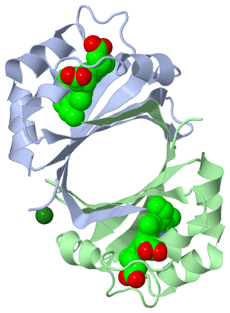
Ligands, Modified Residues, Ions (2, 3)| Asymmetric/Biological Unit (2, 3) |
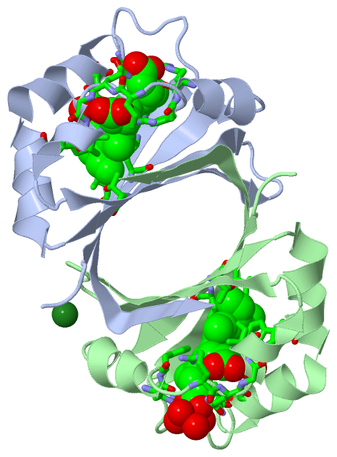
Sites (3, 3)
Asymmetric Unit (3, 3)
|
SS Bonds (0, 0)| (no "SS Bond" information available for 3LGM) |
Cis Peptide Bonds (0, 0)| (no "Cis Peptide Bond" information available for 3LGM) |
SAPs(SNPs)/Variants (0, 0)| (no "SAP(SNP)/Variant" information available for 3LGM) |
PROSITE Motifs (1, 2)
Asymmetric/Biological Unit (1, 2)
|
||||||||||||||||||||||||
Exons (0, 0)| (no "Exon" information available for 3LGM) |
Sequences/Alignments
Asymmetric/Biological UnitChain A from PDB Type:PROTEIN Length:110 aligned with HDOX2_STAAN | Q7A827 from UniProtKB/Swiss-Prot Length:108 Alignment length:110 1 | 8 18 28 38 48 58 68 78 88 98 108 HDOX2_STAAN - --MFMAENRLQLQKGSAEETIERFYNRQGIETIEGFQQMFVTKTLNTEDTDEVKILTIWESEDSFNNWLNSDVFKEAHKNVRLKSDDDGQQSPILSNKVFKYDIGYHYQK 108 SCOP domains d3lgma_ A: Hypothetical protein PG130 (SAV0165) SCOP domains CATH domains 3lgmA00 A:-1-108 [code=3.30.70.900, no name defined] CATH domains Pfam domains -------------------------------------------------------------------------------------------------------------- Pfam domains SAPs(SNPs) -------------------------------------------------------------------------------------------------------------- SAPs(SNPs) PROSITE ---ABM PDB: A:2-93 UniProt: 2-93 --------------- PROSITE Transcript -------------------------------------------------------------------------------------------------------------- Transcript 3lgm A -1 AHMFMAENRLQLQKGSAEETIERFYNRQGIETIEGFQQMFVTKTLNTEDTDEVKILTIWESEDSFNNWLNSDVFKEAHKNVRLKSDDDGQQSPILSNKVFKYDIGYHYQK 108 8 18 28 38 48 58 68 78 88 98 108 Chain B from PDB Type:PROTEIN Length:110 aligned with HDOX2_STAAN | Q7A827 from UniProtKB/Swiss-Prot Length:108 Alignment length:110 1 | 8 18 28 38 48 58 68 78 88 98 108 HDOX2_STAAN - --MFMAENRLQLQKGSAEETIERFYNRQGIETIEGFQQMFVTKTLNTEDTDEVKILTIWESEDSFNNWLNSDVFKEAHKNVRLKSDDDGQQSPILSNKVFKYDIGYHYQK 108 SCOP domains d3lgmb_ B: Hypothetical protein PG130 (SAV0165) SCOP domains CATH domains 3lgmB00 B:-1-108 [code=3.30.70.900, no name defined] CATH domains Pfam domains -------------------------------------------------------------------------------------------------------------- Pfam domains SAPs(SNPs) -------------------------------------------------------------------------------------------------------------- SAPs(SNPs) PROSITE ---ABM PDB: B:2-93 UniProt: 2-93 --------------- PROSITE Transcript -------------------------------------------------------------------------------------------------------------- Transcript 3lgm B -1 AHMFMAENRLQLQKGSAEETIERFYNRQGIETIEGFQQMFVTKTLNTEDTDEVKILTIWESEDSFNNWLNSDVFKEAHKNVRLKSDDDGQQSPILSNKVFKYDIGYHYQK 108 8 18 28 38 48 58 68 78 88 98 108
|
||||||||||||||||||||
SCOP Domains (1, 2)
Asymmetric/Biological Unit
 |
CATH Domains (1, 2)
Asymmetric/Biological Unit
|
Pfam Domains (0, 0)| (no "Pfam Domain" information available for 3LGM) |
Gene Ontology (11, 11)|
Asymmetric/Biological Unit(hide GO term definitions) Chain A,B (HDOX2_STAAN | Q7A827)
|
||||||||||||||||||||||||||||||||||||||||||||||||||||||||||||||||||||||||||||||||||||
Interactive Views
|
|||||||||||||||||||||||||||||||||||||||||||||||||||||||||||||||||||||||||||||||||||||||||||||||||||||||||||||||||||||||||||||||||||||||||||
Still Images
|
||||||||||||||||
Databases
|
||||||||||||||||||||||||||||||||||||||||||||||||||||||||||||||||||||||||||||||||||||||||||||||||||||||||||||||||||||||||||||||||||||||||||||||||||||||||||||||||
Analysis Tools
|
|||||||||||||||||||||||||||||||||||||||||||||||||||||||||||||
Entries Sharing at Least One Protein Chain (UniProt ID)
Related Entries Specified in the PDB File
|
|