|
|
|
|
 Description
|
|
Compounds
|
||||||||||||||||||||||||||||||||||||||||||||
Chains, Units
Summary Information (see also Sequences/Alignments below) |
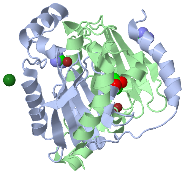
Ligands, Modified Residues, Ions (4, 7)| Asymmetric/Biological Unit (4, 7) |
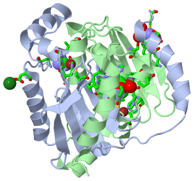
Sites (7, 7)
Asymmetric Unit (7, 7)
|
SS Bonds (0, 0)| (no "SS Bond" information available for 3LAS) |
Cis Peptide Bonds (0, 0)| (no "Cis Peptide Bond" information available for 3LAS) |
SAPs(SNPs)/Variants (0, 0)| (no "SAP(SNP)/Variant" information available for 3LAS) |
PROSITE Motifs (0, 0)| (no "PROSITE Motif" information available for 3LAS) |
Exons (0, 0)| (no "Exon" information available for 3LAS) |
Sequences/Alignments
Asymmetric/Biological UnitChain A from PDB Type:PROTEIN Length:166 aligned with Q8DVY1_STRMU | Q8DVY1 from UniProtKB/TrEMBL Length:166 Alignment length:166 10 20 30 40 50 60 70 80 90 100 110 120 130 140 150 160 Q8DVY1_STRMU 1 MVMSYFDNFIKANQAYVDLHGTAHLPLKPKTRVAIVTCMDSRLHVAPALGLALGDAHILRNAGGRVTDDVIRSLVISEQQLGTSEIVVLHHTDCGAQTFTNAEFTEQLKRDLAVDAGDQDFLPFTDIEESVREDIALLKNSPLIPEDIIISGAIYDVDTGRVREVN 166 SCOP domains d3lasa_ A: automated matches SCOP domains CATH domains ---------------------------------------------------------------------------------------------------------------------------------------------------------------------- CATH domains Pfam domains ---------------------------------------------------------------------------------------------------------------------------------------------------------------------- Pfam domains SAPs(SNPs) ---------------------------------------------------------------------------------------------------------------------------------------------------------------------- SAPs(SNPs) PROSITE ---------------------------------------------------------------------------------------------------------------------------------------------------------------------- PROSITE Transcript ---------------------------------------------------------------------------------------------------------------------------------------------------------------------- Transcript 3las A 1 MVMSYFDNFIKANQAYVDLHGTAHLPLKPKTRVAIVTCMDSRLHVAPALGLALGDAHILRNAGGRVTDDVIRSLVISEQQLGTSEIVVLHHTDCGAQTFTNAEFTEQLKRDLAVDAGDQDFLPFTDIEESVREDIALLKNSPLIPEDIIISGAIYDVDTGRVREVN 166 10 20 30 40 50 60 70 80 90 100 110 120 130 140 150 160 Chain B from PDB Type:PROTEIN Length:166 aligned with Q8DVY1_STRMU | Q8DVY1 from UniProtKB/TrEMBL Length:166 Alignment length:166 10 20 30 40 50 60 70 80 90 100 110 120 130 140 150 160 Q8DVY1_STRMU 1 MVMSYFDNFIKANQAYVDLHGTAHLPLKPKTRVAIVTCMDSRLHVAPALGLALGDAHILRNAGGRVTDDVIRSLVISEQQLGTSEIVVLHHTDCGAQTFTNAEFTEQLKRDLAVDAGDQDFLPFTDIEESVREDIALLKNSPLIPEDIIISGAIYDVDTGRVREVN 166 SCOP domains d3lasb_ B: automated matches SCOP domains CATH domains ---------------------------------------------------------------------------------------------------------------------------------------------------------------------- CATH domains Pfam domains (1) --------------------------------Pro_CA-3lasB01 B:33-162 ---- Pfam domains (1) Pfam domains (2) --------------------------------Pro_CA-3lasB02 B:33-162 ---- Pfam domains (2) SAPs(SNPs) ---------------------------------------------------------------------------------------------------------------------------------------------------------------------- SAPs(SNPs) PROSITE ---------------------------------------------------------------------------------------------------------------------------------------------------------------------- PROSITE Transcript ---------------------------------------------------------------------------------------------------------------------------------------------------------------------- Transcript 3las B 1 MVMSYFDNFIKANQAYVDLHGTAHLPLKPKTRVAIVTCMDSRLHVAPALGLALGDAHILRNAGGRVTDDVIRSLVISEQQLGTSEIVVLHHTDCGAQTFTNAEFTEQLKRDLAVDAGDQDFLPFTDIEESVREDIALLKNSPLIPEDIIISGAIYDVDTGRVREVN 166 10 20 30 40 50 60 70 80 90 100 110 120 130 140 150 160
|
||||||||||||||||||||
SCOP Domains (1, 2)
Asymmetric/Biological Unit
 |
CATH Domains (0, 0)| (no "CATH Domain" information available for 3LAS) |
Pfam Domains (1, 2)
Asymmetric/Biological Unit
|
Gene Ontology (4, 4)|
Asymmetric/Biological Unit(hide GO term definitions) Chain A,B (Q8DVY1_STRMU | Q8DVY1)
|
||||||||||||||||||||||||||||||
Interactive Views
|
|||||||||||||||||||||||||||||||||||||||||||||||||||||||||||||||||||||||||||||||||||||||||||||||||||||||||||||||||||||||||||||||||||||||||||||||||||||||||||||||||||||||||||||||||||||
Still Images
|
||||||||||||||||
Databases
|
||||||||||||||||||||||||||||||||||||||||||||||||||||||||||||||||||||||||||||||||||||||||||||||||||||||||||||||||||||||||||||||||||||||||||||||||||||||||||||||||
Analysis Tools
|
|||||||||||||||||||||||||||||||||||||||||||||||||||||||||||||
Entries Sharing at Least One Protein Chain (UniProt ID)
Related Entries Specified in the PDB File
|
|