|
|
|
|
 Description
|
|
Compounds
|
||||||||||||||||||||||||||||||||||||||||||||||||||||
Chains, Units
Summary Information (see also Sequences/Alignments below) |
Ligands, Modified Residues, Ions (2, 4)| Asymmetric/Biological Unit (2, 4) |
Sites (4, 4)
Asymmetric Unit (4, 4)
|
SS Bonds (0, 0)| (no "SS Bond" information available for 3L6N) |
Cis Peptide Bonds (0, 0)| (no "Cis Peptide Bond" information available for 3L6N) |
SAPs(SNPs)/Variants (0, 0)| (no "SAP(SNP)/Variant" information available for 3L6N) |
PROSITE Motifs (0, 0)| (no "PROSITE Motif" information available for 3L6N) |
Exons (0, 0)| (no "Exon" information available for 3L6N) |
Sequences/Alignments
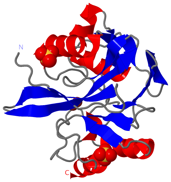
Asymmetric/Biological UnitChain A from PDB Type:PROTEIN Length:218 aligned with A4GRB2_9FLAO | A4GRB2 from UniProtKB/TrEMBL Length:239 Alignment length:218 30 40 50 60 70 80 90 100 110 120 130 140 150 160 170 180 190 200 210 220 230 A4GRB2_9FLAO 21 QVKDFVIEPPIKNNLHIYKTFGVFGGKEYSANSMYLVTKKGVVLFDVPWEKVQYQSLMDTIKKRHNLPVVAVFATHSHDDRAGDLSFFNNKGIKTYATAKTNEFLKKDGKATSTEIIKTGKPYRIGGEEFVVDFLGEGHTADNVVVWFPKYNVLDGGCLVKSNSATDLGYIKEANVEQWPKTINKLKAKYSKATLIIPGHDEWKGGGHVEHTLELLNK 238 SCOP domains d3l6na_ A: automated matches SCOP domains CATH domains 3l6nA00 A:21-238 Metallo-beta-lactamase, chain A CATH domains Pfam domains --------------------------Lactamase_B-3l6nA01 A:47-220 ------------------ Pfam domains SAPs(SNPs) -------------------------------------------------------------------------------------------------------------------------------------------------------------------------------------------------------------------------- SAPs(SNPs) PROSITE -------------------------------------------------------------------------------------------------------------------------------------------------------------------------------------------------------------------------- PROSITE Transcript -------------------------------------------------------------------------------------------------------------------------------------------------------------------------------------------------------------------------- Transcript 3l6n A 21 QVKDFVIEPPIKNNLHIYKTFGVFGGKEYSANSMYLVTKKGVVLFDVPWEKVQYQSLMDTIKKRHNLPVVAVFATHSHDDRAGDLSFFNNKGIKTYATAKTNEFLKKDGKATSTEIIKTGKPYRIGGEEFVVDFLGEGHTADNVVVWFPKYNVLDGGCLVKSNSATDLGYIKEANVEQWPKTINKLKAKYSKATLIIPGHDEWKGGGHVEHTLELLNK 238 30 40 50 60 70 80 90 100 110 120 130 140 150 160 170 180 190 200 210 220 230
|
||||||||||||||||||||
SCOP Domains (1, 1)
Asymmetric/Biological Unit
 |
CATH Domains (1, 1)
Asymmetric/Biological Unit
|
Pfam Domains (1, 1)
Asymmetric/Biological Unit
|
Gene Ontology (4, 4)|
Asymmetric/Biological Unit(hide GO term definitions) Chain A (A4GRB2_9FLAO | A4GRB2)
|
||||||||||||||||||||||||||||||||||||
Interactive Views
|
||||||||||||||||||||||||||||||||||||||||||||||||||||||||||||||||||||||||||||||||||||||||||||||||||||||||||||||||||||||||||||||||||||||||||||||||||
Still Images
|
||||||||||||||||
Databases
|
||||||||||||||||||||||||||||||||||||||||||||||||||||||||||||||||||||||||||||||||||||||||||||||||||||||||||||||||||||||||||||||||||||||||||||||||||||||||||||||||
Analysis Tools
|
|||||||||||||||||||||||||||||||||||||||||||||||||||||||||||||
Entries Sharing at Least One Protein Chain (UniProt ID)
Related Entries Specified in the PDB File
|
|