|
|
|
|
 Description
|
|
Compounds
|
||||||||||||||||||||||||||||||||||||||||||||
Chains, Units
Summary Information (see also Sequences/Alignments below) |
Ligands, Modified Residues, Ions (1, 1)
Asymmetric Unit (1, 1)
|
Sites (0, 0)| (no "Site" information available for 3L60) |
SS Bonds (0, 0)| (no "SS Bond" information available for 3L60) |
Cis Peptide Bonds (1, 1)
Asymmetric Unit
|
||||||||
SAPs(SNPs)/Variants (0, 0)| (no "SAP(SNP)/Variant" information available for 3L60) |
PROSITE Motifs (0, 0)| (no "PROSITE Motif" information available for 3L60) |
Exons (0, 0)| (no "Exon" information available for 3L60) |
Sequences/Alignments
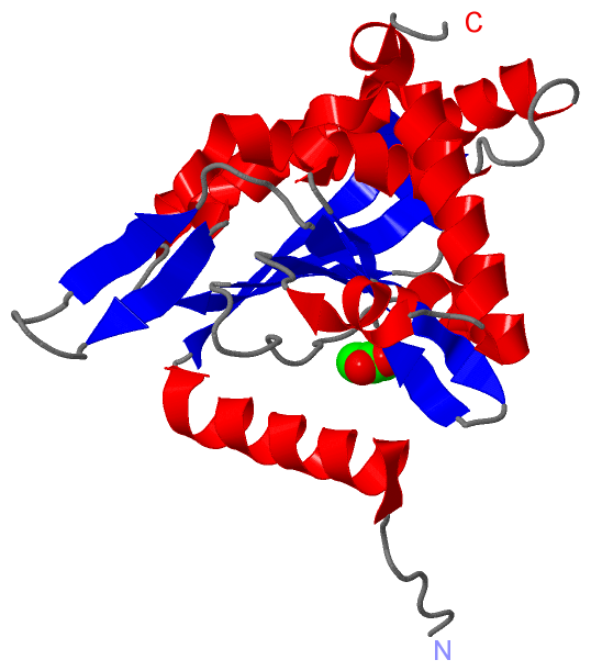
Asymmetric UnitChain A from PDB Type:PROTEIN Length:229 aligned with BKDC_MYCTU | O06159 from UniProtKB/Swiss-Prot Length:393 Alignment length:229 174 184 194 204 214 224 234 244 254 264 274 284 294 304 314 324 334 344 354 364 374 384 BKDC_MYCTU 165 PDVRPVHGVHARMAEKMTLSHKEIPTAKASVEVICAELLRLRDRFVSAAPEITPFALTLRLLVIALKHNVILNSTWVDSGEGPQVHVHRGVHLGFGAATERGLLVPVVTDAQDKNTRELASRVAELITGAREGTLTPAELRGSTFTVSNFGALGVDDGVPVINHPEAAILGLGAIKPRPVVVGGEVVARPTMTLTCVFDHRVVDGAQVAQFMCELRDLIESPETALLDL 393 SCOP domains d3l60a_ A: automated matches SCOP domains CATH domains ------------------------------------------------------------------------------------------------------------------------------------------------------------------------------------------------------------------------------------- CATH domains Pfam domains ------------------------------------------------------------------------------------------------------------------------------------------------------------------------------------------------------------------------------------- Pfam domains SAPs(SNPs) ------------------------------------------------------------------------------------------------------------------------------------------------------------------------------------------------------------------------------------- SAPs(SNPs) PROSITE ------------------------------------------------------------------------------------------------------------------------------------------------------------------------------------------------------------------------------------- PROSITE Transcript ------------------------------------------------------------------------------------------------------------------------------------------------------------------------------------------------------------------------------------- Transcript 3l60 A 165 PDVRPVHGVHARMAEKMTLSHKEIPTAKASVEVICAELLRLRDRFVSAAPEITPFALTLRLLVIALKHNVILNSTWVDSGEGPQVHVHRGVHLGFGAATERGLLVPVVTDAQDKNTRELASRVAELITGAREGTLTPAELRGSTFTVSNFGALGVDDGVPVINHPEAAILGLGAIKPRPVVVGGEVVARPTMTLTCVFDHRVVDGAQVAQFMCELRDLIESPETALLDL 393 174 184 194 204 214 224 234 244 254 264 274 284 294 304 314 324 334 344 354 364 374 384
|
||||||||||||||||||||
SCOP Domains (1, 1)
Asymmetric Unit
 |
CATH Domains (0, 0)| (no "CATH Domain" information available for 3L60) |
Pfam Domains (0, 0)| (no "Pfam Domain" information available for 3L60) |
Gene Ontology (7, 7)|
Asymmetric Unit(hide GO term definitions) Chain A (BKDC_MYCTU | O06159)
|
||||||||||||||||||||||||||||||||||||||||||||||||||||||||||||
Interactive Views
|
||||||||||||||||||||||||||||||||||||||||||||||||||||||||||||||||||||||||||||||||||||||||||||||||||||||||||||||||||||||||||||||||||||||||
Still Images
|
||||||||||||||||
Databases
|
||||||||||||||||||||||||||||||||||||||||||||||||||||||||||||||||||||||||||||||||||||||||||||||||||||||||||||||||||||||||||||||||||||||||||||||||||||||||||||||||
Analysis Tools
|
|||||||||||||||||||||||||||||||||||||||||||||||||||||||||||||
Entries Sharing at Least One Protein Chain (UniProt ID)
Related Entries Specified in the PDB File
|
|