|
|
|
|
 Description
|
|
Compounds
|
||||||||||||||||||||||||||||||||||||||||||
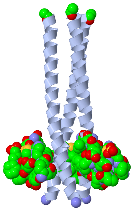
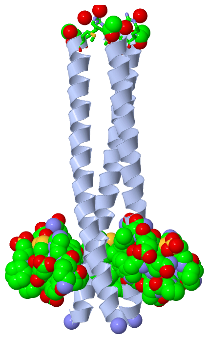
Chains, Units
Summary Information (see also Sequences/Alignments below) |
Ligands, Modified Residues, Ions (13, 20)
Asymmetric Unit (13, 20)
|
Sites (1, 1)
Asymmetric Unit (1, 1)
|
SS Bonds (1, 1)
Asymmetric Unit
|
||||||||
Cis Peptide Bonds (0, 0)| (no "Cis Peptide Bond" information available for 3L36) |
SAPs(SNPs)/Variants (0, 0)| (no "SAP(SNP)/Variant" information available for 3L36) |
PROSITE Motifs (0, 0)| (no "PROSITE Motif" information available for 3L36) |
Exons (0, 0)| (no "Exon" information available for 3L36) |
Sequences/Alignments
Asymmetric Unit
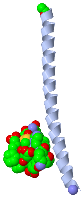
Chain A from PDB Type:PROTEIN Length:47
SCOP domains ----------------------------------------------- SCOP domains
CATH domains ----------------------------------------------- CATH domains
Pfam domains ----------------------------------------------- Pfam domains
SAPs(SNPs) ----------------------------------------------- SAPs(SNPs)
PROSITE ----------------------------------------------- PROSITE
Transcript ----------------------------------------------- Transcript
3l36 A 0 xRMKQIEDKIEEIESKQKKIENEIARIKKLLQLTVWGIKQLQARILx 46
| 9 19 29 39 |
| 46-NH2
0-ACE
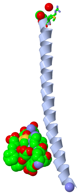
Chain H from PDB Type:OTHER/PROTEIN Length:17
SCOP domains ----------------- SCOP domains
CATH domains ----------------- CATH domains
Pfam domains ----------------- Pfam domains
SAPs(SNPs) ----------------- SAPs(SNPs)
PROSITE ----------------- PROSITE
Transcript ----------------- Transcript
3l36 H 1 xxxxxxxxxxxxxxxxx 17
||||||||10|||||||
||||||||9-DGL||||
1-ACE|||10-DTR|||
2-DLY|| 11-DGN||
3-DHI| 12-DTR|
4-DPR 13-DLE
5-DCY 14-DCY
6-DAS 15-DGL
7-DTY 16-DLE
8-DPR 17-NH2
|
||||||||||||||||||||
SCOP Domains (0, 0)| (no "SCOP Domain" information available for 3L36) |
CATH Domains (0, 0)| (no "CATH Domain" information available for 3L36) |
Pfam Domains (0, 0)| (no "Pfam Domain" information available for 3L36) |
Gene Ontology (0, 0)|
Asymmetric Unit(hide GO term definitions)
(no "Gene Ontology" information available for 3L36)
|
Interactive Views
|
||||||||||||||||||||||||||||||||||||||||||||||||||||||||||||||||||||||||||||||||||||||||||||||||||||||||||||||||||||||||||||||||||||||||||||||||||||||||||||||||||||||||||||||||||||||||||||||||||||||||||||||||||||||||||||
Still Images
|
||||||||||||||||
Databases
|
||||||||||||||||||||||||||||||||||||||||||||||||||||||||||||||||||||||||||||||||||||||||||||||||||||||||||||||||||||||||||||||||||||||||||||||||||||||||||||||||
Analysis Tools
|
|||||||||||||||||||||||||||||||||||||||||||||||||||||||||||||
Entries Sharing at Least One Protein Chain (UniProt ID)
Related Entries Specified in the PDB File
|
|