Show PDB file:   
         Plain Text   HTML   (compressed file size)
QuickSearch:   
by PDB,NDB,UniProt,PROSITE Code or Search Term(s)  
(-)Asymmetric Unit
(-)Asym. Unit - sites
(-)Biological Unit 1
(-)Biol. Unit 1 - sites
collapse expand < >
Image Asymmetric Unit
Asymmetric Unit  (Jmol Viewer)
Image Asym. Unit - sites
Asym. Unit - sites  (Jmol Viewer)
Image Biological Unit 1
Biological Unit 1  (Jmol Viewer)
Image Biol. Unit 1 - sites
Biol. Unit 1 - sites  (Jmol Viewer)

(-) Description

Title :  THE HIGH RESOLUTION STRUCTURE OF C-PHYCOCYANIN FROM GALDIERIA SULPHURARIA
 
Authors :  R. Fromme, B. Thangaraj, C. Vanselow, P. Fromme
Date :  30 Nov 09  (Deposition) - 13 Apr 11  (Release) - 13 Apr 11  (Revision)
Method :  X-RAY DIFFRACTION
Resolution :  1.50
Chains :  Asym. Unit :  A,B
Biol. Unit 1:  A,B  (6x)
Keywords :  Photosystem Ii, Light Harvesting Proteins, Red Algae, Thermostability, Bile Pigment, Chloroplast, Chromophore, Membrane, Phycobilisome, Thylakoid, Transport, Methylation, Phytochrome, Excitation Energy Transfer, Photosynthesis (Keyword Search: [Gene Ontology, PubMed, Web (Google))
 
Reference :  R. Fromme, B. Thangaraj, C. Vanselow, P. Fromme
High Resolution Structure Of C-Phycocyanin From Galdieria Sulphuraria Reveals New Insights Of The Phycobilisome
To Be Published
PubMed: search

(-) Compounds

Molecule 1 - C-PHYCOCYANIN ALPHA CHAIN
    ChainsA
    Organism ScientificGALDIERIA SULPHURARIA
    Organism Taxid130081
 
Molecule 2 - C-PHYCOCYANIN BETA CHAIN
    ChainsB
    Organism ScientificGALDIERIA SULPHURARIA
    Organism Taxid130081

 Structural Features

(-) Chains, Units

  12
Asymmetric Unit AB
Biological Unit 1 (6x)AB

Summary Information (see also Sequences/Alignments below)
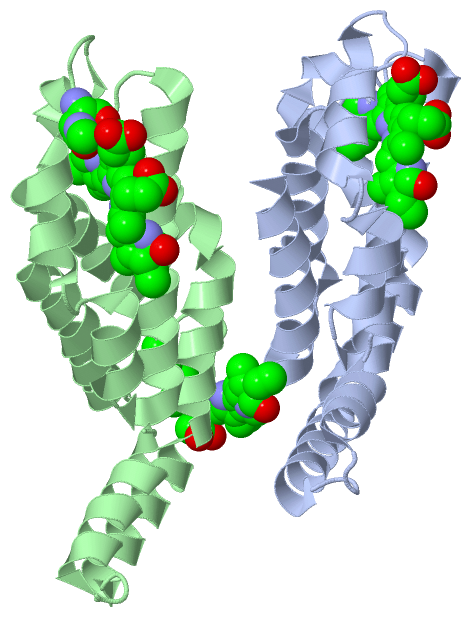
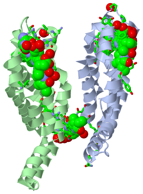
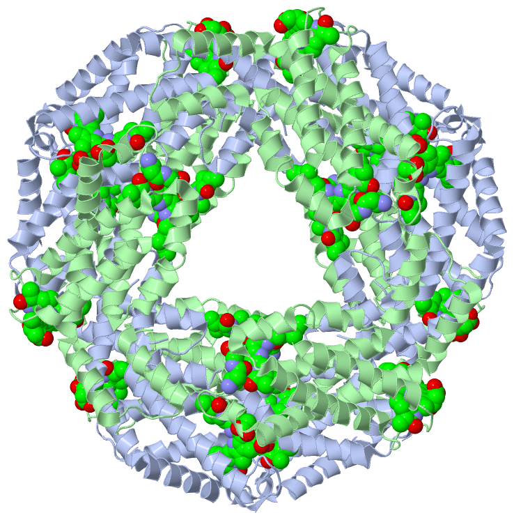
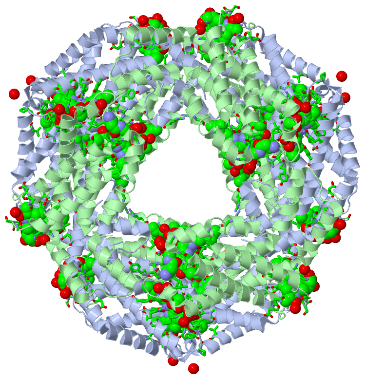
(-) Ligands, Modified Residues, Ions  (2, 4)

Asymmetric Unit (2, 4)
No.NameCountTypeFull Name
1BLA3Ligand/IonBILIVERDINE IX ALPHA
2MEN1Mod. Amino AcidN-METHYL ASPARAGINE
Biological Unit 1 (2, 24)
No.NameCountTypeFull Name
1BLA18Ligand/IonBILIVERDINE IX ALPHA
2MEN6Mod. Amino AcidN-METHYL ASPARAGINE

(-) Sites  (3, 3)

Asymmetric Unit (3, 3)
No.NameEvidenceResiduesDescription
1AC1SOFTWAREVAL A:59 , THR A:66 , PRO A:72 , GLN A:73 , TYR A:74 , ALA A:75 , GLY A:80 , LYS A:83 , CYS A:84 , ARG A:86 , ASP A:87 , TYR A:90 , TYR A:110 , ILE A:118 , PHE A:122 , LEU A:124 , TRP A:128 , TYR A:129 , HOH A:201 , HOH A:204 , HOH A:208 , HOH A:254 , ARG B:57 , ILE B:67 , THR B:75 , ASN B:76 , HOH B:178BINDING SITE FOR RESIDUE BLA A 484
2AC2SOFTWAREASN A:21 , ASN A:28 , ARG A:33 , PRO A:70 , GLN A:145 , ASN A:148 , HOH A:408 , ASN B:35 , LYS B:36 , LEU B:38 , ASP B:39 , VAL B:40 , ALA B:142 , ASN B:143 , ASP B:144 , ILE B:148 , THR B:149 , THR B:150 , GLY B:151 , CYS B:153 , HOH B:191 , HOH B:199 , HOH B:202 , HOH B:250 , HOH B:259 , HOH B:361BINDING SITE FOR RESIDUE BLA B 555
3AC3SOFTWARELEU B:66 , MEN B:72 , ALA B:73 , ARG B:77 , ARG B:78 , ALA B:81 , CYS B:82 , ARG B:84 , ASP B:85 , ILE B:88 , ARG B:108 , LEU B:113 , LEU B:120 , VAL B:122 , PRO B:123 , SER B:126 , HOH B:211 , HOH B:313 , HOH B:498 , HOH B:519 , HOH B:553BINDING SITE FOR RESIDUE BLA B 584

(-) SS Bonds  (0, 0)

(no "SS Bond" information available for 3KVS)

(-) Cis Peptide Bonds  (0, 0)

(no "Cis Peptide Bond" information available for 3KVS)

 Sequence-Structure Mapping

(-) SAPs(SNPs)/Variants  (0, 0)

(no "SAP(SNP)/Variant" information available for 3KVS)

(-) PROSITE Motifs  (0, 0)

(no "PROSITE Motif" information available for 3KVS)

(-) Exons   (0, 0)

(no "Exon" information available for 3KVS)

(-) Sequences/Alignments

Asymmetric Unit
   Reformat: Number of residues per line =  ('0' or empty: single-line sequence representation)
  Number of residues per labelling interval =   
  UniProt sequence: complete  aligned part    
   Show mapping: SCOP domains CATH domains Pfam domains Secondary structure (by author)
SAPs(SNPs) PROSITE motifs Exons
(details for a mapped element are shown in a popup box when the mouse pointer rests over it)
Chain A from PDB  Type:PROTEIN  Length:161
 aligned with PHCA_GALSU | P00306 from UniProtKB/Swiss-Prot  Length:162

    Alignment length:161
                                    11        21        31        41        51        61        71        81        91       101       111       121       131       141       151       161 
           PHCA_GALSU     2 KTPITEAIAAADNQGRFLSNTELQAVNGRYQRAAASLEAARSLTSNAERLINGAAQAVYSKFPYTSQMPGPQYASSAVGKAKCARDIGYYLRMVTYCLVVGGTGPMDEYLIAGLEEINRTFDLSPSWYVEALNYIKANHGLSGQAANEANTYIDYAINALS 162
               SCOP domains d3kvsa_ A: automated matches                                                                                                                                      SCOP domains
               CATH domains ----------------------------------------------------------------------------------------------------------------------------------------------------------------- CATH domains
               Pfam domains ----Phycobilisome-3kvsA01 A:6-162                                                                                                                                 Pfam domains
         Sec.struct. author .hhhhhhhhhhhhh....hhhhhhhhhhhhhhhhhhhhhhhhhhhhhhhhhhhhhhhhhhhhhhhhh........hhhhhhhhhhhhhhhhhhhhhhhhhh.hhhhhhhh..hhhhhhhhh..hhhhhhhhhhhhhhhh..hhhhhhhhhhhhhhhhhhh. Sec.struct. author
                 SAPs(SNPs) ----------------------------------------------------------------------------------------------------------------------------------------------------------------- SAPs(SNPs)
                    PROSITE ----------------------------------------------------------------------------------------------------------------------------------------------------------------- PROSITE
                 Transcript ----------------------------------------------------------------------------------------------------------------------------------------------------------------- Transcript
                 3kvs A   2 KTPITEAIAAADNQGRFLSNTELQAVNGRYQRAAASLEAARSLTSNAQRLINGAAQAVYSKFPYTSQMPGPQYASSAVGKAKCARDIGYYLRMVTYCLVVGGTGPMDEYLIAGLEEINRTFDLSPSWYVEALNYVKSNHGLSGQAANEANTYIDYAINALS 162
                                    11        21        31        41        51        61        71        81        91       101       111       121       131       141       151       161 

Chain B from PDB  Type:PROTEIN  Length:170
 aligned with PHCB_GALSU | P00311 from UniProtKB/Swiss-Prot  Length:172

    Alignment length:170
                                    11        21        31        41        51        61        71        81        91       101       111       121       131       141       151       161       171
           PHCB_GALSU     2 LDAFAKVVAQADARGEFLSNTQLDALSKMVSEGNKRLDVVNRITSNASAIVTNAARALFSEQPQLIQPGGNAYTNRRMAACLRDMEIILRYVSYAIIAGDSSILDDRCLNGLRETYQALGVPGASVAVGIEKMKDSAIAIANDPSGITTGDCSALMAEVGTYFDRAATAV 171
               SCOP domains d3kvsb_ B: automated matches                                                                                                                                               SCOP domains
               CATH domains -------------------------------------------------------------------------------------------------------------------------------------------------------------------------- CATH domains
               Pfam domains ----Phycobilisome-3kvsB01 B:6-171                                                                                                                                          Pfam domains
         Sec.struct. author .hhhhhhhhhhhhh....hhhhhhhhhhhhhhhhhhhhhhhhhhhhhhhhhhhhhhhhhhhhhhhh.......hhhhhhhhhhhhhhhhhhhhhhhhhh.hhhhhhhh..hhhhhhhhhh.hhhhhhhhhhhhhhhhhhhhh.........hhhhhhhhhhhhhhhhhhh Sec.struct. author
                 SAPs(SNPs) -------------------------------------------------------------------------------------------------------------------------------------------------------------------------- SAPs(SNPs)
                    PROSITE -------------------------------------------------------------------------------------------------------------------------------------------------------------------------- PROSITE
                 Transcript -------------------------------------------------------------------------------------------------------------------------------------------------------------------------- Transcript
                 3kvs B   2 LDAFAKVVAQADARGEFLSNTQLDALSKMVSEGNKRLDVVNRITSNASAIVTNAARALFSEQPQLIQPGGnAYTNRRMAACLRDMEIILRYVSYAIIAGDSSVLDDRCLNGLRETYQALGVPGASVAVGVEKMKDSAIAIANDPSGITTGDCSALMAEVGTYFDRAATAV 171
                                    11        21        31        41        51        61        71|       81        91       101       111       121       131       141       151       161       171
                                                                                                 72-MEN                                                                                               

   Legend:   → Mismatch (orange background)
  - → Gap (green background, '-', border residues have a numbering label)
    → Modified Residue (blue background, lower-case, 'x' indicates undefined single-letter code, labelled with number + name)
  x → Chemical Group (purple background, 'x', labelled with number + name, e.g. ACE or NH2)
  extra numbering lines below/above indicate numbering irregularities and modified residue names etc., number ends below/above '|'

 Classification and Annotation

(-) SCOP Domains  (1, 2)

Asymmetric Unit

(-) CATH Domains  (0, 0)

(no "CATH Domain" information available for 3KVS)

(-) Pfam Domains  (1, 2)

Asymmetric Unit
(-)
Clan: Globin (291)

(-) Gene Ontology  (9, 18)

Asymmetric Unit(hide GO term definitions)
Chain A   (PHCA_GALSU | P00306)
biological process
    GO:0055114    oxidation-reduction process    A metabolic process that results in the removal or addition of one or more electrons to or from a substance, with or without the concomitant removal or addition of a proton or protons.
    GO:0015979    photosynthesis    The synthesis by organisms of organic chemical compounds, especially carbohydrates, from carbon dioxide (CO2) using energy obtained from light rather than from the oxidation of chemical compounds.
    GO:0018298    protein-chromophore linkage    The covalent or noncovalent attachment of a chromophore to a protein.
cellular component
    GO:0009507    chloroplast    A chlorophyll-containing plastid with thylakoids organized into grana and frets, or stroma thylakoids, and embedded in a stroma.
    GO:0009535    chloroplast thylakoid membrane    The pigmented membrane of a chloroplast thylakoid. An example of this component is found in Arabidopsis thaliana.
    GO:0016020    membrane    A lipid bilayer along with all the proteins and protein complexes embedded in it an attached to it.
    GO:0030089    phycobilisome    Any of the granules, approximately 32 nm x 48 nm and consisting of highly aggregated phycobiliproteins, that are attached in arrays to the external face of a thylakoid membrane in algae of the phyla Cyanophyta and Rhodophyta, where they function as light-harvesting devices in photosynthesis. Excitation energy in the phycobilisome flows in the sequence: phycoerythrin, phycocyanin, allophycocyanin before passing to the antenna chlorophyll of photosystem II.
    GO:0009536    plastid    Any member of a family of organelles found in the cytoplasm of plants and some protists, which are membrane-bounded and contain DNA. Plant plastids develop from a common type, the proplastid.
    GO:0009579    thylakoid    A membranous cellular structure that bears the photosynthetic pigments in plants, algae, and cyanobacteria. In cyanobacteria thylakoids are of various shapes and are attached to, or continuous with, the plasma membrane. In eukaryotes they are flattened, membrane-bounded disk-like structures located in the chloroplasts; in the chloroplasts of higher plants the thylakoids form dense stacks called grana. Isolated thylakoid preparations can carry out photosynthetic electron transport and the associated phosphorylation.

Chain B   (PHCB_GALSU | P00311)
biological process
    GO:0055114    oxidation-reduction process    A metabolic process that results in the removal or addition of one or more electrons to or from a substance, with or without the concomitant removal or addition of a proton or protons.
    GO:0015979    photosynthesis    The synthesis by organisms of organic chemical compounds, especially carbohydrates, from carbon dioxide (CO2) using energy obtained from light rather than from the oxidation of chemical compounds.
    GO:0018298    protein-chromophore linkage    The covalent or noncovalent attachment of a chromophore to a protein.
cellular component
    GO:0009507    chloroplast    A chlorophyll-containing plastid with thylakoids organized into grana and frets, or stroma thylakoids, and embedded in a stroma.
    GO:0009535    chloroplast thylakoid membrane    The pigmented membrane of a chloroplast thylakoid. An example of this component is found in Arabidopsis thaliana.
    GO:0016020    membrane    A lipid bilayer along with all the proteins and protein complexes embedded in it an attached to it.
    GO:0030089    phycobilisome    Any of the granules, approximately 32 nm x 48 nm and consisting of highly aggregated phycobiliproteins, that are attached in arrays to the external face of a thylakoid membrane in algae of the phyla Cyanophyta and Rhodophyta, where they function as light-harvesting devices in photosynthesis. Excitation energy in the phycobilisome flows in the sequence: phycoerythrin, phycocyanin, allophycocyanin before passing to the antenna chlorophyll of photosystem II.
    GO:0009536    plastid    Any member of a family of organelles found in the cytoplasm of plants and some protists, which are membrane-bounded and contain DNA. Plant plastids develop from a common type, the proplastid.
    GO:0009579    thylakoid    A membranous cellular structure that bears the photosynthetic pigments in plants, algae, and cyanobacteria. In cyanobacteria thylakoids are of various shapes and are attached to, or continuous with, the plasma membrane. In eukaryotes they are flattened, membrane-bounded disk-like structures located in the chloroplasts; in the chloroplasts of higher plants the thylakoids form dense stacks called grana. Isolated thylakoid preparations can carry out photosynthetic electron transport and the associated phosphorylation.

 Visualization

(-) Interactive Views

Asymmetric Unit
  Complete Structure
    Jena3D(integrated viewing of ligand, site, SAP, PROSITE, SCOP information)
    WebMol | AstexViewer[tm]@PDBe
(Java Applets, require no local installation except for Java; loading may be slow)
    STRAP
(Java WebStart application, automatic local installation, requires Java; full application with system access!)
    RasMol
(require local installation)
    Molscript (VRML)
(requires installation of a VRML viewer; select preferred view via VRML and generate a mono or stereo PDF format file)
 
  Ligands, Modified Residues, Ions
    BLA  [ RasMol | Jena3D ]  +environment [ RasMol | Jena3D ]
    MEN  [ RasMol | Jena3D ]  +environment [ RasMol | Jena3D ]
 
  Sites
    AC1  [ RasMol ]  +environment [ RasMol ]
    AC2  [ RasMol ]  +environment [ RasMol ]
    AC3  [ RasMol ]  +environment [ RasMol ]
 
  Cis Peptide Bonds
(no "Cis Peptide Bonds" information available for 3kvs)
 
Biological Unit
  Complete Structure
    Biological Unit 1  [ Jena3D ]

(-) Still Images

Jmol
  protein: cartoon or spacefill or dots and stick; nucleic acid: cartoon and stick; ligands: spacefill; active site: stick
Molscript
  protein, nucleic acid: cartoon; ligands: spacefill; active site: ball and stick

 Databases and Analysis Tools

(-) Databases

Access by PDB/NDB ID
  3kvs
    Family and Domain InformationProDom | SYSTERS
    General Structural InformationGlycoscienceDB | MMDB | NDB | OCA | PDB | PDBe | PDBj | PDBsum | PDBWiki | PQS | PROTEOPEDIA
    Orientation in MembranesOPM
    Protein SurfaceSURFACE
    Secondary StructureDSSP (structure derived) | HSSP (homology derived)
    Structural GenomicsGeneCensus
    Structural NeighboursCE | VAST
    Structure ClassificationCATH | Dali | SCOP
    Validation and Original DataBMRB Data View | BMRB Restraints Grid | EDS | PROCHECK | RECOORD | WHAT_CHECK
 
Access by UniProt ID/Accession number
  PHCA_GALSU | P00306
    Comparative Protein Structure ModelsModBase
    Genomic InformationEnsembl
    Protein-protein InteractionDIP
    Sequence, Family and Domain InformationInterPro | Pfam | SMART | UniProtKB/SwissProt
  PHCB_GALSU | P00311
    Comparative Protein Structure ModelsModBase
    Genomic InformationEnsembl
    Protein-protein InteractionDIP
    Sequence, Family and Domain InformationInterPro | Pfam | SMART | UniProtKB/SwissProt
 
Access by Enzyme Classificator   (EC Number)
  (no 'Enzyme Classificator' available)
    General Enzyme InformationBRENDA | EC-PDB | Enzyme | IntEnz
    PathwayKEGG | MetaCyc
 
Access by Disease Identifier   (MIM ID)
  (no 'MIM ID' available)
    Disease InformationOMIM
 
Access by GenAge ID
  (no 'GenAge ID' available)
    Age Related InformationGenAge

(-) Analysis Tools

Access by PDB/NDB ID
    Domain InformationXDom
    Interatomic Contacts of Structural UnitsCSU
    Ligand-protein ContactsLPC
    Protein CavitiescastP
    Sequence and Secondary StructurePDBCartoon
    Structure AlignmentSTRAP(Java WebStart application, automatic local installation, requires Java; full application with system access!)
    Structure and Sequence BrowserSTING
 
Access by UniProt ID/Accession number
  PHCA_GALSU | P00306
    Protein Disorder PredictionDisEMBL | FoldIndex | GLOBPLOT (for more information see DisProt)
  PHCB_GALSU | P00311
    Protein Disorder PredictionDisEMBL | FoldIndex | GLOBPLOT (for more information see DisProt)

 Related Entries

(-) Entries Sharing at Least One Protein Chain (UniProt ID)

UniProtKB/Swiss-Prot
        PHCA_GALSU | P003061phn 3brp
        PHCB_GALSU | P003111phn 3brp

(-) Related Entries Specified in the PDB File

1f99 CRYSTAL STRUCTURE OF R-PHYCOCYANIN FROM POLYSIPHONIA AT 2.4 A RESOLUTION
1gh0 CRYSTAL STRUCTURE OF C-PHYCOCYANIN FROM SPIRULINA PLATENSIS
1jbo THE 1.45A THREE-DIMENSIONAL STRUCTURE OF C-PHYCOCYANIN FROM THE THERMOPHYLIC CYANOBACTERIUM SYNECHOCOCCUS ELONGATUS
1ktp CRYSTAL STRUCTURE OF C-PHYCOCYANIN OF SYNECHOCOCCUS VULCANUS AT 1.6 ANGSTROMS
1phn STRUCTURE OF PHYCOCYANIN FROM CYANIDIUM CALDARIUM AT 1.65A RESOLUTION
3brp CRYSTAL STRUCTURE OF C-PHYCOCYANIN FROM GALDIERIA SULPHURARIA