Show PDB file:   
         Plain Text   HTML   (compressed file size)
QuickSearch:   
by PDB,NDB,UniProt,PROSITE Code or Search Term(s)  
(-)Asymmetric Unit
(-)Asym. Unit - sites
(-)Biological Unit 1
(-)Biol. Unit 1 - sites
collapse expand < >
Image Asymmetric Unit
Asymmetric Unit  (Jmol Viewer)
Image Asym. Unit - sites
Asym. Unit - sites  (Jmol Viewer)
Image Biological Unit 1
Biological Unit 1  (Jmol Viewer)
Image Biol. Unit 1 - sites
Biol. Unit 1 - sites  (Jmol Viewer)

(-) Description

Title :  STRUCTURE OF THE AMPR EFFECTOR BINDING DOMAIN FROM CITROBACTER FREUNDII
 
Authors :  B. L. Mark, M. D Balcewich
Date :  13 Nov 09  (Deposition) - 26 May 10  (Release) - 28 Jul 10  (Revision)
Method :  X-RAY DIFFRACTION
Resolution :  1.83
Chains :  Asym. Unit :  A
Biol. Unit 1:  A  (2x)
Keywords :  Alpha-Beta Sandwich, Activator, Dna-Binding, Transcription, Transcription Regulation (Keyword Search: [Gene Ontology, PubMed, Web (Google))
 
Reference :  M. D. Balcewich, T. M. Reeve, E. A. Orlikow, L. J. Donald, D. J. Vocadlo, B. L. Mark
Crystal Structure Of The Ampr Effector Binding Domain Provides Insight Into The Molecular Regulation Of Inducible Ampc Beta-Lactamase.
J. Mol. Biol. V. 400 998 2010
PubMed-ID: 20594961  |  Reference-DOI: 10.1016/J.JMB.2010.05.040

(-) Compounds

Molecule 1 - HTH-TYPE TRANSCRIPTIONAL ACTIVATOR AMPR
    ChainsA
    EngineeredYES
    Expression SystemESCHERICHIA COLI
    Expression System PlasmidPET15
    Expression System StrainBL21 (DE3) GOLD HTE
    Expression System Taxid562
    Expression System Vector TypePLASMID
    FragmentUNP RESIDUES 83-291, EFFECTOR BINDING DOMAIN
    GeneAMPR
    Organism ScientificCITROBACTER FREUNDII
    Organism Taxid546

 Structural Features

(-) Chains, Units

  1
Asymmetric Unit A
Biological Unit 1 (2x)A

Summary Information (see also Sequences/Alignments below)

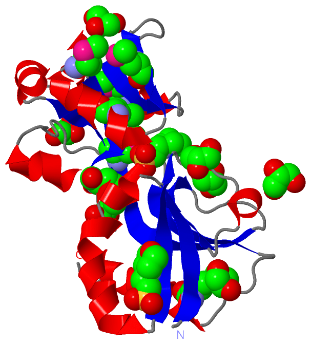
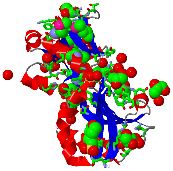
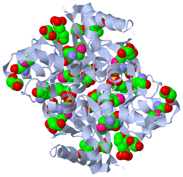
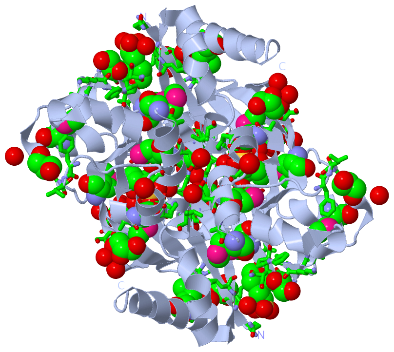
(-) Ligands, Modified Residues, Ions  (3, 16)

Asymmetric Unit (3, 16)
No.NameCountTypeFull Name
1GOL7Ligand/IonGLYCEROL
2MES2Ligand/Ion2-(N-MORPHOLINO)-ETHANESULFONIC ACID
3MSE7Mod. Amino AcidSELENOMETHIONINE
Biological Unit 1 (3, 32)
No.NameCountTypeFull Name
1GOL14Ligand/IonGLYCEROL
2MES4Ligand/Ion2-(N-MORPHOLINO)-ETHANESULFONIC ACID
3MSE14Mod. Amino AcidSELENOMETHIONINE

(-) Sites  (9, 9)

Asymmetric Unit (9, 9)
No.NameEvidenceResiduesDescription
1AC1SOFTWAREHOH A:15 , HOH A:55 , THR A:103 , SER A:191 , SER A:221 , TYR A:264 , HOH A:355 , HOH A:391 , HOH A:401BINDING SITE FOR RESIDUE MES A 302
2AC2SOFTWAREGLN A:93 , ASP A:124 , LEU A:125 , THR A:212 , HIS A:213 , ASN A:214 , VAL A:215 , HOH A:380BINDING SITE FOR RESIDUE MES A 303
3AC3SOFTWAREGLY A:148 , GLY A:149 , GLY A:150 , ALA A:151 , TRP A:152 , HOH A:320 , HOH A:345BINDING SITE FOR RESIDUE GOL A 1
4AC4SOFTWAREHOH A:16 , LYS A:95 , ASP A:142 , ARG A:272 , THR A:275 , HOH A:314 , HOH A:379BINDING SITE FOR RESIDUE GOL A 2
5AC5SOFTWARELEU A:184 , LYS A:185 , PHE A:186 , PRO A:187 , HIS A:213 , ASN A:214BINDING SITE FOR RESIDUE GOL A 3
6AC6SOFTWAREHIS A:130 , ASN A:131 , ARG A:133 , GLU A:139 , TYR A:192 , HOH A:362 , HOH A:374 , HOH A:397BINDING SITE FOR RESIDUE GOL A 4
7AC7SOFTWAREILE A:106 , ASP A:135 , GLU A:226 , ALA A:230 , HIS A:244 , HOH A:336 , HOH A:371BINDING SITE FOR RESIDUE GOL A 5
8AC8SOFTWAREPHE A:255 , LEU A:256 , THR A:257BINDING SITE FOR RESIDUE GOL A 6
9AC9SOFTWARECYS A:161 , SER A:162 , ARG A:240 , MSE A:241 , HOH A:346 , HOH A:348 , HOH A:403BINDING SITE FOR RESIDUE GOL A 7

(-) SS Bonds  (0, 0)

(no "SS Bond" information available for 3KOS)

(-) Cis Peptide Bonds  (0, 0)

(no "Cis Peptide Bond" information available for 3KOS)

 Sequence-Structure Mapping

(-) SAPs(SNPs)/Variants  (0, 0)

(no "SAP(SNP)/Variant" information available for 3KOS)

(-) PROSITE Motifs  (0, 0)

(no "PROSITE Motif" information available for 3KOS)

(-) Exons   (0, 0)

(no "Exon" information available for 3KOS)

(-) Sequences/Alignments

Asymmetric Unit
   Reformat: Number of residues per line =  ('0' or empty: single-line sequence representation)
  Number of residues per labelling interval =   
  UniProt sequence: complete  aligned part    
   Show mapping: SCOP domains CATH domains Pfam domains Secondary structure (by author)
SAPs(SNPs) PROSITE motifs Exons
(details for a mapped element are shown in a popup box when the mouse pointer rests over it)
Chain A from PDB  Type:PROTEIN  Length:200
 aligned with AMPR_CITFR | P12529 from UniProtKB/Swiss-Prot  Length:291

    Alignment length:200
                                                                                                                                                                                                                                291 
                                   102       112       122       132       142       152       162       172       182       192       202       212       222       232       242       252       262       272       282        |-
           AMPR_CITFR    93 QEKLKIGVVGTFAIGCLFPLLSDFKRSYPHIDLHISTHNNRVDPAAEGLDYTIRYGGGAWHDTDAQYLCSALMSPLCSPTLASQIQTPADILKFPLLRSYRRDEWALWMQAAGEAPPSPTHNVMVFDSSVTMLEAAQGGMGVAIAPVRMFTHLLSSERIVQPFLTQIDLGSYWITRLQSRPETPAMREFSRWLTGVLHK-   -
               SCOP domains -------------------------------------------------------------------------------------------------------------------------------------------------------------------------------------------------------- SCOP domains
               CATH domains -------------------------------------------------------------------------------------------------------------------------------------------------------------------------------------------------------- CATH domains
               Pfam domains LysR_substrate-3kosA01 A:93-290                                                                                                                                                                       -- Pfam domains
         Sec.struct. author .eeeeeee.hhhhh..hhhhhhhhhhhhh.eeeeeeee.....hhhh...eeeeee......eeeeeeeeeeeeeee.hhhhhhh..hhhhhh..eeee...hhhhhhhhhhh........eeeee.hhhhhhhhhhh...eeeeehhhhhhhhhh..............eeeeeee......hhhhhhhhhhhhhh... Sec.struct. author
                 SAPs(SNPs) -------------------------------------------------------------------------------------------------------------------------------------------------------------------------------------------------------- SAPs(SNPs)
                    PROSITE -------------------------------------------------------------------------------------------------------------------------------------------------------------------------------------------------------- PROSITE
                 Transcript -------------------------------------------------------------------------------------------------------------------------------------------------------------------------------------------------------- Transcript
                 3kos A  93 QEKLKIGVVGTFAIGCLFPLLSDFKRSYPHIDLHISTHNNRVDPAAEGLDYTIRYGGGAWHDTDAQYLCSALmSPLCSPTLASQIQTPADILKFPLLRSYRRDEWALWmQTVGEAPPSPTHNVmVFDSSVTmLEAAQAGmGVAIAPVRmFTHLLSSERIVQPFLTQIDLGSYWITRLQSRPETPAmREFSRWLTGVLHKT 292
                                   102       112       122       132       142       152       162  |    172       182       192       202       212   |   222 |     232       242       252       262       272     | 282       292
                                                                                                  165-MSE                             201-MSE        216-MSE 224-MSE 232-MSE  241-MSE                              278-MSE          

   Legend:   → Mismatch (orange background)
  - → Gap (green background, '-', border residues have a numbering label)
    → Modified Residue (blue background, lower-case, 'x' indicates undefined single-letter code, labelled with number + name)
  x → Chemical Group (purple background, 'x', labelled with number + name, e.g. ACE or NH2)
  extra numbering lines below/above indicate numbering irregularities and modified residue names etc., number ends below/above '|'

 Classification and Annotation

(-) SCOP Domains  (0, 0)

(no "SCOP Domain" information available for 3KOS)

(-) CATH Domains  (0, 0)

(no "CATH Domain" information available for 3KOS)

(-) Pfam Domains  (1, 1)

Asymmetric Unit
(-)
Clan: PBP (391)

(-) Gene Ontology  (5, 5)

Asymmetric Unit(hide GO term definitions)
Chain A   (AMPR_CITFR | P12529)
molecular function
    GO:0003677    DNA binding    Any molecular function by which a gene product interacts selectively and non-covalently with DNA (deoxyribonucleic acid).
    GO:0003700    transcription factor activity, sequence-specific DNA binding    Interacting selectively and non-covalently with a specific DNA sequence in order to modulate transcription. The transcription factor may or may not also interact selectively with a protein or macromolecular complex.
biological process
    GO:0006355    regulation of transcription, DNA-templated    Any process that modulates the frequency, rate or extent of cellular DNA-templated transcription.
    GO:0006351    transcription, DNA-templated    The cellular synthesis of RNA on a template of DNA.
cellular component
    GO:0005737    cytoplasm    All of the contents of a cell excluding the plasma membrane and nucleus, but including other subcellular structures.

 Visualization

(-) Interactive Views

Asymmetric Unit
  Complete Structure
    Jena3D(integrated viewing of ligand, site, SAP, PROSITE, SCOP information)
    WebMol | AstexViewer[tm]@PDBe
(Java Applets, require no local installation except for Java; loading may be slow)
    STRAP
(Java WebStart application, automatic local installation, requires Java; full application with system access!)
    RasMol
(require local installation)
    Molscript (VRML)
(requires installation of a VRML viewer; select preferred view via VRML and generate a mono or stereo PDF format file)
 
  Ligands, Modified Residues, Ions
    GOL  [ RasMol | Jena3D ]  +environment [ RasMol | Jena3D ]
    MES  [ RasMol | Jena3D ]  +environment [ RasMol | Jena3D ]
    MSE  [ RasMol | Jena3D ]  +environment [ RasMol | Jena3D ]
 
  Sites
    AC1  [ RasMol ]  +environment [ RasMol ]
    AC2  [ RasMol ]  +environment [ RasMol ]
    AC3  [ RasMol ]  +environment [ RasMol ]
    AC4  [ RasMol ]  +environment [ RasMol ]
    AC5  [ RasMol ]  +environment [ RasMol ]
    AC6  [ RasMol ]  +environment [ RasMol ]
    AC7  [ RasMol ]  +environment [ RasMol ]
    AC8  [ RasMol ]  +environment [ RasMol ]
    AC9  [ RasMol ]  +environment [ RasMol ]
 
  Cis Peptide Bonds
(no "Cis Peptide Bonds" information available for 3kos)
 
Biological Unit
  Complete Structure
    Biological Unit 1  [ Jena3D ]

(-) Still Images

Jmol
  protein: cartoon or spacefill or dots and stick; nucleic acid: cartoon and stick; ligands: spacefill; active site: stick
Molscript
  protein, nucleic acid: cartoon; ligands: spacefill; active site: ball and stick

 Databases and Analysis Tools

(-) Databases

Access by PDB/NDB ID
  3kos
    Family and Domain InformationProDom | SYSTERS
    General Structural InformationGlycoscienceDB | MMDB | NDB | OCA | PDB | PDBe | PDBj | PDBsum | PDBWiki | PQS | PROTEOPEDIA
    Orientation in MembranesOPM
    Protein SurfaceSURFACE
    Secondary StructureDSSP (structure derived) | HSSP (homology derived)
    Structural GenomicsGeneCensus
    Structural NeighboursCE | VAST
    Structure ClassificationCATH | Dali | SCOP
    Validation and Original DataBMRB Data View | BMRB Restraints Grid | EDS | PROCHECK | RECOORD | WHAT_CHECK
 
Access by UniProt ID/Accession number
  AMPR_CITFR | P12529
    Comparative Protein Structure ModelsModBase
    Genomic InformationEnsembl
    Protein-protein InteractionDIP
    Sequence, Family and Domain InformationInterPro | Pfam | SMART | UniProtKB/SwissProt
 
Access by Enzyme Classificator   (EC Number)
  (no 'Enzyme Classificator' available)
    General Enzyme InformationBRENDA | EC-PDB | Enzyme | IntEnz
    PathwayKEGG | MetaCyc
 
Access by Disease Identifier   (MIM ID)
  (no 'MIM ID' available)
    Disease InformationOMIM
 
Access by GenAge ID
  (no 'GenAge ID' available)
    Age Related InformationGenAge

(-) Analysis Tools

Access by PDB/NDB ID
    Domain InformationXDom
    Interatomic Contacts of Structural UnitsCSU
    Ligand-protein ContactsLPC
    Protein CavitiescastP
    Sequence and Secondary StructurePDBCartoon
    Structure AlignmentSTRAP(Java WebStart application, automatic local installation, requires Java; full application with system access!)
    Structure and Sequence BrowserSTING
 
Access by UniProt ID/Accession number
  AMPR_CITFR | P12529
    Protein Disorder PredictionDisEMBL | FoldIndex | GLOBPLOT (for more information see DisProt)

 Related Entries

(-) Entries Sharing at Least One Protein Chain (UniProt ID)

UniProtKB/Swiss-Prot
        AMPR_CITFR | P125293kot

(-) Related Entries Specified in the PDB File

3kot