Show PDB file:   
         Plain Text   HTML   (compressed file size)
QuickSearch:   
by PDB,NDB,UniProt,PROSITE Code or Search Term(s)  
(-)Asymmetric Unit
(-)Asym. Unit - sites
(-)Biological Unit 1
(-)Biol. Unit 1 - sites
(-)Biological Unit 2
collapse expand < >
Image Asymmetric Unit
Asymmetric Unit  (Jmol Viewer)
Image Asym. Unit - sites
Asym. Unit - sites  (Jmol Viewer)
Image Biological Unit 1
Biological Unit 1  (Jmol Viewer)
Image Biol. Unit 1 - sites
Biol. Unit 1 - sites  (Jmol Viewer)
Image Biological Unit 2
Biological Unit 2  (Jmol Viewer)

(-) Description

Title :  CRYSTAL STRUCTURE OF PUTATIVE ANTIBIOTIC BIOSYNTHESIS MONOOXYGENASE (NP_810307.1) FROM BACTERIODES THETAIOTAOMICRON VPI-5482 AT 1.30 A RESOLUTION
 
Authors :  Joint Center For Structural Genomics (Jcsg)
Date :  05 Nov 09  (Deposition) - 01 Dec 09  (Release) - 01 Dec 09  (Revision)
Method :  X-RAY DIFFRACTION
Resolution :  1.30
Chains :  Asym. Unit :  A
Biol. Unit 1:  A  (2x)
Biol. Unit 2:  A  (6x)
Keywords :  Putative Antibiotic Biosynthesis Monooxygenase, Structural Genomics, Joint Center For Structural Genomics, Jcsg, Protein Structure Initiative, Psi-2 (Keyword Search: [Gene Ontology, PubMed, Web (Google))
 
Reference :  Joint Center For Structural Genomics (Jcsg)
Crystal Structure Of Putative Antibiotic Biosynthesis Monooxygenase (Np_810307. 1) From Bacteriodes Thetaiotaomicron Vpi-5482 At 1. 30 A Resolution
To Be Published
PubMed: search
(for further references see the PDB file header)

(-) Compounds

Molecule 1 - PUTATIVE ANTIBIOTIC BIOSYNTHESIS MONOOXYGENASE
    ChainsA
    EngineeredYES
    Expression SystemESCHERICHIA COLI
    Expression System PlasmidSPEEDET
    Expression System StrainHK100
    Expression System Taxid562
    Expression System Vector TypePLASMID
    FragmentSEQUENCE DATABASE RESIDUES 28-131
    GeneBT_1394
    Organism ScientificBACTEROIDES THETAIOTAOMICRON
    Organism Taxid818
    SynonymPUTATIVE FLAVOREDOXIN

 Structural Features

(-) Chains, Units

  1
Asymmetric Unit A
Biological Unit 1 (2x)A
Biological Unit 2 (6x)A

Summary Information (see also Sequences/Alignments below)

(-) Ligands, Modified Residues, Ions  (5, 11)

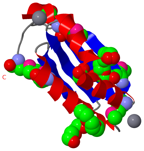
Asymmetric Unit (5, 11)
No.NameCountTypeFull Name
1ACT1Ligand/IonACETATE ION
2CA2Ligand/IonCALCIUM ION
3EDO1Ligand/Ion1,2-ETHANEDIOL
4MSE5Mod. Amino AcidSELENOMETHIONINE
5P6G2Ligand/IonHEXAETHYLENE GLYCOL
Biological Unit 1 (4, 18)
No.NameCountTypeFull Name
1ACT2Ligand/IonACETATE ION
2CA-1Ligand/IonCALCIUM ION
3EDO2Ligand/Ion1,2-ETHANEDIOL
4MSE10Mod. Amino AcidSELENOMETHIONINE
5P6G4Ligand/IonHEXAETHYLENE GLYCOL
Biological Unit 2 (4, 54)
No.NameCountTypeFull Name
1ACT6Ligand/IonACETATE ION
2CA-1Ligand/IonCALCIUM ION
3EDO6Ligand/Ion1,2-ETHANEDIOL
4MSE30Mod. Amino AcidSELENOMETHIONINE
5P6G12Ligand/IonHEXAETHYLENE GLYCOL

(-) Sites  (6, 6)

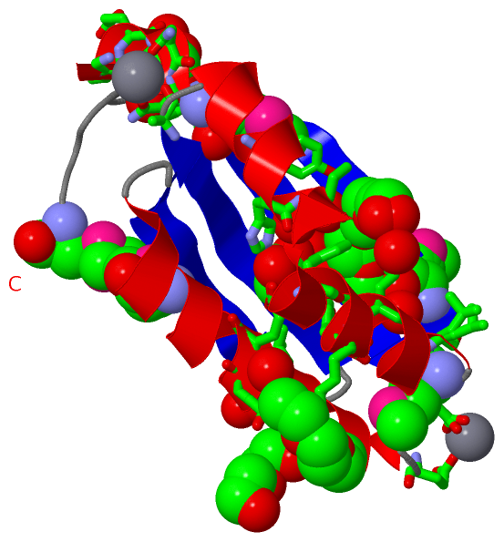
Asymmetric Unit (6, 6)
No.NameEvidenceResiduesDescription
1AC1SOFTWAREASP A:41 , GLU A:43 , GLU A:76 , HOH A:332 , HOH A:395 , HOH A:443BINDING SITE FOR RESIDUE CA A 201
2AC2SOFTWAREGLY A:0 , ASN A:30 , ASN A:31 , ALA A:88 , HOH A:225 , HOH A:356BINDING SITE FOR RESIDUE CA A 202
3AC3SOFTWARELEU A:52 , GLU A:55 , HIS A:96 , PHE A:102 , HOH A:379 , HOH A:397BINDING SITE FOR RESIDUE ACT A 203
4AC4SOFTWAREALA A:28 , GLU A:29 , ASN A:31 , PRO A:123 , HOH A:352 , HOH A:360BINDING SITE FOR RESIDUE EDO A 204
5AC5SOFTWAREALA A:50 , GLU A:54 , LYS A:104 , HOH A:362BINDING SITE FOR RESIDUE P6G A 205
6AC6SOFTWAREILE A:38 , TYR A:93 , ILE A:97 , PHE A:102 , TYR A:105 , LYS A:106 , LEU A:110 , LEU A:116 , HOH A:343 , HOH A:380 , HOH A:382 , HOH A:397BINDING SITE FOR RESIDUE P6G A 206

(-) SS Bonds  (0, 0)

(no "SS Bond" information available for 3KKF)

(-) Cis Peptide Bonds  (0, 0)

(no "Cis Peptide Bond" information available for 3KKF)

 Sequence-Structure Mapping

(-) SAPs(SNPs)/Variants  (0, 0)

(no "SAP(SNP)/Variant" information available for 3KKF)

(-) PROSITE Motifs  (0, 0)

(no "PROSITE Motif" information available for 3KKF)

(-) Exons   (0, 0)

(no "Exon" information available for 3KKF)

(-) Sequences/Alignments

Asymmetric Unit
   Reformat: Number of residues per line =  ('0' or empty: single-line sequence representation)
  Number of residues per labelling interval =   
  UniProt sequence: complete  aligned part    
   Show mapping: SCOP domains CATH domains Pfam domains Secondary structure (by author)
SAPs(SNPs) PROSITE motifs Exons
(details for a mapped element are shown in a popup box when the mouse pointer rests over it)
Chain A from PDB  Type:PROTEIN  Length:105
 aligned with Q8A7Y0_BACTN | Q8A7Y0 from UniProtKB/TrEMBL  Length:131

    Alignment length:105
                                    36        46        56        66        76        86        96       106       116       126     
         Q8A7Y0_BACTN    27 AAENNMVRLSRIIIDPERLEEYNAYLKEEIEVSMRLEPGVLVLYAVAEKERPNHVTILEIYADEAAYKSHIATPHFKKYKEGTLDMVQMLELIDATPLIPGLKMK 131
               SCOP domains --------------------------------------------------------------------------------------------------------- SCOP domains
               CATH domains --------------------------------------------------------------------------------------------------------- CATH domains
               Pfam domains -----ABM-3kkfA01 A:32-108                                                         ----------------------- Pfam domains
         Sec.struct. author hhhhheeeeeeeee...hhhhhhhhhhhhhhhhhhhh..eeeeeeee......eeeeeeee.hhhhhhhhhhhhhhhhhhhhhh..eeeeeeeee.......... Sec.struct. author
                 SAPs(SNPs) --------------------------------------------------------------------------------------------------------- SAPs(SNPs)
                    PROSITE --------------------------------------------------------------------------------------------------------- PROSITE
                 Transcript --------------------------------------------------------------------------------------------------------- Transcript
                 3kkf A   0 GAENNmVRLSRIIIDPERLEEYNAYLKEEIEVSmRLEPGVLVLYAVAEKERPNHVTILEIYADEAAYKSHIATPHFKKYKEGTLDmVQmLELIDATPLIPGLKmK 131
                            ||   |  36        46        56   |    66        76        86        96       106     | 116       126   | 
                            ||   |                          60-MSE                                             112-MSE           130-MSE
                            0|   |                                                                                115-MSE            
                            28   |                                                                                                   
                                32-MSE                                                                                               

   Legend:   → Mismatch (orange background)
  - → Gap (green background, '-', border residues have a numbering label)
    → Modified Residue (blue background, lower-case, 'x' indicates undefined single-letter code, labelled with number + name)
  x → Chemical Group (purple background, 'x', labelled with number + name, e.g. ACE or NH2)
  extra numbering lines below/above indicate numbering irregularities and modified residue names etc., number ends below/above '|'

 Classification and Annotation

(-) SCOP Domains  (0, 0)

(no "SCOP Domain" information available for 3KKF)

(-) CATH Domains  (0, 0)

(no "CATH Domain" information available for 3KKF)

(-) Pfam Domains  (1, 1)

Asymmetric Unit

(-) Gene Ontology  (1, 1)

Asymmetric Unit(hide GO term definitions)
Chain A   (Q8A7Y0_BACTN | Q8A7Y0)
molecular function
    GO:0046872    metal ion binding    Interacting selectively and non-covalently with any metal ion.

 Visualization

(-) Interactive Views

Asymmetric Unit
  Complete Structure
    Jena3D(integrated viewing of ligand, site, SAP, PROSITE, SCOP information)
    WebMol | AstexViewer[tm]@PDBe
(Java Applets, require no local installation except for Java; loading may be slow)
    STRAP
(Java WebStart application, automatic local installation, requires Java; full application with system access!)
    RasMol
(require local installation)
    Molscript (VRML)
(requires installation of a VRML viewer; select preferred view via VRML and generate a mono or stereo PDF format file)
 
  Ligands, Modified Residues, Ions
    ACT  [ RasMol | Jena3D ]  +environment [ RasMol | Jena3D ]
    CA  [ RasMol | Jena3D ]  +environment [ RasMol | Jena3D ]
    EDO  [ RasMol | Jena3D ]  +environment [ RasMol | Jena3D ]
    MSE  [ RasMol | Jena3D ]  +environment [ RasMol | Jena3D ]
    P6G  [ RasMol | Jena3D ]  +environment [ RasMol | Jena3D ]
 
  Sites
    AC1  [ RasMol ]  +environment [ RasMol ]
    AC2  [ RasMol ]  +environment [ RasMol ]
    AC3  [ RasMol ]  +environment [ RasMol ]
    AC4  [ RasMol ]  +environment [ RasMol ]
    AC5  [ RasMol ]  +environment [ RasMol ]
    AC6  [ RasMol ]  +environment [ RasMol ]
 
  Cis Peptide Bonds
(no "Cis Peptide Bonds" information available for 3kkf)
 
Biological Units
  Complete Structure
    Biological Unit 1  [ Jena3D ]
    Biological Unit 2  [ Jena3D ]

(-) Still Images

Jmol
  protein: cartoon or spacefill or dots and stick; nucleic acid: cartoon and stick; ligands: spacefill; active site: stick
Molscript
  protein, nucleic acid: cartoon; ligands: spacefill; active site: ball and stick

 Databases and Analysis Tools

(-) Databases

Access by PDB/NDB ID
  3kkf
    Family and Domain InformationProDom | SYSTERS
    General Structural InformationGlycoscienceDB | MMDB | NDB | OCA | PDB | PDBe | PDBj | PDBsum | PDBWiki | PQS | PROTEOPEDIA
    Orientation in MembranesOPM
    Protein SurfaceSURFACE
    Secondary StructureDSSP (structure derived) | HSSP (homology derived)
    Structural GenomicsGeneCensus
    Structural NeighboursCE | VAST
    Structure ClassificationCATH | Dali | SCOP
    Validation and Original DataBMRB Data View | BMRB Restraints Grid | EDS | PROCHECK | RECOORD | WHAT_CHECK
 
Access by UniProt ID/Accession number
  Q8A7Y0_BACTN | Q8A7Y0
    Comparative Protein Structure ModelsModBase
    Genomic InformationEnsembl
    Protein-protein InteractionDIP
    Sequence, Family and Domain InformationInterPro | Pfam | SMART | UniProtKB/TrEMBL
 
Access by Enzyme Classificator   (EC Number)
  (no 'Enzyme Classificator' available)
    General Enzyme InformationBRENDA | EC-PDB | Enzyme | IntEnz
    PathwayKEGG | MetaCyc
 
Access by Disease Identifier   (MIM ID)
  (no 'MIM ID' available)
    Disease InformationOMIM
 
Access by GenAge ID
  (no 'GenAge ID' available)
    Age Related InformationGenAge

(-) Analysis Tools

Access by PDB/NDB ID
    Domain InformationXDom
    Interatomic Contacts of Structural UnitsCSU
    Ligand-protein ContactsLPC
    Protein CavitiescastP
    Sequence and Secondary StructurePDBCartoon
    Structure AlignmentSTRAP(Java WebStart application, automatic local installation, requires Java; full application with system access!)
    Structure and Sequence BrowserSTING
 
Access by UniProt ID/Accession number
  Q8A7Y0_BACTN | Q8A7Y0
    Protein Disorder PredictionDisEMBL | FoldIndex | GLOBPLOT (for more information see DisProt)

 Related Entries

(-) Entries Sharing at Least One Protein Chain (UniProt ID)

(no "Entries Sharing at Least One Protein Chain" available for 3KKF)

(-) Related Entries Specified in the PDB File

(no "Related Entries Specified in the PDB File" available for 3KKF)