Show PDB file:   
         Plain Text   HTML   (compressed file size)
QuickSearch:   
by PDB,NDB,UniProt,PROSITE Code or Search Term(s)  
(-)Asym./Biol. Unit
(-)Asym./Biol. Unit - sites
collapse expand < >
Image Asym./Biol. Unit
Asym./Biol. Unit  (Jmol Viewer)
Image Asym./Biol. Unit - sites
Asym./Biol. Unit - sites  (Jmol Viewer)

(-) Description

Title :  CATALYTIC FRAGMENT OF CHOLIX TOXIN FROM VIBRIO CHOLERAE IN COMPLEX WITH INHIBITOR GP-D
 
Authors :  R. Jorgensen, P. R Edwards, A. R. Merrill
Date :  31 Oct 09  (Deposition) - 27 Oct 10  (Release) - 27 Oct 10  (Revision)
Method :  X-RAY DIFFRACTION
Resolution :  1.29
Chains :  Asym./Biol. Unit :  A
Keywords :  Adp-Ribosyl Transferase, Alpha-Beta Complex, Diphthamide, Transferase, Toxin (Keyword Search: [Gene Ontology, PubMed, Web (Google))
 
Reference :  R. Jorgensen, P. R. Edwards, A. R. Merrill
Structure Function Analysis Of Soluble Inhibitors Of Cholix Toxin From Vibrio Cholerae
To Be Published
PubMed: search

(-) Compounds

Molecule 1 - CHOLIX TOXIN
    ChainsA
    EC Number2.4.2.36
    EngineeredYES
    Expression SystemESCHERICHIA COLI
    Expression System PlasmidPET28+
    Expression System StrainER2566
    Expression System Taxid562
    Expression System Vector TypePLASMID
    FragmentCATALYTIC DOMAIN, RESIDUES 459-665
    GeneCHXA, TOXA
    Organism ScientificVIBRIO CHOLERAE
    Organism Taxid666
    StrainTP

 Structural Features

(-) Chains, Units

  1
Asymmetric/Biological Unit A

Summary Information (see also Sequences/Alignments below)
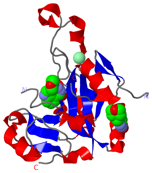
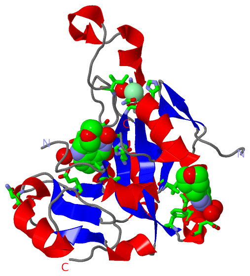
(-) Ligands, Modified Residues, Ions  (2, 3)

Asymmetric/Biological Unit (2, 3)
No.NameCountTypeFull Name
1CL1Ligand/IonCHLORIDE ION
2G9D2Ligand/Ion3-(MORPHOLIN-4-YLMETHYL)-1,5-DIHYDRO-6H-[1,2]DIAZEPINO[4,5,6-CD]INDOL-6-ONE

(-) Sites  (3, 3)

Asymmetric Unit (3, 3)
No.NameEvidenceResiduesDescription
1AC1SOFTWAREHOH A:36 , HOH A:221 , HIS A:460 , GLY A:461 , TYR A:493 , VAL A:494 , VAL A:500 , ALA A:501 , TYR A:504 , GLU A:581BINDING SITE FOR RESIDUE G9D A 1
2AC2SOFTWAREHOH A:135 , HOH A:341 , LEU A:449 , GLU A:452 , TYR A:454 , ASN A:550 , VAL A:593BINDING SITE FOR RESIDUE G9D A 635
3AC3SOFTWAREHOH A:10 , ASN A:463 , HIS A:464 , ILE A:507 , ARG A:525BINDING SITE FOR RESIDUE CL A 636

(-) SS Bonds  (0, 0)

(no "SS Bond" information available for 3KI0)

(-) Cis Peptide Bonds  (1, 1)

Asymmetric/Biological Unit
No.Residues
1Leu A:563 -Pro A:564

 Sequence-Structure Mapping

(-) SAPs(SNPs)/Variants  (0, 0)

(no "SAP(SNP)/Variant" information available for 3KI0)

(-) PROSITE Motifs  (0, 0)

(no "PROSITE Motif" information available for 3KI0)

(-) Exons   (0, 0)

(no "Exon" information available for 3KI0)

(-) Sequences/Alignments

Asymmetric/Biological Unit
   Reformat: Number of residues per line =  ('0' or empty: single-line sequence representation)
  Number of residues per labelling interval =   
  UniProt sequence: complete  aligned part    
   Show mapping: SCOP domains CATH domains Pfam domains Secondary structure (by author)
SAPs(SNPs) PROSITE motifs Exons
(details for a mapped element are shown in a popup box when the mouse pointer rests over it)
Chain A from PDB  Type:PROTEIN  Length:199
 aligned with CHXA_VIBCL | Q5EK40 from UniProtKB/Swiss-Prot  Length:666

    Alignment length:211
                                   459       469       479       489       499       509       519       529       539       549       559       569       579       589       599       609       619       629       639       649       659 
           CHXA_VIBCL   450 GRSYLPENRAVITPQGVTNWTYQELEATHQALTREGYVFVGYHGTNHVAAQTIVNRIAPVPRGNNTENEEKWGGLYVATHAEVAHGYARIKEGTGEYGLPTRAERDARGVMLRVYIPRASLERFYRTNTPLENAEEHITQVIGHSLPLRNEAFTGPESAGGEDETVIGWDMAIHAVAIPSTIPGNAYEELAIDEEAVAKEQSISTKPPYKE 660
               SCOP domains d     3ki0a_ A: automated matches                                                                                                                                                                                   SCOP domains
               CATH domains ------------------------------------------------------------------------------------------------------------------------------------------------------------------------------------------------------------------- CATH domains
               Pfam domains ------------------------------------------------------------------------------------------------------------------------------------------------------------------------------------------------------------------- Pfam domains
         Sec.struct. author .-----....eee..eee..hhhhhhhhhhhhhhh.eeeeeeeeehhhhhhhhhhh....-------.......eee..hhhhhhh........hhhhh.hhhhhhh..eeeeeeee.hhhh.eee...hhhhhhhhhhhhhh........eeeee......eeeeehhhhhh..eeeeeeee.........hhhhhhhhhhhh....... Sec.struct. author
                 SAPs(SNPs) ------------------------------------------------------------------------------------------------------------------------------------------------------------------------------------------------------------------- SAPs(SNPs)
                    PROSITE ------------------------------------------------------------------------------------------------------------------------------------------------------------------------------------------------------------------- PROSITE
                 Transcript ------------------------------------------------------------------------------------------------------------------------------------------------------------------------------------------------------------------- Transcript
                 3ki0 A 423 G-----SHMAVITPQGVTNWTYQELEATHQALTREGYVFVGYHGTNHVAAQTIVNRIAPV-------NEEKWGGLYVATHAEVAHGYARIKEGTGEYGLPTRAERDARGVMLRVYIPRASLERFYRTNTPLENAEEHITQVIGHSLPLRNEAFTGPESAGGEDETVIGWDMAIHAVAIPSTIPGNAYEELAIDEEAVAKEQSISTKPPYKE 628
                            |     |427       437       447       457       467       477       487       497       507       517       527       537       547       557       567       577       587       597       607       617       627 
                            |   424                                                  477     485                                                                                                                                               
                          423                                                                                                                                                                                                                  

   Legend:   → Mismatch (orange background)
  - → Gap (green background, '-', border residues have a numbering label)
    → Modified Residue (blue background, lower-case, 'x' indicates undefined single-letter code, labelled with number + name)
  x → Chemical Group (purple background, 'x', labelled with number + name, e.g. ACE or NH2)
  extra numbering lines below/above indicate numbering irregularities and modified residue names etc., number ends below/above '|'

 Classification and Annotation

(-) SCOP Domains  (1, 1)

Asymmetric/Biological Unit

(-) CATH Domains  (0, 0)

(no "CATH Domain" information available for 3KI0)

(-) Pfam Domains  (0, 0)

(no "Pfam Domain" information available for 3KI0)

(-) Gene Ontology  (3, 3)

Asymmetric/Biological Unit(hide GO term definitions)
Chain A   (CHXA_VIBCL | Q5EK40)
molecular function
    GO:0047286    NAD+-diphthamide ADP-ribosyltransferase activity    Catalysis of the reaction: peptide diphthamide + NAD+ = peptide N-(ADP-D-ribosyl)diphthamide + niacinamide.
    GO:0016740    transferase activity    Catalysis of the transfer of a group, e.g. a methyl group, glycosyl group, acyl group, phosphorus-containing, or other groups, from one compound (generally regarded as the donor) to another compound (generally regarded as the acceptor). Transferase is the systematic name for any enzyme of EC class 2.
    GO:0016757    transferase activity, transferring glycosyl groups    Catalysis of the transfer of a glycosyl group from one compound (donor) to another (acceptor).

 Visualization

(-) Interactive Views

Asymmetric/Biological Unit
  Complete Structure
    Jena3D(integrated viewing of ligand, site, SAP, PROSITE, SCOP information)
    WebMol | AstexViewer[tm]@PDBe
(Java Applets, require no local installation except for Java; loading may be slow)
    STRAP
(Java WebStart application, automatic local installation, requires Java; full application with system access!)
    RasMol
(require local installation)
    Molscript (VRML)
(requires installation of a VRML viewer; select preferred view via VRML and generate a mono or stereo PDF format file)
 
  Ligands, Modified Residues, Ions
    CL  [ RasMol | Jena3D ]  +environment [ RasMol | Jena3D ]
    G9D  [ RasMol | Jena3D ]  +environment [ RasMol | Jena3D ]
 
  Sites
    AC1  [ RasMol ]  +environment [ RasMol ]
    AC2  [ RasMol ]  +environment [ RasMol ]
    AC3  [ RasMol ]  +environment [ RasMol ]
 
  Cis Peptide Bonds
    Leu A:563 - Pro A:564   [ RasMol ]  
 

(-) Still Images

Jmol
  protein: cartoon or spacefill or dots and stick; nucleic acid: cartoon and stick; ligands: spacefill; active site: stick
Molscript
  protein, nucleic acid: cartoon; ligands: spacefill; active site: ball and stick

 Databases and Analysis Tools

(-) Databases

Access by PDB/NDB ID
  3ki0
    Family and Domain InformationProDom | SYSTERS
    General Structural InformationGlycoscienceDB | MMDB | NDB | OCA | PDB | PDBe | PDBj | PDBsum | PDBWiki | PQS | PROTEOPEDIA
    Orientation in MembranesOPM
    Protein SurfaceSURFACE
    Secondary StructureDSSP (structure derived) | HSSP (homology derived)
    Structural GenomicsGeneCensus
    Structural NeighboursCE | VAST
    Structure ClassificationCATH | Dali | SCOP
    Validation and Original DataBMRB Data View | BMRB Restraints Grid | EDS | PROCHECK | RECOORD | WHAT_CHECK
 
Access by UniProt ID/Accession number
  CHXA_VIBCL | Q5EK40
    Comparative Protein Structure ModelsModBase
    Genomic InformationEnsembl
    Protein-protein InteractionDIP
    Sequence, Family and Domain InformationInterPro | Pfam | SMART | UniProtKB/SwissProt
 
Access by Enzyme Classificator   (EC Number)
  2.4.2.36
    General Enzyme InformationBRENDA | EC-PDB | Enzyme | IntEnz
    PathwayKEGG | MetaCyc
 
Access by Disease Identifier   (MIM ID)
  (no 'MIM ID' available)
    Disease InformationOMIM
 
Access by GenAge ID
  (no 'GenAge ID' available)
    Age Related InformationGenAge

(-) Analysis Tools

Access by PDB/NDB ID
    Domain InformationXDom
    Interatomic Contacts of Structural UnitsCSU
    Ligand-protein ContactsLPC
    Protein CavitiescastP
    Sequence and Secondary StructurePDBCartoon
    Structure AlignmentSTRAP(Java WebStart application, automatic local installation, requires Java; full application with system access!)
    Structure and Sequence BrowserSTING
 
Access by UniProt ID/Accession number
  CHXA_VIBCL | Q5EK40
    Protein Disorder PredictionDisEMBL | FoldIndex | GLOBPLOT (for more information see DisProt)

 Related Entries

(-) Entries Sharing at Least One Protein Chain (UniProt ID)

UniProtKB/Swiss-Prot
        CHXA_VIBCL | Q5EK402q5t 2q6m 3ess 3ki1 3ki2 3ki3 3ki4 3ki5 3ki6 3ki7 3ny6 3q9o

(-) Related Entries Specified in the PDB File

2q5t FULL-LENGTH CHOLIX TOXIN FROM VIBRIO CHOLERAE
2q6m CATALYTIC FRAGMENT OF CHOLIX TOXIN FROM VIBRIO CHOLERAE IN COMPLEX WITH THE PJ34 INHIBITOR
3ess CATALYTIC FRAGMENT OF CHOLIX TOXIN FROM VIBRIO CHOLERAE IN COMPLEX WITH THE 1,8-NAPHTHALIMIDE INHIBITOR
3ki1 CATALYTIC FRAGMENT OF CHOLIX TOXIN FROM VIBRIO CHOLERAE IN COMPLEX WITH INHIBITOR GP-F
3ki2 CATALYTIC FRAGMENT OF CHOLIX TOXIN FROM VIBRIO CHOLERAE IN COMPLEX WITH INHIBITOR GP-G
3ki3 CATALYTIC FRAGMENT OF CHOLIX TOXIN FROM VIBRIO CHOLERAE IN COMPLEX WITH INHIBITOR GP-H
3ki4 CATALYTIC FRAGMENT OF CHOLIX TOXIN FROM VIBRIO CHOLERAE IN COMPLEX WITH INHIBITOR GP-P
3ki5 CATALYTIC FRAGMENT OF CHOLIX TOXIN FROM VIBRIO CHOLERAE IN COMPLEX WITH INHIBITOR GP-M
3ki6 CATALYTIC FRAGMENT OF CHOLIX TOXIN FROM VIBRIO CHOLERAE IN COMPLEX WITH INHIBITOR GP-L
3ki7 CATALYTIC FRAGMENT OF CHOLIX TOXIN FROM VIBRIO CHOLERAE IN COMPLEX WITH INHIBITOR GP-I