|
|
|
|
 Description
|
|
Compounds
|
||||||||||||||||||||||||||||||||||||||||||||||||
Chains, Units
Summary Information (see also Sequences/Alignments below) |
Ligands, Modified Residues, Ions (0, 0)| (no "Ligand,Modified Residues,Ions" information available for 3KH9) |
Sites (0, 0)| (no "Site" information available for 3KH9) |
SS Bonds (1, 1)
Asymmetric/Biological Unit
|
||||||||
Cis Peptide Bonds (1, 1)
Asymmetric/Biological Unit
|
||||||||
SAPs(SNPs)/Variants (0, 0)| (no "SAP(SNP)/Variant" information available for 3KH9) |
PROSITE Motifs (2, 2)
Asymmetric/Biological Unit (2, 2)
|
||||||||||||||||||||||||||||||||
Exons (0, 0)| (no "Exon" information available for 3KH9) |
Sequences/Alignments
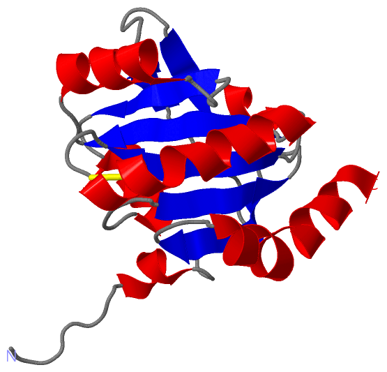
Asymmetric/Biological UnitChain A from PDB Type:PROTEIN Length:155 aligned with DSBE_PSEAE | Q9I3N1 from UniProtKB/Swiss-Prot Length:180 Alignment length:155 24 23 | | | 30 40 50 60 70 80 90 100 110 120 130 140 150 160 170 DSBE_PSEAE 22 RG-LWLDPSELPSALIGKPFPAFDLPSVQDPARRLTEADLKGKPALVNVWGTWCPSCRVEHPELTRLAEQGVVIYGINYKDDNAAAIKWLNELHNPYLLSISDADGTLGLDLGVYGAPETYLIDKQGIIRHKIVGVVDQKVWREQLAPLYQQLLD 175 SCOP domains ----------------------------------------------------------------------------------------------------------------------------------------------------------- SCOP domains CATH domains ----------------------------------------------------------------------------------------------------------------------------------------------------------- CATH domains Pfam domains --------------Redoxin-3kh9A01 A:35-172 --- Pfam domains SAPs(SNPs) ----------------------------------------------------------------------------------------------------------------------------------------------------------- SAPs(SNPs) PROSITE (1) -------------THIOREDOXIN_2 PDB: A:34-175 UniProt: 34-175 PROSITE (1) PROSITE (2) ---------------------------------------------THIOREDOXIN_1 ------------------------------------------------------------------------------------------- PROSITE (2) Transcript ----------------------------------------------------------------------------------------------------------------------------------------------------------- Transcript 3kh9 A 21 RGSHMLDPSELPSALIGKPFPAFDLPSVQDPARRLTEADLKGKPALVNVWGTWCPSCRVEHPELTRLAEQGVVIYGINYKDDNAAAIKWLNELHNPYLLSISDADGTLGLDLGVYGAPETYLIDKQGIIRHKIVGVVDQKVWREQLAPLYQQLLD 175 30 40 50 60 70 80 90 100 110 120 130 140 150 160 170
|
||||||||||||||||||||
SCOP Domains (0, 0)| (no "SCOP Domain" information available for 3KH9) |
CATH Domains (0, 0)| (no "CATH Domain" information available for 3KH9) |
Pfam Domains (1, 1)| Asymmetric/Biological Unit |
Gene Ontology (9, 9)|
Asymmetric/Biological Unit(hide GO term definitions) Chain A (DSBE_PSEAE | Q9I3N1)
|
||||||||||||||||||||||||||||||||||||||||||||||||||||||||||||||||||||||||
Interactive Views
|
|||||||||||||||||||||||||||||||||||||||||||||||||||||||||||||||||||||||||||||||||||||||||||||||||||||||||||||||||||||
Still Images
|
||||||||||||||||
Databases
|
||||||||||||||||||||||||||||||||||||||||||||||||||||||||||||||||||||||||||||||||||||||||||||||||||||||||||||||||||||||||||||||||||||||||||||||||||||||||||||||||
Analysis Tools
|
|||||||||||||||||||||||||||||||||||||||||||||||||||||||||||||
Entries Sharing at Least One Protein Chain (UniProt ID)
Related Entries Specified in the PDB File
|
|