Show PDB file:   
         Plain Text   HTML   (compressed file size)
QuickSearch:   
by PDB,NDB,UniProt,PROSITE Code or Search Term(s)  
(-)Asymmetric Unit
(-)Asym. Unit - sites
(-)Biological Unit 1
(-)Biol. Unit 1 - sites
(-)Biological Unit 2
collapse expand < >
Image Asymmetric Unit
Asymmetric Unit  (Jmol Viewer)
Image Asym. Unit - sites
Asym. Unit - sites  (Jmol Viewer)
Image Biological Unit 1
Biological Unit 1  (Jmol Viewer)
Image Biol. Unit 1 - sites
Biol. Unit 1 - sites  (Jmol Viewer)
Image Biological Unit 2
Biological Unit 2  (Jmol Viewer)

(-) Description

Title :  CRYSTAL STRUCTURE OF THE SDCEN/SKMLCK COMPLEX
 
Authors :  L. Radu, L. Assairi, Y. Blouquit, D. Durand, S. Miron, J. B. Charbonnier C. T. Craescu
Date :  27 Oct 09  (Deposition) - 26 Jan 11  (Release) - 26 Jan 11  (Revision)
Method :  X-RAY DIFFRACTION
Resolution :  2.60
Chains :  Asym. Unit :  A,B,C,D
Biol. Unit 1:  A,B  (1x)
Biol. Unit 2:  C,D  (1x)
Keywords :  Centrin, Myosin Light Chain Kinase, Cell Cycle, Cell Division, Mitosis, Calmodulin-Binding, Cell Cycle-Calcium-Binding Protein Complex (Keyword Search: [Gene Ontology, PubMed, Web (Google))
 
Reference :  L. Radu, S. Miron, D. Durand, L. Assairi, Y. Blouquit, J. B. Charbonnie
Structural Features Of The Complexes Formed By Scherffelia Dubia Centrin
To Be Published
PubMed: search

(-) Compounds

Molecule 1 - CALTRACTIN
    ChainsA, C
    EngineeredYES
    Expression SystemESCHERICHIA COLI
    Expression System PlasmidPET24A(+)
    Expression System StrainBL21(DE3)
    Expression System Taxid562
    Expression System Vector TypePLASMID
    FragmentUNP RESIDUES 20-168 OUT OF 168
    Organism ScientificSCHERFFELIA DUBIA
    Organism Taxid3190
    SynonymCENTRIN
 
Molecule 2 - MYOSIN LIGHT CHAIN KINASE 2, SKELETAL/CARDIAC MUSCLE
    ChainsB, D
    EngineeredYES
    FragmentUNP RESIDUES 566-587 OUT OF 596
    Other DetailsTHE PEPTIDE WAS CHEMICALLY SYNTHESIZED. THIS SEQUENCE OCCURS IN HOMO SAPIENS
    SynonymMLCK2
    SyntheticYES

 Structural Features

(-) Chains, Units

  1234
Asymmetric Unit ABCD
Biological Unit 1 (1x)AB  
Biological Unit 2 (1x)  CD

Summary Information (see also Sequences/Alignments below)

(-) Ligands, Modified Residues, Ions  (1, 8)

Asymmetric Unit (1, 8)
No.NameCountTypeFull Name
1CA8Ligand/IonCALCIUM ION
Biological Unit 1 (0, 0)
No.NameCountTypeFull Name
1CA-1Ligand/IonCALCIUM ION
Biological Unit 2 (0, 0)
No.NameCountTypeFull Name
1CA-1Ligand/IonCALCIUM ION

(-) Sites  (8, 8)

Asymmetric Unit (8, 8)
No.NameEvidenceResiduesDescription
1AC1SOFTWAREASP A:37 , ASP A:39 , SER A:41 , THR A:43 , GLU A:48 , HOH A:193BINDING SITE FOR RESIDUE CA A 301
2AC2SOFTWAREASP A:73 , ASP A:75 , SER A:77 , THR A:79 , ASP A:81 , GLU A:84BINDING SITE FOR RESIDUE CA A 302
3AC3SOFTWAREASP A:146 , ASP A:148 , ASP A:150 , GLU A:152 , GLU A:157 , HOH A:178BINDING SITE FOR RESIDUE CA A 304
4AC4SOFTWAREHOH A:17 , ASP A:110 , ASP A:112 , THR A:114 , LYS A:116 , ASN A:121BINDING SITE FOR RESIDUE CA A 303
5AC5SOFTWAREHOH C:12 , ASP C:73 , ASP C:75 , SER C:77 , THR C:79 , GLU C:84BINDING SITE FOR RESIDUE CA C 302
6AC6SOFTWAREHOH C:19 , ASP C:146 , ASP C:148 , ASP C:150 , GLU C:152 , GLU C:157BINDING SITE FOR RESIDUE CA C 304
7AC7SOFTWAREASP C:37 , ASP C:39 , SER C:41 , THR C:43 , GLU C:48 , HOH C:177BINDING SITE FOR RESIDUE CA C 301
8AC8SOFTWAREASP C:110 , ASP C:112 , THR C:114 , LYS C:116 , ASN C:121 , HOH C:187BINDING SITE FOR RESIDUE CA C 303

(-) SS Bonds  (0, 0)

(no "SS Bond" information available for 3KF9)

(-) Cis Peptide Bonds  (0, 0)

(no "Cis Peptide Bond" information available for 3KF9)

 Sequence-Structure Mapping

(-) SAPs(SNPs)/Variants  (0, 0)

(no "SAP(SNP)/Variant" information available for 3KF9)

(-) PROSITE Motifs  (2, 14)

Asymmetric Unit (2, 14)
 PROSITEUniProtKBPDB
No.IDACDescriptionIDLocationCountLocation
1EF_HAND_2PS50222 EF-hand calcium-binding domain profile.CATR_SCHDU24-59
 
60-95
 
97-132
 
133-168
 
  8A:24-59
C:24-59
A:60-95
C:60-95
A:97-132
C:97-132
A:133-168
C:133-168
2EF_HAND_1PS00018 EF-hand calcium-binding domain.CATR_SCHDU37-49
 
73-85
 
146-158
 
  6A:37-49
C:37-49
A:73-85
C:73-85
A:146-158
C:146-158
Biological Unit 1 (2, 7)
 PROSITEUniProtKBPDB
No.IDACDescriptionIDLocationCountLocation
1EF_HAND_2PS50222 EF-hand calcium-binding domain profile.CATR_SCHDU24-59
 
60-95
 
97-132
 
133-168
 
  4A:24-59
-
A:60-95
-
A:97-132
-
A:133-168
-
2EF_HAND_1PS00018 EF-hand calcium-binding domain.CATR_SCHDU37-49
 
73-85
 
146-158
 
  3A:37-49
-
A:73-85
-
A:146-158
-
Biological Unit 2 (2, 7)
 PROSITEUniProtKBPDB
No.IDACDescriptionIDLocationCountLocation
1EF_HAND_2PS50222 EF-hand calcium-binding domain profile.CATR_SCHDU24-59
 
60-95
 
97-132
 
133-168
 
  4-
C:24-59
-
C:60-95
-
C:97-132
-
C:133-168
2EF_HAND_1PS00018 EF-hand calcium-binding domain.CATR_SCHDU37-49
 
73-85
 
146-158
 
  3-
C:37-49
-
C:73-85
-
C:146-158

(-) Exons   (0, 0)

(no "Exon" information available for 3KF9)

(-) Sequences/Alignments

Asymmetric Unit
   Reformat: Number of residues per line =  ('0' or empty: single-line sequence representation)
  Number of residues per labelling interval =   
  UniProt sequence: complete  aligned part    
   Show mapping: SCOP domains CATH domains Pfam domains Secondary structure (by author)
SAPs(SNPs) PROSITE motifs Exons
(details for a mapped element are shown in a popup box when the mouse pointer rests over it)
Chain A from PDB  Type:PROTEIN  Length:149
 aligned with CATR_SCHDU | Q06827 from UniProtKB/Swiss-Prot  Length:168

    Alignment length:149
                                    29        39        49        59        69        79        89        99       109       119       129       139       149       159         
           CATR_SCHDU    20 GLTEEQKQEIREAFDLFDTDGSGTIDAKELKVAMRALGFEPKKEEIKKMIADIDKDGSGTIDFEEFLQMMTAKMGERDSREEIMKAFRLFDDDETGKISFKNLKRVAKELGENMTDEELQEMIDEADRDGDGEVNEEEFFRIMKKTSLF 168
               SCOP domains d3kf9a_ A: automated matches                                                                                                                          SCOP domains
               CATH domains ----------------------------------------------------------------------------------------------------------------------------------------------------- CATH domains
               Pfam domains ----------------------------------------------------------------------------------------------------------------------------------------------------- Pfam domains
         Sec.struct. author ..hhhhhhhhhhhhhhhh.....eehhhhhhhhhhhh....hhhhhhhhhhhhh......eehhhhhhhh....hhhhhhhhhhhhhhhhh.......hhhhhhhhhhhh....hhhhhhhhhhhhhh......hhhhhhhhh.hhhhh Sec.struct. author
                 SAPs(SNPs) ----------------------------------------------------------------------------------------------------------------------------------------------------- SAPs(SNPs)
                PROSITE (1) ----EF_HAND_2  PDB: A:24-59             EF_HAND_2  PDB: A:60-95             -EF_HAND_2  PDB: A:97-132            EF_HAND_2  PDB: A:133-168            PROSITE (1)
                PROSITE (2) -----------------EF_HAND_1    -----------------------EF_HAND_1    ------------------------------------------------------------EF_HAND_1    ---------- PROSITE (2)
                 Transcript ----------------------------------------------------------------------------------------------------------------------------------------------------- Transcript
                 3kf9 A  20 GLTEEQKQEIREAFDLFDTDGSGTIDAKELKVAMRALGFEPKKEEIKKMIADIDKDGSGTIDFEEFLQMMTAKMGERDSREEIMKAFRLFDDDETGKISFKNLKRVAKELGENMTDEELQEMIDEADRDGDGEVNEEEFFRIMKKTSLF 168
                                    29        39        49        59        69        79        89        99       109       119       129       139       149       159         

Chain B from PDB  Type:PROTEIN  Length:22
 aligned with MYLK2_HUMAN | Q9H1R3 from UniProtKB/Swiss-Prot  Length:596

    Alignment length:22
                                   575       585  
          MYLK2_HUMAN   566 KRRWKKNFIAVSAANRFKKISS 587
               SCOP domains ---------------------- SCOP domains
               CATH domains ---------------------- CATH domains
               Pfam domains ---------------------- Pfam domains
         Sec.struct. author hhhhhhhhhhhhhhhhhhhhhh Sec.struct. author
                 SAPs(SNPs) ---------------------- SAPs(SNPs)
                    PROSITE ---------------------- PROSITE
                 Transcript ---------------------- Transcript
                 3kf9 B   1 KRRWKKNFIAVSAANRFKKISS  22
                                    10        20  

Chain C from PDB  Type:PROTEIN  Length:149
 aligned with CATR_SCHDU | Q06827 from UniProtKB/Swiss-Prot  Length:168

    Alignment length:149
                                    29        39        49        59        69        79        89        99       109       119       129       139       149       159         
           CATR_SCHDU    20 GLTEEQKQEIREAFDLFDTDGSGTIDAKELKVAMRALGFEPKKEEIKKMIADIDKDGSGTIDFEEFLQMMTAKMGERDSREEIMKAFRLFDDDETGKISFKNLKRVAKELGENMTDEELQEMIDEADRDGDGEVNEEEFFRIMKKTSLF 168
               SCOP domains d3kf9c_ C: automated matches                                                                                                                          SCOP domains
               CATH domains ----------------------------------------------------------------------------------------------------------------------------------------------------- CATH domains
           Pfam domains (1) --------EF_hand_5-3kf9C01 C:28-89                                     -----------------------EF_hand_6-3kf9C03 C:113-165                          --- Pfam domains (1)
           Pfam domains (2) --------EF_hand_5-3kf9C02 C:28-89                                     -----------------------EF_hand_6-3kf9C04 C:113-165                          --- Pfam domains (2)
           Pfam domains (3) --------------------------------------------efhand-3kf9C05 C:64-92       ---------------------------------------------------------------------------- Pfam domains (3)
           Pfam domains (4) --------------------------------------------efhand-3kf9C06 C:64-92       ---------------------------------------------------------------------------- Pfam domains (4)
         Sec.struct. author ..hhhhhhhhhhhhhhhh.....eehhhhhhhhhhhhh...hhhhhhhhhhhhh......eehhhhhhhh....hhhhhhhhhhhhhhhhh.....eehhhhhhhhhhhh....hhhhhhhhhhhhh......eehhhhhhhh.hhhhh Sec.struct. author
                 SAPs(SNPs) ----------------------------------------------------------------------------------------------------------------------------------------------------- SAPs(SNPs)
                PROSITE (1) ----EF_HAND_2  PDB: C:24-59             EF_HAND_2  PDB: C:60-95             -EF_HAND_2  PDB: C:97-132            EF_HAND_2  PDB: C:133-168            PROSITE (1)
                PROSITE (2) -----------------EF_HAND_1    -----------------------EF_HAND_1    ------------------------------------------------------------EF_HAND_1    ---------- PROSITE (2)
                 Transcript ----------------------------------------------------------------------------------------------------------------------------------------------------- Transcript
                 3kf9 C  20 GLTEEQKQEIREAFDLFDTDGSGTIDAKELKVAMRALGFEPKKEEIKKMIADIDKDGSGTIDFEEFLQMMTAKMGERDSREEIMKAFRLFDDDETGKISFKNLKRVAKELGENMTDEELQEMIDEADRDGDGEVNEEEFFRIMKKTSLF 168
                                    29        39        49        59        69        79        89        99       109       119       129       139       149       159         

Chain D from PDB  Type:PROTEIN  Length:21
 aligned with MYLK2_HUMAN | Q9H1R3 from UniProtKB/Swiss-Prot  Length:596

    Alignment length:21
                                   576       586 
          MYLK2_HUMAN   567 RRWKKNFIAVSAANRFKKISS 587
               SCOP domains --------------------- SCOP domains
               CATH domains --------------------- CATH domains
               Pfam domains --------------------- Pfam domains
         Sec.struct. author hhhhhhhhhhhhhhhhhhhhh Sec.struct. author
                 SAPs(SNPs) --------------------- SAPs(SNPs)
                    PROSITE --------------------- PROSITE
                 Transcript --------------------- Transcript
                 3kf9 D   2 RRWKKNFIAVSAANRFKKISS  22
                                    11        21 

   Legend:   → Mismatch (orange background)
  - → Gap (green background, '-', border residues have a numbering label)
    → Modified Residue (blue background, lower-case, 'x' indicates undefined single-letter code, labelled with number + name)
  x → Chemical Group (purple background, 'x', labelled with number + name, e.g. ACE or NH2)
  extra numbering lines below/above indicate numbering irregularities and modified residue names etc., number ends below/above '|'

 Classification and Annotation

(-) SCOP Domains  (1, 2)

Asymmetric Unit

(-) CATH Domains  (0, 0)

(no "CATH Domain" information available for 3KF9)

(-) Pfam Domains  (3, 6)

Asymmetric Unit
(-)
Clan: EF_hand (270)

(-) Gene Ontology  (29, 29)

Asymmetric Unit(hide GO term definitions)
Chain A,C   (CATR_SCHDU | Q06827)
molecular function
    GO:0005509    calcium ion binding    Interacting selectively and non-covalently with calcium ions (Ca2+).
    GO:0046872    metal ion binding    Interacting selectively and non-covalently with any metal ion.
biological process
    GO:0007049    cell cycle    The progression of biochemical and morphological phases and events that occur in a cell during successive cell replication or nuclear replication events. Canonically, the cell cycle comprises the replication and segregation of genetic material followed by the division of the cell, but in endocycles or syncytial cells nuclear replication or nuclear division may not be followed by cell division.
    GO:0051301    cell division    The process resulting in division and partitioning of components of a cell to form more cells; may or may not be accompanied by the physical separation of a cell into distinct, individually membrane-bounded daughter cells.

Chain B,D   (MYLK2_HUMAN | Q9H1R3)
molecular function
    GO:0005524    ATP binding    Interacting selectively and non-covalently with ATP, adenosine 5'-triphosphate, a universally important coenzyme and enzyme regulator.
    GO:0005516    calmodulin binding    Interacting selectively and non-covalently with calmodulin, a calcium-binding protein with many roles, both in the calcium-bound and calcium-free states.
    GO:0004683    calmodulin-dependent protein kinase activity    Catalysis of the reactions: ATP + a protein serine = ADP + protein serine phosphate; and ATP + a protein threonine = ADP + protein threonine phosphate. These reactions require the presence of calcium-bound calmodulin.
    GO:0016301    kinase activity    Catalysis of the transfer of a phosphate group, usually from ATP, to a substrate molecule.
    GO:0004687    myosin light chain kinase activity    Catalysis of the reaction: ATP + myosin-light-chain = ADP + myosin-light-chain phosphate.
    GO:0000166    nucleotide binding    Interacting selectively and non-covalently with a nucleotide, any compound consisting of a nucleoside that is esterified with (ortho)phosphate or an oligophosphate at any hydroxyl group on the ribose or deoxyribose.
    GO:0005515    protein binding    Interacting selectively and non-covalently with any protein or protein complex (a complex of two or more proteins that may include other nonprotein molecules).
    GO:0004672    protein kinase activity    Catalysis of the phosphorylation of an amino acid residue in a protein, usually according to the reaction: a protein + ATP = a phosphoprotein + ADP.
    GO:0004674    protein serine/threonine kinase activity    Catalysis of the reactions: ATP + protein serine = ADP + protein serine phosphate, and ATP + protein threonine = ADP + protein threonine phosphate.
    GO:0016740    transferase activity    Catalysis of the transfer of a group, e.g. a methyl group, glycosyl group, acyl group, phosphorus-containing, or other groups, from one compound (generally regarded as the donor) to another compound (generally regarded as the acceptor). Transferase is the systematic name for any enzyme of EC class 2.
biological process
    GO:0060048    cardiac muscle contraction    Muscle contraction of cardiac muscle tissue.
    GO:0055008    cardiac muscle tissue morphogenesis    The process in which the anatomical structures of cardiac muscle tissue are generated and organized.
    GO:0007274    neuromuscular synaptic transmission    The process of synaptic transmission from a neuron to a muscle, across a synapse.
    GO:0018107    peptidyl-threonine phosphorylation    The phosphorylation of peptidyl-threonine to form peptidyl-O-phospho-L-threonine.
    GO:0016310    phosphorylation    The process of introducing a phosphate group into a molecule, usually with the formation of a phosphoric ester, a phosphoric anhydride or a phosphoric amide.
    GO:0010628    positive regulation of gene expression    Any process that increases the frequency, rate or extent of gene expression. Gene expression is the process in which a gene's coding sequence is converted into a mature gene product or products (proteins or RNA). This includes the production of an RNA transcript as well as any processing to produce a mature RNA product or an mRNA or circRNA (for protein-coding genes) and the translation of that mRNA or circRNA into protein. Protein maturation is included when required to form an active form of a product from an inactive precursor form.
    GO:0046777    protein autophosphorylation    The phosphorylation by a protein of one or more of its own amino acid residues (cis-autophosphorylation), or residues on an identical protein (trans-autophosphorylation).
    GO:0006468    protein phosphorylation    The process of introducing a phosphate group on to a protein.
    GO:0032971    regulation of muscle filament sliding    Any process that modulates the frequency, rate or extent of muscle filament sliding.
    GO:0035914    skeletal muscle cell differentiation    The process in which a relatively unspecialized cell acquires specialized features of a skeletal muscle cell, a somatic cell located in skeletal muscle.
    GO:0014816    skeletal muscle satellite cell differentiation    The process in which a relatively unspecialized cell acquires specialized features of a satellite cell.
    GO:0006941    striated muscle contraction    A process in which force is generated within striated muscle tissue, resulting in the shortening of the muscle. Force generation involves a chemo-mechanical energy conversion step that is carried out by the actin/myosin complex activity, which generates force through ATP hydrolysis. Striated muscle is a type of muscle in which the repeating units (sarcomeres) of the contractile myofibrils are arranged in registry throughout the cell, resulting in transverse or oblique striations observable at the level of the light microscope.
cellular component
    GO:0005737    cytoplasm    All of the contents of a cell excluding the plasma membrane and nucleus, but including other subcellular structures.
    GO:0005634    nucleus    A membrane-bounded organelle of eukaryotic cells in which chromosomes are housed and replicated. In most cells, the nucleus contains all of the cell's chromosomes except the organellar chromosomes, and is the site of RNA synthesis and processing. In some species, or in specialized cell types, RNA metabolism or DNA replication may be absent.
    GO:0030017    sarcomere    The repeating unit of a myofibril in a muscle cell, composed of an array of overlapping thick and thin filaments between two adjacent Z discs.
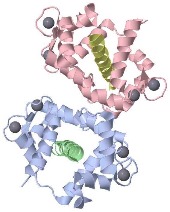
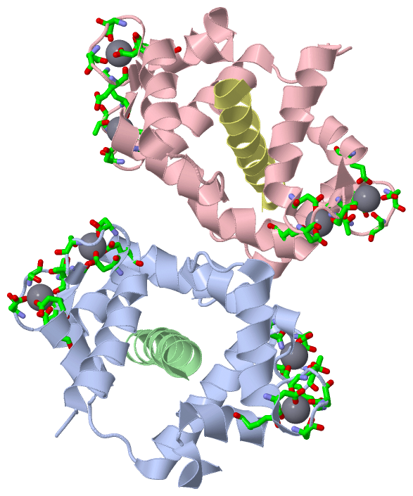
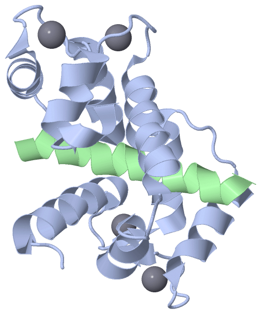
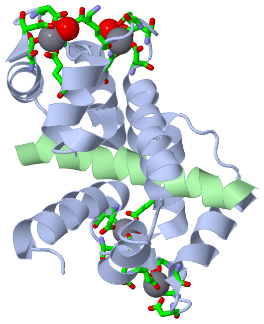
 Visualization

(-) Interactive Views

Asymmetric Unit
  Complete Structure
    Jena3D(integrated viewing of ligand, site, SAP, PROSITE, SCOP information)
    WebMol | AstexViewer[tm]@PDBe
(Java Applets, require no local installation except for Java; loading may be slow)
    STRAP
(Java WebStart application, automatic local installation, requires Java; full application with system access!)
    RasMol
(require local installation)
    Molscript (VRML)
(requires installation of a VRML viewer; select preferred view via VRML and generate a mono or stereo PDF format file)
 
  Ligands, Modified Residues, Ions
    CA  [ RasMol | Jena3D ]  +environment [ RasMol | Jena3D ]
 
  Sites
    AC1  [ RasMol ]  +environment [ RasMol ]
    AC2  [ RasMol ]  +environment [ RasMol ]
    AC3  [ RasMol ]  +environment [ RasMol ]
    AC4  [ RasMol ]  +environment [ RasMol ]
    AC5  [ RasMol ]  +environment [ RasMol ]
    AC6  [ RasMol ]  +environment [ RasMol ]
    AC7  [ RasMol ]  +environment [ RasMol ]
    AC8  [ RasMol ]  +environment [ RasMol ]
 
  Cis Peptide Bonds
(no "Cis Peptide Bonds" information available for 3kf9)
 
Biological Units
  Complete Structure
    Biological Unit 1  [ Jena3D ]
    Biological Unit 2  [ Jena3D ]

(-) Still Images

Jmol
  protein: cartoon or spacefill or dots and stick; nucleic acid: cartoon and stick; ligands: spacefill; active site: stick
Molscript
  protein, nucleic acid: cartoon; ligands: spacefill; active site: ball and stick

 Databases and Analysis Tools

(-) Databases

Access by PDB/NDB ID
  3kf9
    Family and Domain InformationProDom | SYSTERS
    General Structural InformationGlycoscienceDB | MMDB | NDB | OCA | PDB | PDBe | PDBj | PDBsum | PDBWiki | PQS | PROTEOPEDIA
    Orientation in MembranesOPM
    Protein SurfaceSURFACE
    Secondary StructureDSSP (structure derived) | HSSP (homology derived)
    Structural GenomicsGeneCensus
    Structural NeighboursCE | VAST
    Structure ClassificationCATH | Dali | SCOP
    Validation and Original DataBMRB Data View | BMRB Restraints Grid | EDS | PROCHECK | RECOORD | WHAT_CHECK
 
Access by UniProt ID/Accession number
  CATR_SCHDU | Q06827
    Comparative Protein Structure ModelsModBase
    Genomic InformationEnsembl
    Protein-protein InteractionDIP
    Sequence, Family and Domain InformationInterPro | Pfam | SMART | UniProtKB/SwissProt
  MYLK2_HUMAN | Q9H1R3
    Comparative Protein Structure ModelsModBase
    Genomic InformationEnsembl
    Protein-protein InteractionDIP
    Sequence, Family and Domain InformationInterPro | Pfam | SMART | UniProtKB/SwissProt
 
Access by Enzyme Classificator   (EC Number)
  (no 'Enzyme Classificator' available)
    General Enzyme InformationBRENDA | EC-PDB | Enzyme | IntEnz
    PathwayKEGG | MetaCyc
 
Access by Disease Identifier   (MIM ID)
  (no 'MIM ID' available)
    Disease InformationOMIM
 
Access by GenAge ID
  (no 'GenAge ID' available)
    Age Related InformationGenAge

(-) Analysis Tools

Access by PDB/NDB ID
    Domain InformationXDom
    Interatomic Contacts of Structural UnitsCSU
    Ligand-protein ContactsLPC
    Protein CavitiescastP
    Sequence and Secondary StructurePDBCartoon
    Structure AlignmentSTRAP(Java WebStart application, automatic local installation, requires Java; full application with system access!)
    Structure and Sequence BrowserSTING
 
Access by UniProt ID/Accession number
  CATR_SCHDU | Q06827
    Protein Disorder PredictionDisEMBL | FoldIndex | GLOBPLOT (for more information see DisProt)
  MYLK2_HUMAN | Q9H1R3
    Protein Disorder PredictionDisEMBL | FoldIndex | GLOBPLOT (for more information see DisProt)

 Related Entries

(-) Entries Sharing at Least One Protein Chain (UniProt ID)

UniProtKB/Swiss-Prot
        MYLK2_HUMAN | Q9H1R32lv6

(-) Related Entries Specified in the PDB File

(no "Related Entries Specified in the PDB File" available for 3KF9)