Show PDB file:   
         Plain Text   HTML   (compressed file size)
QuickSearch:   
by PDB,NDB,UniProt,PROSITE Code or Search Term(s)  
(-)Asymmetric Unit
(-)Biological Unit 1
(-)Biological Unit 2
collapse expand < >
Image Asymmetric Unit
Asymmetric Unit  (Jmol Viewer)
Image Biological Unit 1
Biological Unit 1  (Jmol Viewer)
Image Biological Unit 2
Biological Unit 2  (Jmol Viewer)

(-) Description

Title :  PRY-SPRY DOMAIN OF HUMAN TRIM72
 
Authors :  E. Y. Park, O. -B. Kwon, B. -C. Jeong, H. K. Song
Date :  20 Oct 09  (Deposition) - 22 Dec 09  (Release) - 22 Dec 09  (Revision)
Method :  X-RAY DIFFRACTION
Resolution :  1.50
Chains :  Asym. Unit :  A
Biol. Unit 1:  A  (1x)
Biol. Unit 2:  A  (2x)
Keywords :  B30. 2, Gustavus, Spry, Trim21, Tripartite Motif, Trim72, Pry, High Resolution, Mg53 (Keyword Search: [Gene Ontology, PubMed, Web (Google))
 
Reference :  E. Y. Park, O. -B. Kwon, B. -C. Jeong, J. -S. Yi, C. S. Lee, Y. -G. Ko, H. K. Song
Crystal Structure Of Pry-Spry Domain Of Human Trim72
Proteins 2009
PubMed-ID: 19967786  |  Reference-DOI: 10.1002/PROT.22647
(for further references see the PDB file header)

(-) Compounds

Molecule 1 - TRIPARTITE MOTIF-CONTAINING PROTEIN 72
    ChainsA
    EngineeredYES
    Expression SystemESCHERICHIA COLI
    Expression System Taxid562
    Expression System Vector TypePLASMID
    FragmentPRY-SPRY DOMAIN, UNP RESIDUES 278-470
    GenePRY-SPRY
    Organism CommonHUMAN
    Organism ScientificHOMO SAPIENS
    Organism Taxid9606
    SynonymMITSUGUMIN-53, MG53

 Structural Features

(-) Chains, Units

  1
Asymmetric Unit A
Biological Unit 1 (1x)A
Biological Unit 2 (2x)A

Summary Information (see also Sequences/Alignments below)

(-) Ligands, Modified Residues, Ions  (0, 0)

(no "Ligand,Modified Residues,Ions" information available for 3KB5)

(-) Sites  (0, 0)

(no "Site" information available for 3KB5)

(-) SS Bonds  (0, 0)

(no "SS Bond" information available for 3KB5)

(-) Cis Peptide Bonds  (2, 2)

Asymmetric Unit
No.Residues
1Ser A:405 -Pro A:406
2Arg A:447 -Pro A:448

 Sequence-Structure Mapping

(-) SAPs(SNPs)/Variants  (0, 0)

(no "SAP(SNP)/Variant" information available for 3KB5)

(-) PROSITE Motifs  (0, 0)

(no "PROSITE Motif" information available for 3KB5)

(-) Exons   (0, 0)

(no "Exon" information available for 3KB5)

(-) Sequences/Alignments

Asymmetric Unit
   Reformat: Number of residues per line =  ('0' or empty: single-line sequence representation)
  Number of residues per labelling interval =   
  UniProt sequence: complete  aligned part    
   Show mapping: SCOP domains CATH domains Pfam domains Secondary structure (by author)
SAPs(SNPs) PROSITE motifs Exons
(details for a mapped element are shown in a popup box when the mouse pointer rests over it)
Chain A from PDB  Type:PROTEIN  Length:193
 aligned with TRI72_HUMAN | Q6ZMU5 from UniProtKB/Swiss-Prot  Length:477

    Alignment length:193
                                   287       297       307       317       327       337       347       357       367       377       387       397       407       417       427       437       447       457       467   
          TRI72_HUMAN   278 RKMFRALMPALEELTFDPSSAHPSLVVSSSGRRVECSEQKAPPAGEDPRQFDKAVAVVAHQQLSEGEHYWEVDVGDKPRWALGVIAAEAPRRGRLHAVPSQGLWLLGLREGKILEAHVEAKEPRALRSPERRPTRIGLYLSFGDGVLSFYDASDADALVPLFAFHERLPRPVYPFFDVCWHDKGKNAQPLLLV 470
               SCOP domains ------------------------------------------------------------------------------------------------------------------------------------------------------------------------------------------------- SCOP domains
               CATH domains ------------------------------------------------------------------------------------------------------------------------------------------------------------------------------------------------- CATH domains
               Pfam domains -------------PRY-3kb5A01 A:291-340                             -SPRY-3kb5A02 A:342-470                                                                                                            Pfam domains
         Sec.struct. author hhhhhhhh................eeee....eeee...................eee........eeeeeee......eeeeeee...........hhhh.eeeeeee...eeee.......ee.........eeeeeee....eeeeee.......eeeeee.......eeeeee............eee. Sec.struct. author
                 SAPs(SNPs) ------------------------------------------------------------------------------------------------------------------------------------------------------------------------------------------------- SAPs(SNPs)
                    PROSITE ------------------------------------------------------------------------------------------------------------------------------------------------------------------------------------------------- PROSITE
                 Transcript ------------------------------------------------------------------------------------------------------------------------------------------------------------------------------------------------- Transcript
                 3kb5 A 278 RKMFRALMPALEELTFDPSSAHPSLVVSSSGRRVECSEQKAPPAGEDPRQFDKAVAVVAHQQLSEGEHYWEVDVGDKPRWALGVIAAEAPRRGRLHAVPSQGLWLLGLREGKILEAHVEAKEPRALRSPERRPTRIGLYLSFGDGVLSFYDASDADALVPLFAFHERLPRPVYPFFDVCWHDKGKNAQPLLLV 470
                                   287       297       307       317       327       337       347       357       367       377       387       397       407       417       427       437       447       457       467   

   Legend:   → Mismatch (orange background)
  - → Gap (green background, '-', border residues have a numbering label)
    → Modified Residue (blue background, lower-case, 'x' indicates undefined single-letter code, labelled with number + name)
  x → Chemical Group (purple background, 'x', labelled with number + name, e.g. ACE or NH2)
  extra numbering lines below/above indicate numbering irregularities and modified residue names etc., number ends below/above '|'

 Classification and Annotation

(-) SCOP Domains  (0, 0)

(no "SCOP Domain" information available for 3KB5)

(-) CATH Domains  (0, 0)

(no "CATH Domain" information available for 3KB5)

(-) Pfam Domains  (2, 2)

Asymmetric Unit

(-) Gene Ontology  (24, 24)

Asymmetric Unit(hide GO term definitions)
Chain A   (TRI72_HUMAN | Q6ZMU5)
molecular function
    GO:0046872    metal ion binding    Interacting selectively and non-covalently with any metal ion.
    GO:0001786    phosphatidylserine binding    Interacting selectively and non-covalently with phosphatidylserine, a class of glycophospholipids in which a phosphatidyl group is esterified to the hydroxyl group of L-serine.
    GO:0005515    protein binding    Interacting selectively and non-covalently with any protein or protein complex (a complex of two or more proteins that may include other nonprotein molecules).
    GO:0031624    ubiquitin conjugating enzyme binding    Interacting selectively and non-covalently with a ubiquitin conjugating enzyme, any of the E2 proteins.
    GO:0061630    ubiquitin protein ligase activity    Catalysis of the transfer of ubiquitin to a substrate protein via the reaction X-ubiquitin + S -> X + S-ubiquitin, where X is either an E2 or E3 enzyme, the X-ubiquitin linkage is a thioester bond, and the S-ubiquitin linkage is an amide bond: an isopeptide bond between the C-terminal glycine of ubiquitin and the epsilon-amino group of lysine residues in the substrate or, in the linear extension of ubiquitin chains, a peptide bond the between the C-terminal glycine and N-terminal methionine of ubiquitin residues.
    GO:0008270    zinc ion binding    Interacting selectively and non-covalently with zinc (Zn) ions.
biological process
    GO:0006887    exocytosis    A process of secretion by a cell that results in the release of intracellular molecules (e.g. hormones, matrix proteins) contained within a membrane-bounded vesicle. Exocytosis can occur either by full fusion, when the vesicle collapses into the plasma membrane, or by a kiss-and-run mechanism that involves the formation of a transient contact, a pore, between a granule (for exemple of chromaffin cells) and the plasma membrane. The latter process most of the time leads to only partial secretion of the granule content. Exocytosis begins with steps that prepare vesicles for fusion with the membrane (tethering and docking) and ends when molecules are secreted from the cell.
    GO:0006936    muscle contraction    A process in which force is generated within muscle tissue, resulting in a change in muscle geometry. Force generation involves a chemo-mechanical energy conversion step that is carried out by the actin/myosin complex activity, which generates force through ATP hydrolysis.
    GO:0007517    muscle organ development    The process whose specific outcome is the progression of the muscle over time, from its formation to the mature structure. The muscle is an organ consisting of a tissue made up of various elongated cells that are specialized to contract and thus to produce movement and mechanical work.
    GO:0003012    muscle system process    A organ system process carried out at the level of a muscle. Muscle tissue is composed of contractile cells or fibers.
    GO:0046627    negative regulation of insulin receptor signaling pathway    Any process that stops, prevents, or reduces the frequency, rate or extent of insulin receptor signaling.
    GO:0043569    negative regulation of insulin-like growth factor receptor signaling pathway    Any process that stops, prevents, or reduces the frequency, rate or extent of insulin-like growth factor receptor signaling.
    GO:0010832    negative regulation of myotube differentiation    Any process that decreases the frequency, rate or extent of myotube differentiation. Myotube differentiation is the process in which a relatively unspecialized cell acquires specialized features of a myotube cell. Myotubes are multinucleated cells that are formed when proliferating myoblasts exit the cell cycle, differentiate and fuse.
    GO:0001778    plasma membrane repair    The resealing of a cell plasma membrane after cellular wounding due to, for instance, mechanical stress.
    GO:0043161    proteasome-mediated ubiquitin-dependent protein catabolic process    The chemical reactions and pathways resulting in the breakdown of a protein or peptide by hydrolysis of its peptide bonds, initiated by the covalent attachment of ubiquitin, and mediated by the proteasome.
    GO:0051260    protein homooligomerization    The process of creating protein oligomers, compounds composed of a small number, usually between three and ten, of identical component monomers. Oligomers may be formed by the polymerization of a number of monomers or the depolymerization of a large protein polymer.
    GO:0016567    protein ubiquitination    The process in which one or more ubiquitin groups are added to a protein.
    GO:0006810    transport    The directed movement of substances (such as macromolecules, small molecules, ions) or cellular components (such as complexes and organelles) into, out of or within a cell, or between cells, or within a multicellular organism by means of some agent such as a transporter, pore or motor protein.
cellular component
    GO:0031410    cytoplasmic vesicle    A vesicle found in the cytoplasm of a cell.
    GO:0030659    cytoplasmic vesicle membrane    The lipid bilayer surrounding a cytoplasmic vesicle.
    GO:0005622    intracellular    The living contents of a cell; the matter contained within (but not including) the plasma membrane, usually taken to exclude large vacuoles and masses of secretory or ingested material. In eukaryotes it includes the nucleus and cytoplasm.
    GO:0016020    membrane    A lipid bilayer along with all the proteins and protein complexes embedded in it an attached to it.
    GO:0005886    plasma membrane    The membrane surrounding a cell that separates the cell from its external environment. It consists of a phospholipid bilayer and associated proteins.
    GO:0042383    sarcolemma    The outer membrane of a muscle cell, consisting of the plasma membrane, a covering basement membrane (about 100 nm thick and sometimes common to more than one fiber), and the associated loose network of collagen fibers.
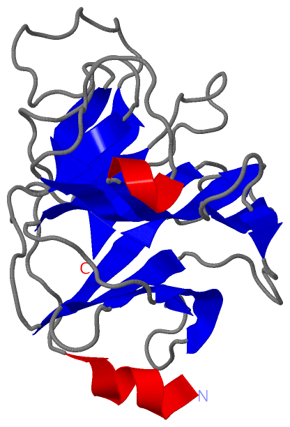
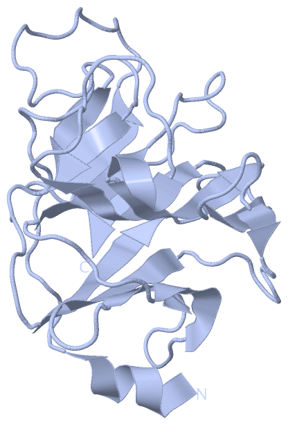
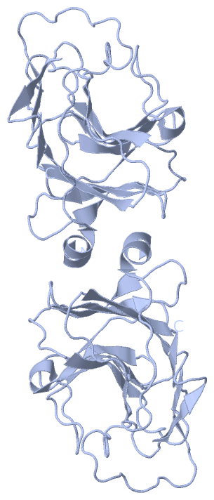
 Visualization

(-) Interactive Views

Asymmetric Unit
  Complete Structure
    Jena3D(integrated viewing of ligand, site, SAP, PROSITE, SCOP information)
    WebMol | AstexViewer[tm]@PDBe
(Java Applets, require no local installation except for Java; loading may be slow)
    STRAP
(Java WebStart application, automatic local installation, requires Java; full application with system access!)
    RasMol
(require local installation)
    Molscript (VRML)
(requires installation of a VRML viewer; select preferred view via VRML and generate a mono or stereo PDF format file)
 
  Ligands, Modified Residues, Ions
(no "Ligands, Modified Residues, Ions" information available for 3kb5)
 
  Sites
(no "Sites" information available for 3kb5)
 
  Cis Peptide Bonds
    Arg A:447 - Pro A:448   [ RasMol ]  
    Ser A:405 - Pro A:406   [ RasMol ]  
 
Biological Units
  Complete Structure
    Biological Unit 1  [ Jena3D ]
    Biological Unit 2  [ Jena3D ]

(-) Still Images

Jmol
  protein: cartoon or spacefill or dots and stick; nucleic acid: cartoon and stick; ligands: spacefill; active site: stick
Molscript
  protein, nucleic acid: cartoon; ligands: spacefill; active site: ball and stick

 Databases and Analysis Tools

(-) Databases

Access by PDB/NDB ID
  3kb5
    Family and Domain InformationProDom | SYSTERS
    General Structural InformationGlycoscienceDB | MMDB | NDB | OCA | PDB | PDBe | PDBj | PDBsum | PDBWiki | PQS | PROTEOPEDIA
    Orientation in MembranesOPM
    Protein SurfaceSURFACE
    Secondary StructureDSSP (structure derived) | HSSP (homology derived)
    Structural GenomicsGeneCensus
    Structural NeighboursCE | VAST
    Structure ClassificationCATH | Dali | SCOP
    Validation and Original DataBMRB Data View | BMRB Restraints Grid | EDS | PROCHECK | RECOORD | WHAT_CHECK
 
Access by UniProt ID/Accession number
  TRI72_HUMAN | Q6ZMU5
    Comparative Protein Structure ModelsModBase
    Genomic InformationEnsembl
    Protein-protein InteractionDIP
    Sequence, Family and Domain InformationInterPro | Pfam | SMART | UniProtKB/SwissProt
 
Access by Enzyme Classificator   (EC Number)
  (no 'Enzyme Classificator' available)
    General Enzyme InformationBRENDA | EC-PDB | Enzyme | IntEnz
    PathwayKEGG | MetaCyc
 
Access by Disease Identifier   (MIM ID)
  (no 'MIM ID' available)
    Disease InformationOMIM
 
Access by GenAge ID
  (no 'GenAge ID' available)
    Age Related InformationGenAge

(-) Analysis Tools

Access by PDB/NDB ID
    Domain InformationXDom
    Interatomic Contacts of Structural UnitsCSU
    Ligand-protein ContactsLPC
    Protein CavitiescastP
    Sequence and Secondary StructurePDBCartoon
    Structure AlignmentSTRAP(Java WebStart application, automatic local installation, requires Java; full application with system access!)
    Structure and Sequence BrowserSTING
 
Access by UniProt ID/Accession number
  TRI72_HUMAN | Q6ZMU5
    Protein Disorder PredictionDisEMBL | FoldIndex | GLOBPLOT (for more information see DisProt)

 Related Entries

(-) Entries Sharing at Least One Protein Chain (UniProt ID)

(no "Entries Sharing at Least One Protein Chain" available for 3KB5)

(-) Related Entries Specified in the PDB File

(no "Related Entries Specified in the PDB File" available for 3KB5)