|
|
|
|
 Description
|
|
Compounds
|
||||||||||||||||||||||||||||||||||||||||||||||||||||||||||||||
Chains, Units
Summary Information (see also Sequences/Alignments below) |
Ligands, Modified Residues, Ions (0, 0)| (no "Ligand,Modified Residues,Ions" information available for 3K7U) |
Sites (0, 0)| (no "Site" information available for 3K7U) |
SS Bonds (1, 1)
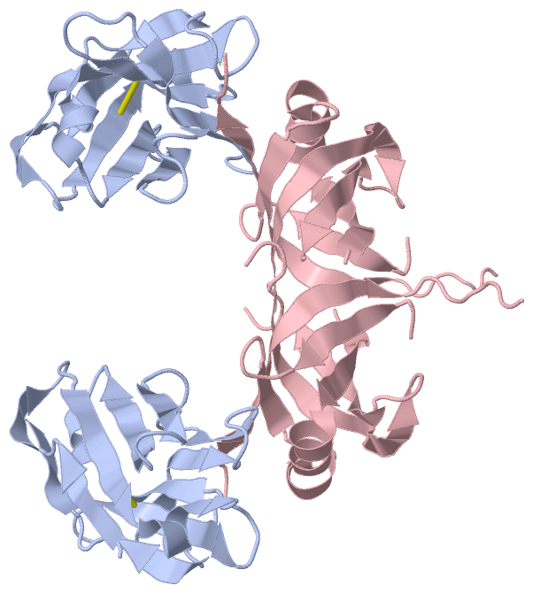
Asymmetric Unit
|
||||||||
Cis Peptide Bonds (2, 2)
Asymmetric Unit
|
||||||||||||
SAPs(SNPs)/Variants (0, 0)| (no "SAP(SNP)/Variant" information available for 3K7U) |
PROSITE Motifs (0, 0)| (no "PROSITE Motif" information available for 3K7U) |
Exons (0, 0)| (no "Exon" information available for 3K7U) |
Sequences/Alignments
Asymmetric Unit
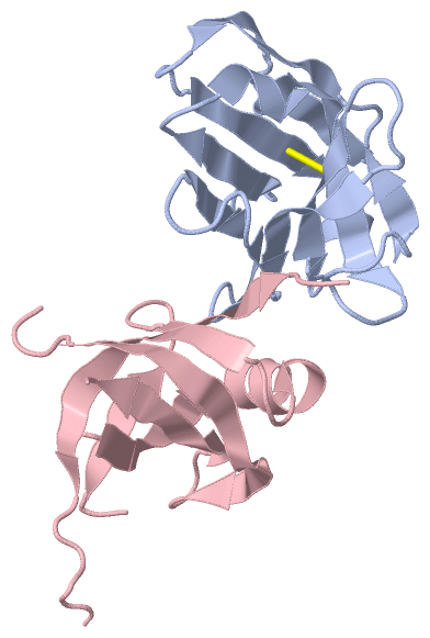
Chain A from PDB Type:PROTEIN Length:119
SCOP domains d3k7ua_ A: automated matches SCOP domains
CATH domains ----------------------------------------------------------------------------------------------------------------------- CATH domains
Pfam domains ----------------------------------------------------------------------------------------------------------------------- Pfam domains
SAPs(SNPs) ----------------------------------------------------------------------------------------------------------------------- SAPs(SNPs)
PROSITE ----------------------------------------------------------------------------------------------------------------------- PROSITE
Transcript ----------------------------------------------------------------------------------------------------------------------- Transcript
3k7u A 3 QLQESGGGLVQAGGSLTLSCAASGRTFSNNAMGWFRQAPGKEREFVAAISWTGGLLFYADSVNGRFTISRDNAKRTVTLQMNSLKPEDTAVYYCAARPQGDYVTAHYDYWGQGTQVTVS 121
12 22 32 42 52 62 72 82 92 102 112
Chain C from PDB Type:PROTEIN Length:98 aligned with Q38B90_TRYB2 | Q38B90 from UniProtKB/TrEMBL Length:164 Alignment length:114 30 40 50 60 70 80 90 100 110 120 130 Q38B90_TRYB2 21 SVNSVTLVGVVHDIQSGFVYEDAVTQFTLTTTSIDTTHPTQEVVVEKDHHTIRCFGELFSAEVKQKVKEGNVVCVNGRLRLSPQLEPSCNKHFYFPYIQVQPPHGQVAVIHGDR 134 SCOP domains ------------------------------------------------------------------------------------------------------------------ SCOP domains CATH domains ------------------------------------------------------------------------------------------------------------------ CATH domains Pfam domains ------------------------------------------------------------------------------------------------------------------ Pfam domains SAPs(SNPs) ------------------------------------------------------------------------------------------------------------------ SAPs(SNPs) PROSITE ------------------------------------------------------------------------------------------------------------------ PROSITE Transcript ------------------------------------------------------------------------------------------------------------------ Transcript 3k7u C 21 SVNSVTLVGVVHDIQSGFVYEDAVTQFTLTTTSI-----------EKDHHTIRCFGELFSAEVKQKVKEGNVVCVNGRLRLSPQLEPS-----YFPYIQVQPPHGQVAVIHGDR 134 30 40 50 | - | 70 80 90 100 | - | 120 130 54 66 108 114
|
||||||||||||||||||||
SCOP Domains (1, 1)
Asymmetric Unit
 |
CATH Domains (0, 0)| (no "CATH Domain" information available for 3K7U) |
Pfam Domains (0, 0)| (no "Pfam Domain" information available for 3K7U) |
Gene Ontology (7, 7)|
Asymmetric Unit(hide GO term definitions) Chain C (Q38B90_TRYB2 | Q38B90)
|
||||||||||||||||||||||||||||||||||||||||||||||||||||||||||||
Interactive Views
|
||||||||||||||||||||||||||||||||||||||||||||||||||||||||||||||||||||||||||||||||||||||||||||||||||||||||||||||||||||||||||||||||||||||||||||||
Still Images
|
||||||||||||||||
Databases
|
||||||||||||||||||||||||||||||||||||||||||||||||||||||||||||||||||||||||||||||||||||||||||||||||||||||||||||||||||||||||||||||||||||||||||||||||||||||||||||||||
Analysis Tools
|
|||||||||||||||||||||||||||||||||||||||||||||||||||||||||||||
Entries Sharing at Least One Protein Chain (UniProt ID)
Related Entries Specified in the PDB File
|
|