| molecular function |
|---|
| | GO:0005524 | | ATP binding | | Interacting selectively and non-covalently with ATP, adenosine 5'-triphosphate, a universally important coenzyme and enzyme regulator. |
| | GO:0016301 | | kinase activity | | Catalysis of the transfer of a phosphate group, usually from ATP, to a substrate molecule. |
| | GO:0000287 | | magnesium ion binding | | Interacting selectively and non-covalently with magnesium (Mg) ions. |
| | GO:0030145 | | manganese ion binding | | Interacting selectively and non-covalently with manganese (Mn) ions. |
| | GO:0000166 | | nucleotide binding | | Interacting selectively and non-covalently with a nucleotide, any compound consisting of a nucleoside that is esterified with (ortho)phosphate or an oligophosphate at any hydroxyl group on the ribose or deoxyribose. |
| | GO:0005515 | | protein binding | | Interacting selectively and non-covalently with any protein or protein complex (a complex of two or more proteins that may include other nonprotein molecules). |
| | GO:0004672 | | protein kinase activity | | Catalysis of the phosphorylation of an amino acid residue in a protein, usually according to the reaction: a protein + ATP = a phosphoprotein + ADP. |
| | GO:0004674 | | protein serine/threonine kinase activity | | Catalysis of the reactions: ATP + protein serine = ADP + protein serine phosphate, and ATP + protein threonine = ADP + protein threonine phosphate. |
| | GO:0004712 | | protein serine/threonine/tyrosine kinase activity | | Catalysis of the reactions: ATP + a protein serine = ADP + protein serine phosphate; ATP + a protein threonine = ADP + protein threonine phosphate; and ATP + a protein tyrosine = ADP + protein tyrosine phosphate. |
| | GO:0004713 | | protein tyrosine kinase activity | | Catalysis of the reaction: ATP + a protein tyrosine = ADP + protein tyrosine phosphate. |
| | GO:0016740 | | transferase activity | | Catalysis of the transfer of a group, e.g. a methyl group, glycosyl group, acyl group, phosphorus-containing, or other groups, from one compound (generally regarded as the donor) to another compound (generally regarded as the acceptor). Transferase is the systematic name for any enzyme of EC class 2. |
| | GO:0043130 | | ubiquitin binding | | Interacting selectively and non-covalently with ubiquitin, a protein that when covalently bound to other cellular proteins marks them for proteolytic degradation. |
| biological process |
|---|
| | GO:0006915 | | apoptotic process | | A programmed cell death process which begins when a cell receives an internal (e.g. DNA damage) or external signal (e.g. an extracellular death ligand), and proceeds through a series of biochemical events (signaling pathway phase) which trigger an execution phase. The execution phase is the last step of an apoptotic process, and is typically characterized by rounding-up of the cell, retraction of pseudopodes, reduction of cellular volume (pyknosis), chromatin condensation, nuclear fragmentation (karyorrhexis), plasma membrane blebbing and fragmentation of the cell into apoptotic bodies. When the execution phase is completed, the cell has died. |
| | GO:0006974 | | cellular response to DNA damage stimulus | | Any process that results in a change in state or activity of a cell (in terms of movement, secretion, enzyme production, gene expression, etc.) as a result of a stimulus indicating damage to its DNA from environmental insults or errors during metabolism. |
| | GO:0042771 | | intrinsic apoptotic signaling pathway in response to DNA damage by p53 class mediator | | A series of molecular signals in which an intracellular signal is conveyed to trigger the apoptotic death of a cell. The pathway is induced by the cell cycle regulator phosphoprotein p53, or an equivalent protein, in response to the detection of DNA damage, and ends when the execution phase of apoptosis is triggered. |
| | GO:0051534 | | negative regulation of NFAT protein import into nucleus | | Any process that stops, prevents, or reduces the frequency, rate or extent of the movement of an NFAT protein from the cytoplasm to the nucleus. |
| | GO:0018108 | | peptidyl-tyrosine phosphorylation | | The phosphorylation of peptidyl-tyrosine to form peptidyl-O4'-phospho-L-tyrosine. |
| | GO:0016310 | | phosphorylation | | The process of introducing a phosphate group into a molecule, usually with the formation of a phosphoric ester, a phosphoric anhydride or a phosphoric amide. |
| | GO:0045725 | | positive regulation of glycogen biosynthetic process | | Any process that activates or increases the frequency, rate or extent of the chemical reactions and pathways resulting in the formation of glycogen. |
| | GO:0006468 | | protein phosphorylation | | The process of introducing a phosphate group on to a protein. |
| | GO:1901796 | | regulation of signal transduction by p53 class mediator | | Any process that modulates the frequency, rate or extent of signal transduction by p53 class mediator. |
| | GO:0007224 | | smoothened signaling pathway | | A series of molecular signals generated as a consequence of activation of the transmembrane protein Smoothened. |
| cellular component |
|---|
| | GO:0005737 | | cytoplasm | | All of the contents of a cell excluding the plasma membrane and nucleus, but including other subcellular structures. |
| | GO:0030529 | | intracellular ribonucleoprotein complex | | An intracellular macromolecular complex containing both protein and RNA molecules. |
| | GO:0005654 | | nucleoplasm | | That part of the nuclear content other than the chromosomes or the nucleolus. |
| | GO:0005634 | | nucleus | | A membrane-bounded organelle of eukaryotic cells in which chromosomes are housed and replicated. In most cells, the nucleus contains all of the cell's chromosomes except the organellar chromosomes, and is the site of RNA synthesis and processing. In some species, or in specialized cell types, RNA metabolism or DNA replication may be absent. |
| | GO:0000151 | | ubiquitin ligase complex | | A protein complex that includes a ubiquitin-protein ligase and enables ubiquitin protein ligase activity. The complex also contains other proteins that may confer substrate specificity on the complex. |
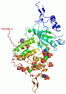
 Description
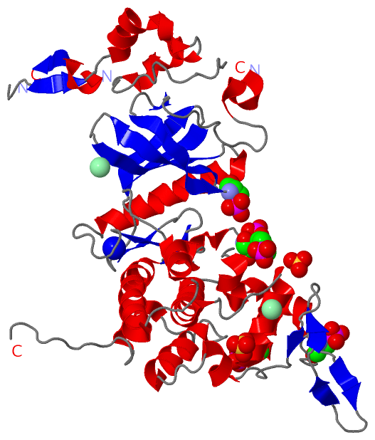
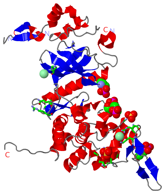