|
|
|
|
 Description
|
|
Compounds
|
||||||||||||||||||||||||||||||||||||||||||||||||||||||||
Chains, Units
Summary Information (see also Sequences/Alignments below) |
Ligands, Modified Residues, Ions (3, 5)
Asymmetric Unit (3, 5)
|
Sites (5, 5)
Asymmetric Unit (5, 5)
|
SS Bonds (6, 6)
Asymmetric Unit
|
||||||||||||||||||||||||||||
Cis Peptide Bonds (2, 2)
Asymmetric Unit
|
||||||||||||
SAPs(SNPs)/Variants (0, 0)| (no "SAP(SNP)/Variant" information available for 3K1E) |
PROSITE Motifs (0, 0)| (no "PROSITE Motif" information available for 3K1E) |
Exons (0, 0)| (no "Exon" information available for 3K1E) |
Sequences/Alignments
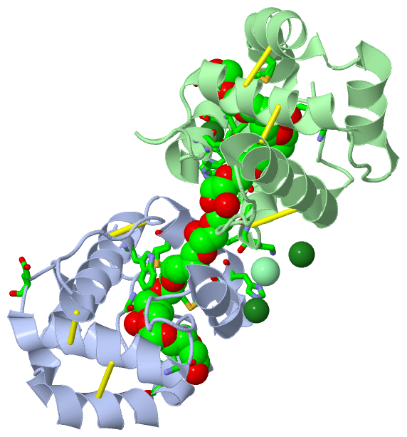
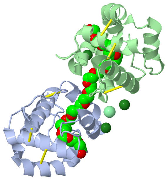
Asymmetric UnitChain A from PDB Type:PROTEIN Length:124 aligned with Q6Y2R8_AEDAE | Q6Y2R8 from UniProtKB/TrEMBL Length:143 Alignment length:124 29 39 49 59 69 79 89 99 109 119 129 139 Q6Y2R8_AEDAE 20 VTPRRDAEYPPPEFLEAMKPLREICIKKTGVTEEAIIEFSDGKVHEDENLKCYMNCLFHEAKVVDDTGHVHLEKLHDALPDSMHDIALHMGKRCLYPEGENLCEKAFWLHKCWKESDPKHYFLI 143 SCOP domains d3k1ea_ A: automated matches SCOP domains CATH domains ---------------------------------------------------------------------------------------------------------------------------- CATH domains Pfam domains ---------------------------------------------------------------------------------------------------------------------------- Pfam domains SAPs(SNPs) ---------------------------------------------------------------------------------------------------------------------------- SAPs(SNPs) PROSITE ---------------------------------------------------------------------------------------------------------------------------- PROSITE Transcript ---------------------------------------------------------------------------------------------------------------------------- Transcript 3k1e A 2 VTPRRDAEYPPPEFLEAMKPLREICIKKTGVTEEAIIEFSDGKVHEDENLKCYMNCLFHEAKVVDDTGHVHLEKLHDALPDSMHDIALHMGKRCLYPEGENLCEKAFWLHKCWKESDPKHYFLI 125 11 21 31 41 51 61 71 81 91 101 111 121 Chain B from PDB Type:PROTEIN Length:116 aligned with Q6Y2R8_AEDAE | Q6Y2R8 from UniProtKB/TrEMBL Length:143 Alignment length:116 37 47 57 67 77 87 97 107 117 127 137 Q6Y2R8_AEDAE 28 YPPPEFLEAMKPLREICIKKTGVTEEAIIEFSDGKVHEDENLKCYMNCLFHEAKVVDDTGHVHLEKLHDALPDSMHDIALHMGKRCLYPEGENLCEKAFWLHKCWKESDPKHYFLI 143 SCOP domains d3k1eb_ B: automated matches SCOP domains CATH domains -------------------------------------------------------------------------------------------------------------------- CATH domains Pfam domains (1) PBP_GOBP-3k1eB01 B:10-119 ------ Pfam domains (1) Pfam domains (2) PBP_GOBP-3k1eB02 B:10-119 ------ Pfam domains (2) SAPs(SNPs) -------------------------------------------------------------------------------------------------------------------- SAPs(SNPs) PROSITE -------------------------------------------------------------------------------------------------------------------- PROSITE Transcript -------------------------------------------------------------------------------------------------------------------- Transcript 3k1e B 10 YPPPEFLEAMKPLREICIKKTGVTEEAIIEFSDGKVHEDENLKCYMNCLFHEAKVVDDTGHVHLEKLHDALPDSMHDIALHMGKRCLYPEGENLCEKAFWLHKCWKESDPKHYFLI 125 19 29 39 49 59 69 79 89 99 109 119
|
||||||||||||||||||||
SCOP Domains (1, 2)
Asymmetric Unit
 |
CATH Domains (0, 0)| (no "CATH Domain" information available for 3K1E) |
Pfam Domains (1, 2)
Asymmetric Unit
|
Gene Ontology (2, 2)|
Asymmetric Unit(hide GO term definitions) Chain A,B (Q6Y2R8_AEDAE | Q6Y2R8)
|
||||||||||||||||||
Interactive Views
|
|||||||||||||||||||||||||||||||||||||||||||||||||||||||||||||||||||||||||||||||||||||||||||||||||||||||||||||||||||||||||||||||||||||||||||||||||||||||||||||||||||||||||||||||||||||||||||||||
Still Images
|
||||||||||||||||
Databases
|
||||||||||||||||||||||||||||||||||||||||||||||||||||||||||||||||||||||||||||||||||||||||||||||||||||||||||||||||||||||||||||||||||||||||||||||||||||||||||||||||
Analysis Tools
|
|||||||||||||||||||||||||||||||||||||||||||||||||||||||||||||
Entries Sharing at Least One Protein Chain (UniProt ID)
Related Entries Specified in the PDB File
|
|