Show PDB file:   
         Plain Text   HTML   (compressed file size)
QuickSearch:   
by PDB,NDB,UniProt,PROSITE Code or Search Term(s)  
(-)Asymmetric Unit
(-)Asym. Unit - sites
(-)Biological Unit 1
(-)Biol. Unit 1 - sites
(-)Biological Unit 2
(-)Biological Unit 3
collapse expand < >
Image Asymmetric Unit
Asymmetric Unit  (Jmol Viewer)
Image Asym. Unit - sites
Asym. Unit - sites  (Jmol Viewer)
Image Biological Unit 1
Biological Unit 1  (Jmol Viewer)
Image Biol. Unit 1 - sites
Biol. Unit 1 - sites  (Jmol Viewer)
Image Biological Unit 2
Biological Unit 2  (Jmol Viewer)
Image Biological Unit 3
Biological Unit 3  (Jmol Viewer)

(-) Description

Title :  CRYSTAL STRUCTURE OF XG34, AN EVOLVED XYLOGLUCAN BINDING CBM
 
Authors :  C. Divne, T. -C. Tan, H. Brumer, F. Gullfot
Date :  21 Sep 09  (Deposition) - 27 Oct 09  (Release) - 13 Jul 11  (Revision)
Method :  X-RAY DIFFRACTION
Resolution :  1.60
Chains :  Asym. Unit :  A,B,C
Biol. Unit 1:  A  (1x)
Biol. Unit 2:  B  (1x)
Biol. Unit 3:  C  (1x)
Keywords :  Cbm, Xyloglucan Binding, Calcium Binding, Xylan Degradation, Hydrolase, Carbohydrate-Binding Domain (Keyword Search: [Gene Ontology, PubMed, Web (Google))
 
Reference :  F. Gullfot, T. C. Tan, L. Von Schantz, E. N. Karlsson, M. Ohlin, H. Brumer, C. Divne
The Crystal Structure Of Xg-34, An Evolved Xyloglucan-Specific Carbohydrate-Binding Module.
Proteins V. 78 785 2009
PubMed-ID: 19950365  |  Reference-DOI: 10.1002/PROT.22642

(-) Compounds

Molecule 1 - XYLANASE
    ChainsA, B, C
    EngineeredYES
    Expression SystemESCHERICHIA COLI
    Expression System PlasmidPET-22B(+)
    Expression System StrainBL21(DE3)
    Expression System Taxid562
    Expression System Vector TypePLASMID
    GeneXG-34
    MutationYES
    Organism CommonRHODOTHERMUS OBAMENSIS
    Organism ScientificRHODOTHERMUS MARINUS
    Organism Taxid29549

 Structural Features

(-) Chains, Units

  123
Asymmetric Unit ABC
Biological Unit 1 (1x)A  
Biological Unit 2 (1x) B 
Biological Unit 3 (1x)  C

Summary Information (see also Sequences/Alignments below)
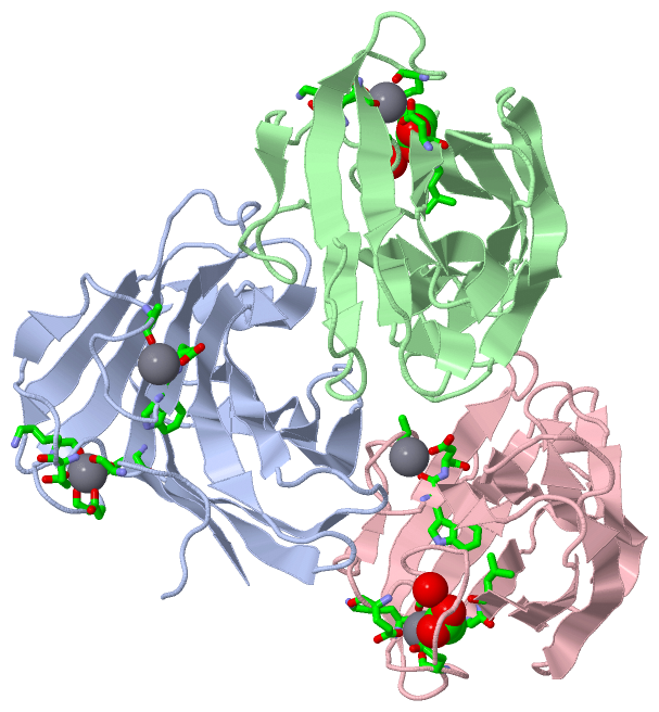
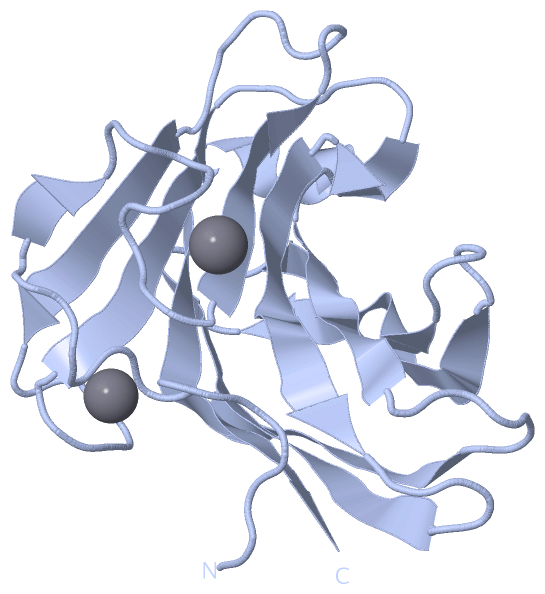
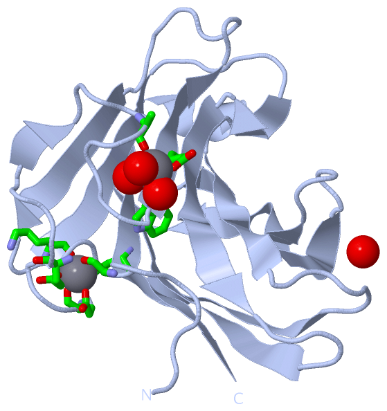
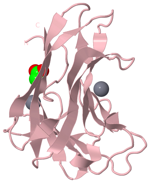
(-) Ligands, Modified Residues, Ions  (2, 7)

Asymmetric Unit (2, 7)
No.NameCountTypeFull Name
1ACT2Ligand/IonACETATE ION
2CA5Ligand/IonCALCIUM ION
Biological Unit 1 (0, 0)
No.NameCountTypeFull Name
1ACT-1Ligand/IonACETATE ION
2CA-1Ligand/IonCALCIUM ION
Biological Unit 2 (1, 1)
No.NameCountTypeFull Name
1ACT1Ligand/IonACETATE ION
2CA-1Ligand/IonCALCIUM ION
Biological Unit 3 (1, 1)
No.NameCountTypeFull Name
1ACT1Ligand/IonACETATE ION
2CA-1Ligand/IonCALCIUM ION

(-) Sites  (7, 7)

Asymmetric Unit (7, 7)
No.NameEvidenceResiduesDescription
1AC1SOFTWAREGLY A:9 , GLU A:11 , GLU A:52 , LYS A:55 , ASP A:160BINDING SITE FOR RESIDUE CA A 301
2AC2SOFTWAREALA A:22 , TRP A:28 , ASP A:29 , HOH A:1093 , HOH A:1140 , HOH A:1228BINDING SITE FOR RESIDUE CA A 302
3AC3SOFTWAREGLY B:9 , GLU B:11 , GLU B:52 , LYS B:55 , ASP B:160BINDING SITE FOR RESIDUE CA B 301
4AC4SOFTWARELEU B:162 , HOH B:1040BINDING SITE FOR RESIDUE ACT B 401
5AC5SOFTWAREGLY C:9 , GLU C:11 , GLU C:52 , LYS C:55 , ASP C:160BINDING SITE FOR RESIDUE CA C 301
6AC6SOFTWAREHOH A:1125 , HOH B:1019 , ALA C:22 , TRP C:28 , ASP C:29 , HOH C:1029 , HOH C:1039BINDING SITE FOR RESIDUE CA C 302
7AC7SOFTWAREGLY C:9 , GLY C:161 , LEU C:162 , HOH C:1044BINDING SITE FOR RESIDUE ACT C 401

(-) SS Bonds  (0, 0)

(no "SS Bond" information available for 3JXS)

(-) Cis Peptide Bonds  (3, 3)

Asymmetric Unit
No.Residues
1Leu A:76 -Pro A:77
2Leu B:76 -Pro B:77
3Leu C:76 -Pro C:77

 Sequence-Structure Mapping

(-) SAPs(SNPs)/Variants  (0, 0)

(no "SAP(SNP)/Variant" information available for 3JXS)

(-) PROSITE Motifs  (0, 0)

(no "PROSITE Motif" information available for 3JXS)

(-) Exons   (0, 0)

(no "Exon" information available for 3JXS)

(-) Sequences/Alignments

Asymmetric Unit
   Reformat: Number of residues per line =  ('0' or empty: single-line sequence representation)
  Number of residues per labelling interval =   
  UniProt sequence: complete  aligned part    
   Show mapping: SCOP domains CATH domains Pfam domains Secondary structure (by author)
SAPs(SNPs) PROSITE motifs Exons
(details for a mapped element are shown in a popup box when the mouse pointer rests over it)
Chain A from PDB  Type:PROTEIN  Length:166
 aligned with Q7WTN6_RHOMR | Q7WTN6 from UniProtKB/TrEMBL  Length:997

    Alignment length:166
                                   220       230       240       250       260       270       280       290       300       310       320       330       340       350       360       370      
         Q7WTN6_RHOMR   211 ELVANINGGFESTPAGVVTDLAEGVEGWDLNVGSSVTNPPVFEVLETSDAPEGNKVLAVTVNGVGNNPWDIEATAFPVNVRPGVTYTYTIWARAEQDGAVVSFTVGNQSFQEYGRLHEQQITTEWQPFTFEFTVSDQETVIRAPIHFGYAANVGNTIYIDGLAIVD 376
               SCOP domains d3jxsa_ A: automated matches                                                                                                                                           SCOP domains
               CATH domains 3jxsA00 A:1-166 Galactose-binding domain-like                                                                                                                          CATH domains
               Pfam domains ---------------------------------------------------------------------------------------------------------------------------------------------------------------------- Pfam domains
         Sec.struct. author ...............ee..........eeeee........eeeeee........eeeeeee.....hhh.eeeeeeeee.....eeeeeeeeee....eeeeeeee.....eeeeeeeeee....eeeeeeee......eeeeeee..hhhhh.eeeeeeeeeee. Sec.struct. author
                 SAPs(SNPs) ---------------------------------------------------------------------------------------------------------------------------------------------------------------------- SAPs(SNPs)
                    PROSITE ---------------------------------------------------------------------------------------------------------------------------------------------------------------------- PROSITE
                 Transcript ---------------------------------------------------------------------------------------------------------------------------------------------------------------------- Transcript
                 3jxs A   1 MLVANINGGFESTPAGVVTDLAEGVEGWDLNVGSSVTNPPVFEVLETSDAPEGNKVLAVTVNGVGNNPYDIHATALPVNVRPGVTYTYTIWARAEQDGAVVSFTVGNQSHDDYGRLHEQQITTEWQPFTFEFTVSDQETVIRAPIHFGFAANVGNTIYIDGLAIVD 166
                                    10        20        30        40        50        60        70        80        90       100       110       120       130       140       150       160      

Chain B from PDB  Type:PROTEIN  Length:166
 aligned with Q7WTN6_RHOMR | Q7WTN6 from UniProtKB/TrEMBL  Length:997

    Alignment length:166
                                   220       230       240       250       260       270       280       290       300       310       320       330       340       350       360       370      
         Q7WTN6_RHOMR   211 ELVANINGGFESTPAGVVTDLAEGVEGWDLNVGSSVTNPPVFEVLETSDAPEGNKVLAVTVNGVGNNPWDIEATAFPVNVRPGVTYTYTIWARAEQDGAVVSFTVGNQSFQEYGRLHEQQITTEWQPFTFEFTVSDQETVIRAPIHFGYAANVGNTIYIDGLAIVD 376
               SCOP domains d3jxsb_ B: automated matches                                                                                                                                           SCOP domains
               CATH domains 3jxsB00 B:1-166 Galactose-binding domain-like                                                                                                                          CATH domains
               Pfam domains ---------------------------------------------------------------------------------------------------------------------------------------------------------------------- Pfam domains
         Sec.struct. author ...............ee..........eeeee........eeeeee........eeeeeee.....hhh.eeeeeeeee.....eeeeeeeeee....eeeeeeee.....eeeeeeeeee....eeeeeeee......eeeeeee..hhhhh.eeeeeeeeeee. Sec.struct. author
                 SAPs(SNPs) ---------------------------------------------------------------------------------------------------------------------------------------------------------------------- SAPs(SNPs)
                    PROSITE ---------------------------------------------------------------------------------------------------------------------------------------------------------------------- PROSITE
                 Transcript ---------------------------------------------------------------------------------------------------------------------------------------------------------------------- Transcript
                 3jxs B   1 MLVANINGGFESTPAGVVTDLAEGVEGWDLNVGSSVTNPPVFEVLETSDAPEGNKVLAVTVNGVGNNPYDIHATALPVNVRPGVTYTYTIWARAEQDGAVVSFTVGNQSHDDYGRLHEQQITTEWQPFTFEFTVSDQETVIRAPIHFGFAANVGNTIYIDGLAIVD 166
                                    10        20        30        40        50        60        70        80        90       100       110       120       130       140       150       160      

Chain C from PDB  Type:PROTEIN  Length:166
 aligned with Q7WTN6_RHOMR | Q7WTN6 from UniProtKB/TrEMBL  Length:997

    Alignment length:166
                                   220       230       240       250       260       270       280       290       300       310       320       330       340       350       360       370      
         Q7WTN6_RHOMR   211 ELVANINGGFESTPAGVVTDLAEGVEGWDLNVGSSVTNPPVFEVLETSDAPEGNKVLAVTVNGVGNNPWDIEATAFPVNVRPGVTYTYTIWARAEQDGAVVSFTVGNQSFQEYGRLHEQQITTEWQPFTFEFTVSDQETVIRAPIHFGYAANVGNTIYIDGLAIVD 376
               SCOP domains d3jxsc_ C: automated matches                                                                                                                                           SCOP domains
               CATH domains 3jxsC00 C:1-166 Galactose-binding domain-like                                                                                                                          CATH domains
               Pfam domains ---------------------------------------------------------------------------------------------------------------------------------------------------------------------- Pfam domains
         Sec.struct. author ...............ee..........eeeee........eeeeee........eeeeee......hhh.eeeeeeeee.....eeeeeeeeee....eeeeeeee.....eeeeeeeeee....eeeeeeee......eeeeeee..hhhhh..eeeeeeeeee. Sec.struct. author
                 SAPs(SNPs) ---------------------------------------------------------------------------------------------------------------------------------------------------------------------- SAPs(SNPs)
                    PROSITE ---------------------------------------------------------------------------------------------------------------------------------------------------------------------- PROSITE
                 Transcript ---------------------------------------------------------------------------------------------------------------------------------------------------------------------- Transcript
                 3jxs C   1 MLVANINGGFESTPAGVVTDLAEGVEGWDLNVGSSVTNPPVFEVLETSDAPEGNKVLAVTVNGVGNNPYDIHATALPVNVRPGVTYTYTIWARAEQDGAVVSFTVGNQSHDDYGRLHEQQITTEWQPFTFEFTVSDQETVIRAPIHFGFAANVGNTIYIDGLAIVD 166
                                    10        20        30        40        50        60        70        80        90       100       110       120       130       140       150       160      

   Legend:   → Mismatch (orange background)
  - → Gap (green background, '-', border residues have a numbering label)
    → Modified Residue (blue background, lower-case, 'x' indicates undefined single-letter code, labelled with number + name)
  x → Chemical Group (purple background, 'x', labelled with number + name, e.g. ACE or NH2)
  extra numbering lines below/above indicate numbering irregularities and modified residue names etc., number ends below/above '|'

 Classification and Annotation

(-) SCOP Domains  (1, 3)

Asymmetric Unit

(-) CATH Domains  (1, 3)

Asymmetric Unit
(-)
Class: Mainly Beta (13760)

(-) Pfam Domains  (0, 0)

(no "Pfam Domain" information available for 3JXS)

(-) Gene Ontology  (7, 7)

Asymmetric Unit(hide GO term definitions)
Chain A,B,C   (Q7WTN6_RHOMR | Q7WTN6)
molecular function
    GO:0016787    hydrolase activity    Catalysis of the hydrolysis of various bonds, e.g. C-O, C-N, C-C, phosphoric anhydride bonds, etc. Hydrolase is the systematic name for any enzyme of EC class 3.
    GO:0016798    hydrolase activity, acting on glycosyl bonds    Catalysis of the hydrolysis of any glycosyl bond.
    GO:0004553    hydrolase activity, hydrolyzing O-glycosyl compounds    Catalysis of the hydrolysis of any O-glycosyl bond.
    GO:0046872    metal ion binding    Interacting selectively and non-covalently with any metal ion.
biological process
    GO:0005975    carbohydrate metabolic process    The chemical reactions and pathways involving carbohydrates, any of a group of organic compounds based of the general formula Cx(H2O)y. Includes the formation of carbohydrate derivatives by the addition of a carbohydrate residue to another molecule.
    GO:0008152    metabolic process    The chemical reactions and pathways, including anabolism and catabolism, by which living organisms transform chemical substances. Metabolic processes typically transform small molecules, but also include macromolecular processes such as DNA repair and replication, and protein synthesis and degradation.
    GO:0045493    xylan catabolic process    The chemical reactions and pathways resulting in the breakdown of xylan, a polymer containing a beta-1,4-linked D-xylose backbone.

 Visualization

(-) Interactive Views

Asymmetric Unit
  Complete Structure
    Jena3D(integrated viewing of ligand, site, SAP, PROSITE, SCOP information)
    WebMol | AstexViewer[tm]@PDBe
(Java Applets, require no local installation except for Java; loading may be slow)
    STRAP
(Java WebStart application, automatic local installation, requires Java; full application with system access!)
    RasMol
(require local installation)
    Molscript (VRML)
(requires installation of a VRML viewer; select preferred view via VRML and generate a mono or stereo PDF format file)
 
  Ligands, Modified Residues, Ions
    ACT  [ RasMol | Jena3D ]  +environment [ RasMol | Jena3D ]
    CA  [ RasMol | Jena3D ]  +environment [ RasMol | Jena3D ]
 
  Sites
    AC1  [ RasMol ]  +environment [ RasMol ]
    AC2  [ RasMol ]  +environment [ RasMol ]
    AC3  [ RasMol ]  +environment [ RasMol ]
    AC4  [ RasMol ]  +environment [ RasMol ]
    AC5  [ RasMol ]  +environment [ RasMol ]
    AC6  [ RasMol ]  +environment [ RasMol ]
    AC7  [ RasMol ]  +environment [ RasMol ]
 
  Cis Peptide Bonds
    Leu A:76 - Pro A:77   [ RasMol ]  
    Leu B:76 - Pro B:77   [ RasMol ]  
    Leu C:76 - Pro C:77   [ RasMol ]  
 
Biological Units
  Complete Structure
    Biological Unit 1  [ Jena3D ]
    Biological Unit 2  [ Jena3D ]
    Biological Unit 3  [ Jena3D ]

(-) Still Images

Jmol
  protein: cartoon or spacefill or dots and stick; nucleic acid: cartoon and stick; ligands: spacefill; active site: stick
Molscript
  protein, nucleic acid: cartoon; ligands: spacefill; active site: ball and stick

 Databases and Analysis Tools

(-) Databases

Access by PDB/NDB ID
  3jxs
    Family and Domain InformationProDom | SYSTERS
    General Structural InformationGlycoscienceDB | MMDB | NDB | OCA | PDB | PDBe | PDBj | PDBsum | PDBWiki | PQS | PROTEOPEDIA
    Orientation in MembranesOPM
    Protein SurfaceSURFACE
    Secondary StructureDSSP (structure derived) | HSSP (homology derived)
    Structural GenomicsGeneCensus
    Structural NeighboursCE | VAST
    Structure ClassificationCATH | Dali | SCOP
    Validation and Original DataBMRB Data View | BMRB Restraints Grid | EDS | PROCHECK | RECOORD | WHAT_CHECK
 
Access by UniProt ID/Accession number
  Q7WTN6_RHOMR | Q7WTN6
    Comparative Protein Structure ModelsModBase
    Genomic InformationEnsembl
    Protein-protein InteractionDIP
    Sequence, Family and Domain InformationInterPro | Pfam | SMART | UniProtKB/TrEMBL
 
Access by Enzyme Classificator   (EC Number)
  (no 'Enzyme Classificator' available)
    General Enzyme InformationBRENDA | EC-PDB | Enzyme | IntEnz
    PathwayKEGG | MetaCyc
 
Access by Disease Identifier   (MIM ID)
  (no 'MIM ID' available)
    Disease InformationOMIM
 
Access by GenAge ID
  (no 'GenAge ID' available)
    Age Related InformationGenAge

(-) Analysis Tools

Access by PDB/NDB ID
    Domain InformationXDom
    Interatomic Contacts of Structural UnitsCSU
    Ligand-protein ContactsLPC
    Protein CavitiescastP
    Sequence and Secondary StructurePDBCartoon
    Structure AlignmentSTRAP(Java WebStart application, automatic local installation, requires Java; full application with system access!)
    Structure and Sequence BrowserSTING
 
Access by UniProt ID/Accession number
  Q7WTN6_RHOMR | Q7WTN6
    Protein Disorder PredictionDisEMBL | FoldIndex | GLOBPLOT (for more information see DisProt)

 Related Entries

(-) Entries Sharing at Least One Protein Chain (UniProt ID)

UniProtKB/TrEMBL
        Q7WTN6_RHOMR | Q7WTN62y64 2y6g 2y6h 2y6j 2y6k 2y6l 5dpn

(-) Related Entries Specified in the PDB File

1k45 SOLUTION STRUCTURE OF CBM4-2 WHICH IS THE PARENTAL MOLECULE OF XG34