|
|
|
|
 Description
|
|
Compounds
|
||||||||||||||||||||||||||||||||||||||||||||||||||||||||
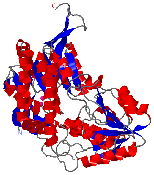
Chains, Units
Summary Information (see also Sequences/Alignments below) |
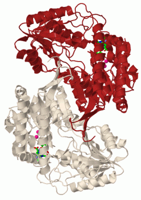
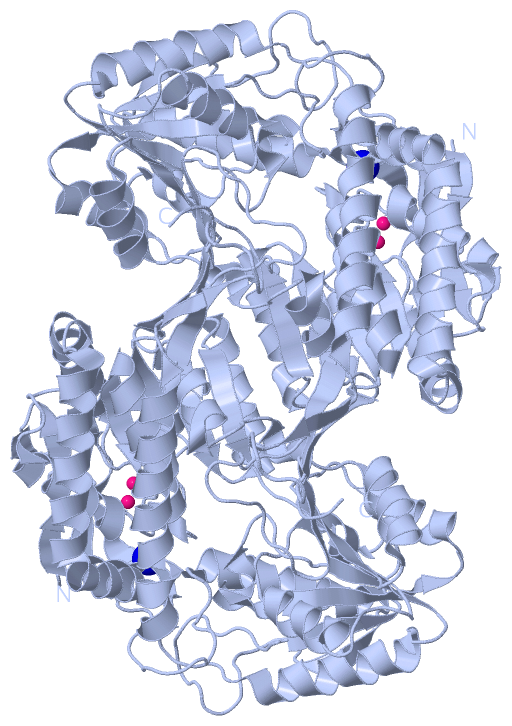
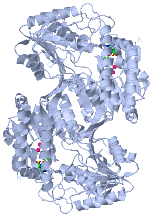
Ligands, Modified Residues, Ions (2, 3)| Asymmetric Unit (2, 3) Biological Unit 1 (1, 4) |
Sites (1, 1)
Asymmetric Unit (1, 1)
|
SS Bonds (0, 0)| (no "SS Bond" information available for 3I44) |
Cis Peptide Bonds (0, 0)| (no "Cis Peptide Bond" information available for 3I44) |
SAPs(SNPs)/Variants (0, 0)| (no "SAP(SNP)/Variant" information available for 3I44) |
PROSITE Motifs (0, 0)| (no "PROSITE Motif" information available for 3I44) |
Exons (0, 0)| (no "Exon" information available for 3I44) |
Sequences/Alignments
Asymmetric UnitChain A from PDB Type:PROTEIN Length:476 aligned with A0A0H3M4W2_B | A0A0H3M4W2 from UniProtKB/TrEMBL Length:476 Alignment length:476 10 20 30 40 50 60 70 80 90 100 110 120 130 140 150 160 170 180 190 200 210 220 230 240 250 260 270 280 290 300 310 320 330 340 350 360 370 380 390 400 410 420 430 440 450 460 470 A0A0H3M4W2_B 1 MLNKRKFYINGLWDDPSTPHDLYVIDPSTEEACAVISLGSTRDADKAINAAKKAFQTWKTTSPHERLGFVEKILEIYEKRSSDMAKTISMEMGAPIDMALNAQTATGSSHIRNFIKAYKEFSFQEALIEGNEQAILHYDAIGVVGLITPWNWPMNQVTLKVIPALLAGCTMVLKPSEIAPLSAMLFAEILDEAALPSGVFNLINGDGANVGSYLSAHPDLGMISFTGSTRAGKDISKNASNTLKRVCLELGGKGANIIFADADIDALQRGVRHCFYNSGQSCNAPTRMLVEQAIYDKAIKTAKDIAEKTQVGPGHQTGNHIGPVVSKEQYDKIQDLIQSGIDEGATLVTGGTGLPMGMERGYYVRPTVFADVKPHMRIFREEIFGPVLSLLPFNTEDEAVTLANDTEYGLTNYIQSQDRSKCRRIAAQVRSGMVEVNGHELPGGSYFGGVKFSGRAREGGLWGIKEFLDTKAISYW 476 SCOP domains d3i44a_ A: automated matches SCOP domains CATH domains -----------------------------------------------------------------------------------------------------------------------------------------------------------------------------------------------------------------------------------------------------------3i44A02 A:252-442 Aldehyde Dehydrogenase; Chain A, domain 2 ---------------------------------- CATH domains Pfam domains -------------------------------------------------------------------------------------------------------------------------------------------------------------------------------------------------------------------------------------------------------------------------------------------------------------------------------------------------------------------------------------------------------------------------------------------------------------------------------------------- Pfam domains SAPs(SNPs) -------------------------------------------------------------------------------------------------------------------------------------------------------------------------------------------------------------------------------------------------------------------------------------------------------------------------------------------------------------------------------------------------------------------------------------------------------------------------------------------- SAPs(SNPs) PROSITE -------------------------------------------------------------------------------------------------------------------------------------------------------------------------------------------------------------------------------------------------------------------------------------------------------------------------------------------------------------------------------------------------------------------------------------------------------------------------------------------- PROSITE Transcript -------------------------------------------------------------------------------------------------------------------------------------------------------------------------------------------------------------------------------------------------------------------------------------------------------------------------------------------------------------------------------------------------------------------------------------------------------------------------------------------- Transcript 3i44 A 1 MLNKRKFYINGLWDDPSTPHDLYVIDPSTEEACAVISLGSTRDADKAINAAKKAFQTWKTTSPHERLGFVEKILEIYEKRSSDMAKTISMEMGAPIDMALNAQTATGSSHIRNFIKAYKEFSFQEALIEGNEQAILHYDAIGVVGLITPWNWPMNQVTLKVIPALLAGCTMVLKPSEIAPLSAMLFAEILDEAALPSGVFNLINGDGANVGSYLSAHPDLEMISFTGSTRAGKDISKNASNTLKRVCLELGGKGANIIFADADIDALQRGVRHCFYNSGQSCNAPTRMLVEQAIYDKAIKTAKDIAEKTQVGPGHQTGNHIGPVVSKEQYDKIQDLIQSGIDEGATLVTGGTGLPMGMERGYYVRPTVFADVKPHMRIFREEIFGPVLSLLPFNTEDEAVTLANDTEYGLTNYIQSQDRSKCRRIAAQVRSGMVEVNGHELPGGSYFGGVKFSGRAREGGLWGIKEFLDTKAISYW 476 10 20 30 40 50 60 70 80 90 100 110 120 130 140 150 160 170 180 190 200 210 220 230 240 250 260 270 280 290 300 310 320 330 340 350 360 370 380 390 400 410 420 430 440 450 460 470
|
||||||||||||||||||||
SCOP Domains (1, 1)
Asymmetric Unit
 |
CATH Domains (1, 1)
Asymmetric Unit
|
Pfam Domains (0, 0)| (no "Pfam Domain" information available for 3I44) |
Gene Ontology (0, 0)|
Asymmetric Unit(hide GO term definitions)
(no "Gene Ontology" information available for 3I44)
|
Interactive Views
|
|||||||||||||||||||||||||||||||||||||||||||||||||||||||||||||||||||||||||||||||||||||||||||||||||||||||||||||||||||||||||||||||||||||||||||||||
Still Images
|
||||||||||||||||
Databases
|
||||||||||||||||||||||||||||||||||||||||||||||||||||||||||||||||||||||||||||||||||||||||||||||||||||||||||||||||||||||||||||||||||||||||||||||||||||||||||||||||
Analysis Tools
|
|||||||||||||||||||||||||||||||||||||||||||||||||||||||||||||
Entries Sharing at Least One Protein Chain (UniProt ID)
Related Entries Specified in the PDB File
|
|