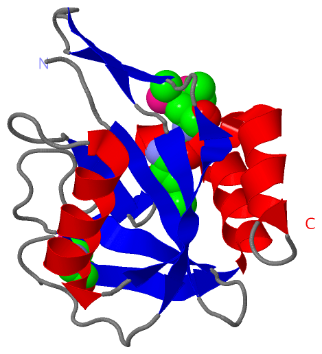
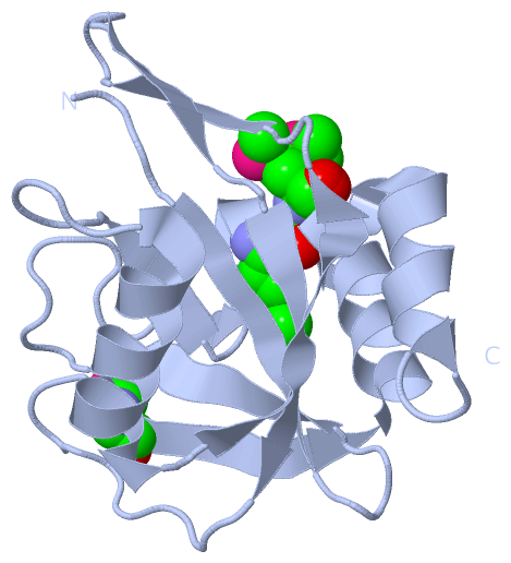
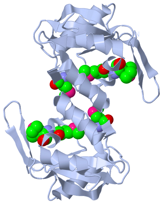
Show PDB file:   
         Plain Text   HTML   (compressed file size)
QuickSearch:   
by PDB,NDB,UniProt,PROSITE Code or Search Term(s)  
(-)Asymmetric Unit
(-)Biological Unit 1
(-)Biological Unit 2
collapse expand < >
Image Asymmetric Unit
Asymmetric Unit  (Jmol Viewer)
Image Biological Unit 1
Biological Unit 1  (Jmol Viewer)
Image Biological Unit 2
Biological Unit 2  (Jmol Viewer)

(-) Description

Title :  CRYSTAL STRUCTURE OF ISOPENTENYL-DIPHOSPHATE DELTA-ISOMERASE FROM SALMONELLA ENTERICASE
 
Authors :  Y. Kim, M. Zhou, S. Peterson, W. F. Anderson, A. Joachimiak
Date :  22 Jun 09  (Deposition) - 30 Jun 09  (Release) - 13 Jul 11  (Revision)
Method :  X-RAY DIFFRACTION
Resolution :  1.52
Chains :  Asym. Unit :  A
Biol. Unit 1:  A  (1x)
Biol. Unit 2:  A  (2x)
Keywords :  Alpha-Beta Structure, Isomerase, Isoprene Biosynthesis, Magnesium, Manganese, Metal-Binding, Structural Genomics, Center For Structural Genomics Of Infectious Diseases, Csgid (Keyword Search: [Gene Ontology, PubMed, Web (Google))
 
Reference :  Y. Kim, M. Zhou, S. Peterson, W. F. Anderson, A. Joachimiak
Crystal Structure Of Isopentenyl-Diphosphate Delta-Isomeras From Salmonella Entericase
To Be Published
PubMed: search

(-) Compounds

Molecule 1 - ISOPENTENYL-DIPHOSPHATE DELTA-ISOMERASE
    ChainsA
    EC Number5.3.3.2
    EngineeredYES
    Expression SystemESCHERICHIA COLI
    Expression System PlasmidPMCSG7
    Expression System StrainBL21 MAGIC
    Expression System Taxid562
    Expression System Vector TypePLASMID
    GeneIDI
    IppDMAPP ISOMERASE
    Organism ScientificSALMONELLA ENTERICA SUBSP. ENTERICA SEROVAR TYPHIMURIUM
    Organism Taxid99287
    StrainLT2
    SynonymIPP ISOMERASE, ISOPENTENYL PYROPHOSPHATE ISOMERASE,

 Structural Features

(-) Chains, Units

  1
Asymmetric Unit A
Biological Unit 1 (1x)A
Biological Unit 2 (2x)A

Summary Information (see also Sequences/Alignments below)

(-) Ligands, Modified Residues, Ions  (1, 3)

Asymmetric Unit (1, 3)
No.NameCountTypeFull Name
1MSE3Mod. Amino AcidSELENOMETHIONINE
Biological Unit 1 (1, 3)
No.NameCountTypeFull Name
1MSE3Mod. Amino AcidSELENOMETHIONINE
Biological Unit 2 (1, 6)
No.NameCountTypeFull Name
1MSE6Mod. Amino AcidSELENOMETHIONINE

(-) Sites  (0, 0)

(no "Site" information available for 3HYQ)

(-) SS Bonds  (0, 0)

(no "SS Bond" information available for 3HYQ)

(-) Cis Peptide Bonds  (0, 0)

(no "Cis Peptide Bond" information available for 3HYQ)

 Sequence-Structure Mapping

(-) SAPs(SNPs)/Variants  (0, 0)

(no "SAP(SNP)/Variant" information available for 3HYQ)

(-) PROSITE Motifs  (0, 0)

(no "PROSITE Motif" information available for 3HYQ)

(-) Exons   (0, 0)

(no "Exon" information available for 3HYQ)

(-) Sequences/Alignments

Asymmetric Unit
   Reformat: Number of residues per line =  ('0' or empty: single-line sequence representation)
  Number of residues per labelling interval =   
  UniProt sequence: complete  aligned part    
   Show mapping: SCOP domains CATH domains Pfam domains Secondary structure (by author)
SAPs(SNPs) PROSITE motifs Exons
(details for a mapped element are shown in a popup box when the mouse pointer rests over it)
Chain A from PDB  Type:PROTEIN  Length:151
 aligned with IDI_SALTY | Q8ZM82 from UniProtKB/Swiss-Prot  Length:181

    Alignment length:151
                                    40        50        60        70        80        90       100       110       120       130       140       150       160       170       180 
            IDI_SALTY    31 LHLAFSCWLFNEDGQLLVTRRSLSKKAWPGVWTNSVCGHPQQGETTEEAIIRRCRFELGVEITDLTPVYPHFSYRATDPNGIVENEVCPVFAARATSVLQVNSEEVMDYQWSEFKSVWKSLLATPWAFSPWMVMQASDEQARERLLNYCQR 181
               SCOP domains d3hyqa_ A: Isopentenyl diphosphate isomerase                                                                                                            SCOP domains
               CATH domains 3hyqA00 A:31-181 Nucleoside Triphosphate Pyrophosphohydrolase                                                                                           CATH domains
               Pfam domains ------------------------------------------------------------------------------------------------------------------------------------------------------- Pfam domains
         Sec.struct. author ....eeeeee.....eeeeee..........ee..eee......hhhhhhhhhhhhhhh..eeeeeeee...eeeee.....eeeee..eeeeee..........eeeeeeehhhhhhhhhhhhhhhhhhhhhhhhhhhhhhhhhhh.... Sec.struct. author
                 SAPs(SNPs) ------------------------------------------------------------------------------------------------------------------------------------------------------- SAPs(SNPs)
                    PROSITE ------------------------------------------------------------------------------------------------------------------------------------------------------- PROSITE
                 Transcript ------------------------------------------------------------------------------------------------------------------------------------------------------- Transcript
                 3hyq A  31 LHLAFSCWLFNEDGQLLVTRRSLSKKAWPGVWTNSVCGHPQQGETTEEAIIRRCRFELGVEITDLTPVYPHFSYRATDPNGIVENEVCPVFAARATSVLQVNSEEVmDYQWSEFKSVWKSLLATPWAFSPWmVmQASDEQARERLLNYCQR 181
                                    40        50        60        70        80        90       100       110       120       130      |140       150       160 | |   170       180 
                                                                                                                                    137-MSE                  162-MSE               
                                                                                                                                                               164-MSE             

   Legend:   → Mismatch (orange background)
  - → Gap (green background, '-', border residues have a numbering label)
    → Modified Residue (blue background, lower-case, 'x' indicates undefined single-letter code, labelled with number + name)
  x → Chemical Group (purple background, 'x', labelled with number + name, e.g. ACE or NH2)
  extra numbering lines below/above indicate numbering irregularities and modified residue names etc., number ends below/above '|'

 Classification and Annotation

(-) SCOP Domains  (1, 1)

Asymmetric Unit

(-) CATH Domains  (1, 1)

Asymmetric Unit
(-)
Class: Alpha Beta (26913)

(-) Pfam Domains  (0, 0)

(no "Pfam Domain" information available for 3HYQ)

(-) Gene Ontology  (7, 7)

Asymmetric Unit(hide GO term definitions)
Chain A   (IDI_SALTY | Q8ZM82)
molecular function
    GO:0016787    hydrolase activity    Catalysis of the hydrolysis of various bonds, e.g. C-O, C-N, C-C, phosphoric anhydride bonds, etc. Hydrolase is the systematic name for any enzyme of EC class 3.
    GO:0016853    isomerase activity    Catalysis of the geometric or structural changes within one molecule. Isomerase is the systematic name for any enzyme of EC class 5.
    GO:0004452    isopentenyl-diphosphate delta-isomerase activity    Catalysis of the reaction: isopentenyl diphosphate = dimethylallyl diphosphate.
    GO:0046872    metal ion binding    Interacting selectively and non-covalently with any metal ion.
biological process
    GO:0050992    dimethylallyl diphosphate biosynthetic process    The chemical reactions and pathways resulting in the formation of dimethylallyl diphosphate.
    GO:0008299    isoprenoid biosynthetic process    The chemical reactions and pathways resulting in the formation of any isoprenoid compound, isoprene (2-methylbuta-1,3-diene) or compounds containing or derived from linked isoprene (3-methyl-2-butenylene) residues.
cellular component
    GO:0005737    cytoplasm    All of the contents of a cell excluding the plasma membrane and nucleus, but including other subcellular structures.

 Visualization

(-) Interactive Views

Asymmetric Unit
  Complete Structure
    Jena3D(integrated viewing of ligand, site, SAP, PROSITE, SCOP information)
    WebMol | AstexViewer[tm]@PDBe
(Java Applets, require no local installation except for Java; loading may be slow)
    STRAP
(Java WebStart application, automatic local installation, requires Java; full application with system access!)
    RasMol
(require local installation)
    Molscript (VRML)
(requires installation of a VRML viewer; select preferred view via VRML and generate a mono or stereo PDF format file)
 
  Ligands, Modified Residues, Ions
    MSE  [ RasMol | Jena3D ]  +environment [ RasMol | Jena3D ]
 
  Sites
(no "Sites" information available for 3hyq)
 
  Cis Peptide Bonds
(no "Cis Peptide Bonds" information available for 3hyq)
 
Biological Units
  Complete Structure
    Biological Unit 1  [ Jena3D ]
    Biological Unit 2  [ Jena3D ]

(-) Still Images

Jmol
  protein: cartoon or spacefill or dots and stick; nucleic acid: cartoon and stick; ligands: spacefill; active site: stick
Molscript
  protein, nucleic acid: cartoon; ligands: spacefill; active site: ball and stick

 Databases and Analysis Tools

(-) Databases

Access by PDB/NDB ID
  3hyq
    Family and Domain InformationProDom | SYSTERS
    General Structural InformationGlycoscienceDB | MMDB | NDB | OCA | PDB | PDBe | PDBj | PDBsum | PDBWiki | PQS | PROTEOPEDIA
    Orientation in MembranesOPM
    Protein SurfaceSURFACE
    Secondary StructureDSSP (structure derived) | HSSP (homology derived)
    Structural GenomicsGeneCensus
    Structural NeighboursCE | VAST
    Structure ClassificationCATH | Dali | SCOP
    Validation and Original DataBMRB Data View | BMRB Restraints Grid | EDS | PROCHECK | RECOORD | WHAT_CHECK
 
Access by UniProt ID/Accession number
  IDI_SALTY | Q8ZM82
    Comparative Protein Structure ModelsModBase
    Genomic InformationEnsembl
    Protein-protein InteractionDIP
    Sequence, Family and Domain InformationInterPro | Pfam | SMART | UniProtKB/SwissProt
 
Access by Enzyme Classificator   (EC Number)
  5.3.3.2
    General Enzyme InformationBRENDA | EC-PDB | Enzyme | IntEnz
    PathwayKEGG | MetaCyc
 
Access by Disease Identifier   (MIM ID)
  (no 'MIM ID' available)
    Disease InformationOMIM
 
Access by GenAge ID
  (no 'GenAge ID' available)
    Age Related InformationGenAge

(-) Analysis Tools

Access by PDB/NDB ID
    Domain InformationXDom
    Interatomic Contacts of Structural UnitsCSU
    Ligand-protein ContactsLPC
    Protein CavitiescastP
    Sequence and Secondary StructurePDBCartoon
    Structure AlignmentSTRAP(Java WebStart application, automatic local installation, requires Java; full application with system access!)
    Structure and Sequence BrowserSTING
 
Access by UniProt ID/Accession number
  IDI_SALTY | Q8ZM82
    Protein Disorder PredictionDisEMBL | FoldIndex | GLOBPLOT (for more information see DisProt)

 Related Entries

(-) Entries Sharing at Least One Protein Chain (UniProt ID)

(no "Entries Sharing at Least One Protein Chain" available for 3HYQ)

(-) Related Entries Specified in the PDB File

(no "Related Entries Specified in the PDB File" available for 3HYQ)